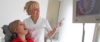
Dental articulator with face bow: what is it?
Caries, bruxism, bad heredity can cause tooth loss.
To restore a beautiful smile, dentures are installed. Each person has a different jaw structure and teeth arrangement. Therefore, at the beginning of prosthetics, the doctor receives the necessary data about the patient’s jaw system. For this, articulators are used that reproduce the movement of the lower jaw. If you additionally need to determine the position of the upper jaw, use a face bow. A dental articulator with a face bow allows the doctor to understand and reproduce the trajectory of jaw movement and make a high-quality prosthesis.
Face bows are actively used by orthopedists and orthodontists. Such devices simplify and speed up work and allow prosthetics to be performed at a high level.
In orthopedics, this design is used for:
- Determination of the position of the jaws relative to the bones of the skull.
- Bite designations.
- Creating a jaw model with the correct orientation and trajectory of movement.
- Determination of the rotational axis of the condyle.
- Transferring the rotational axis of the lower jaw and the position of the upper jaw into the articulator.
In the orthodontic field, a facebow is used in conjunction with braces to correct bites and straighten teeth.
The design is used in the following cases:
- The front teeth are very crowded.
- It is necessary to correct the bite and improve the process of aligning the dentition.
- After eliminating crowding, you need to ensure the correct positioning of the molars.
Why do you need a facebow?
It is used if the patient is missing at least 30% of his teeth. The arch allows you to make impressions of the upper and lower jaws, and then correctly compare them to see exactly how the teeth are located in the oral cavity. The procedure goes like this:
This manipulation is performed during the main treatment of the oral cavity, aimed at restoring chewing function. It is important for the doctor not only to install dentures in place of missing teeth, but to recreate the correct trajectory of the lower jaw when chewing, so that it is subsequently comfortable. Each person has individual characteristics of the functioning of the jaws, and it is the face bow that allows them to be calculated in the best possible way.
Why is facebow treatment effective?
This form of research is aimed at the most individualized result in the typical correction of oral problems. With the help of a facebow, the doctor will be able to:
- recreate the most accurate shape of teeth;
- take into account all the anatomical features of the temporomandibular joints;
- identify facial landmarks and create designs that will correspond to them.
The latter is especially important: with the help of a facial bow and an articulator, the doctor can create even a very complex design that will give the best effect. At the same time, every detail of the future prosthesis or artificial teeth will be ideally comfortable for the patient - he will be able to chew even hard food without worrying about complications.
What can a patient expect?
The use of these tools is advisable in different types of treatment. When used correctly, it is possible not only to restore normal chewing, but also:
Before starting treatment, the doctor conducts a thorough examination. He evaluates the characteristics of the closure of the teeth and studies the specifics of the movement of the lower jaw. In such a diagnosis, the facial bow and articulator become the main guidelines for creating a perfect smile, and most importantly, they allow you to restore all chewing functions without any special costs for the patient.
Both instruments are used in many dental practices, but only the most qualified centers can guarantee accurate solutions. To receive effective treatment and achieve an ideal result, you should contact professionals who have modern diagnostic and therapeutic techniques. You can find them in a dental office that offers correction of any dysfunction of the jaws.
An underbite is not only unsightly, but can also lead to serious problems with swallowing, chewing and speaking.
To correct improperly grown teeth, dentists use various devices, such as a facebow and an articulator, the use of which is practiced in orthodontics, orthopedics and other branches of medicine.
What indicators influence the cost of the device?
The price of the structure is influenced by the following indicators:
- type of fixation device: head or neck;
- the condition of the patient's oral cavity;
- patient's age.
How much does it cost to install a device such as a dental articulator with a facebow? The price is determined after a thorough examination of the patient and examination of X-ray images.
An orthodontic facebow offers versatility but requires additional fitting. Before installing an orthodontic arch, a panoramic photograph of the oral cavity and an x-ray of both jaws are taken.
The price of a facebow for children and adults varies. On average, the cost of treatment using a face bow ranges from 2,500 to 9,000 rubles.
Indications for installation
A facebow is a metal device for correcting malocclusion, which is usually used in conjunction with a braces system. The device consists of two brackets, one of which is attached to the braces in the patient’s mouth, and the other on the back of the head or neck. Yes, the metal arc does not look very aesthetically pleasing, but its high efficiency is worth turning a blind eye to this drawback. To prevent the structure from putting pressure on the head, its back part is made of elastic fabric and soft pads.
The orthodontist prescribes wearing a facebow in the following cases:
- when it is necessary to make room for the front teeth by moving the others back;
- to align the anterior dentition to avoid premature movement of the lateral teeth;
- in the case of tooth extraction due to severe crowding - to maintain the correct position of the entire row;
- to control the correct formation of the jaw in children and adolescents.
A metal structure can slow down the development of such an anomaly as prognathia - protrusion of the upper jaw. Moreover, the sooner correction is made, the better. After all, if in adolescence you do not install braces and a facial arch for the teeth, then in the future the correction of prognathia will not be possible without surgical intervention or removal of healthy teeth.
You need to wear a facebow for 8-10 hours a day, and you don’t have to wear it to school or for a walk. The device can be worn at night, and while the child is sleeping, the design will do its job. At the same time, it is not recommended to forget about the healing arc, otherwise its effect will not live up to expectations. In general, children wear it from several months to a year, and during this time good results are achieved.
Modern dentistry uses face bows with three types of fastening: on the head, neck, and a combined version. The orthodontist determines which type is more suitable for a particular person.
Indications for installation of face bows are:
- The lateral molars need to be moved back to make room for the front molars to fit correctly.
- It is necessary to form the jaw of a child or teenager.
- The front row is aligned and the movement of the lateral teeth must be prevented.
- It is necessary to maintain the correct position of the chewing elements when removing the front teeth.
Advantages and disadvantages
Any medical device is designed to solve a specific problem and has a number of advantages. Dental structures also have disadvantages, which the doctor must inform the patient about before starting treatment.
Advantages of using an articulator with a facebow:
- Chewing function is completely restored.
- The ability to check and straighten the inclination of the teeth relative to the movement in the joint in the incisal (lateral) direction.
- Ensuring harmonious jaw development in adolescents and children.
- Prostheses created using articulator data are ideal for a person and are aesthetically pleasing.
- The number of trips to the dental clinic to install a prosthesis is reduced.
- The load on the dentition is distributed rationally. This extends the life of the prosthesis.
- Ensuring the aesthetically correct position of the anterior dentition relative to the lips, nose and eyes.
- The facebow articulator is comfortable to wear. Therefore, the patient quickly gets used to it.
- No contraindications for use.
Dental articulator with facebow
The face arc design has the following disadvantages:
- The system can provoke problems with eating, diction, and sleep.
- An unaesthetic appearance causes psychological discomfort.
- In case of complex occlusion pathologies, the arch turns out to be ineffective.
- Sharp parts of the device can damage the mucous membrane, causing inflammation, swelling, and bleeding of the gums.
What should you eat during orthodontic treatment with braces?
Hard, hard foods (for example, nuts, crackers, carrots, large pieces of meat, seeds) contribute to increased work of the arch, so immediately after installing the braces system they can cause discomfort as a result of the load. But when eating solid food, there is a risk that the bracket may come off the tooth due to sudden movement.
To avoid this, you need to take fruits and vegetables cut into slices, eat nut kernels not whole, but finely crushed, and eat meat in the form of cutlets or cut into small pieces. Categorically exclude crackers and chips from the diet. Do not drink sugary carbonated drinks, as they have a detrimental effect on tooth enamel (they contribute to the dissolution of the protective shell of the tooth, increasing the risk of caries).
We suggest you read: How long does it take for facial neuritis to go away?
Severe crowding of teeth and lack of space at the initial stage of bite correction
Sometimes even a small arch can create significant stress, and in some cases the bracket can come off the tooth. If the brush is used carelessly (to clean the spaces between the tooth and the arch), if the brush is abruptly removed from the interdental space, the bracket can come off. In case of violation of the diet, consumption of hard foods, which you need to abstain from while wearing braces.
You need to contact a specialist as quickly as possible who will help you once again fix the bracket that has come unstuck, since the bracket system is a single whole, so the breakdown of even one link slows down the operation of the entire system.
If tooth brushing is insufficient, soft plaque and food debris accumulate on the enamel around the braces. Because of this, the enamel becomes very vulnerable, and conditions are created that promote the proliferation of microbes that cause caries. The gums can also become inflamed, which is manifested by swelling and bleeding of the gum mucosa when brushing your teeth.
The phenomena of inflammation are aggravated by the accumulation of plaque. Because of this, it is difficult to carry out thorough dental hygiene measures and contributes to the development of caries. In the area where food debris and soft plaque accumulate in the greatest quantities, after a few weeks a patch of bright white enamel without shine may appear.
This picture represents the initial reversible form of caries. It can be seen on the front tooth surface around braces and in the cervical area. If measures are not taken to improve oral hygiene, then after a few weeks it will be possible to detect destroyed areas of hard tissue.
Features of installation and use
To make a facebow, an impression of the teeth is made. To do this, a special material is applied to the bite fork and inserted into the oral cavity. Press it against the teeth of the upper jaw and hold it in this position until the material hardens. Then the device is removed. The impression is sent to a dental technician, who creates a facial arch structure.
The use of a face bow has the following features:
- Before installation, it is necessary to eliminate enamel defects and check the integrity of crowns and fillings.
- If there are pathological changes in the tissues around the tooth (inflammation, ulcers), then the possibility of using a face bow is agreed with the periodontist.
- The system should be worn 12-14 hours a day.
- During vigorous activity, it is better to remove the system to avoid facial injury.
- The device should be worn while sleeping, watching TV, or reading books.
- If a person is allergic to the structure, it is necessary to be examined by an allergist.
- It is better for children and teenagers to wear cervical facebows (they are the safest).
- Every six months it is worth visiting the dental clinic so that the doctor can sanitize the oral cavity.
- If the patient's gums begin to swell and bleed while using the device, it is better to make an appointment with a dentist. It is likely that the pressure of the structure needs to be changed.
- You need to wear a dental articulator with a facebow for 4 months to a year.
Orthodontic facebow with bite fork
It is recommended to read the instructions before using the device. If you follow the rules for wearing face bows, you can quickly achieve the desired result in correcting your bite and straightening your teeth.
When identifying problems or malocclusions in a patient, the orthodontist faces several important tasks:
- determine the optimal period of use of the face bow;
- install a bracket system compatible with the fixation device;
- select an individual facebow size;
- choose where it is best to fix the structure - on the neck or back of the head;
- instruct the patient on how to properly use the facebow;
- determine the correct traction force.
Also, owners of a face bow should follow several important rules:
- do not eat while wearing the structure;
- do not engage in sports or active games;
- When dressing at night, sleep on your back so as not to damage the device itself and the oral cavity.
Parents whose children need to wear an arch need to closely monitor their child so that he does not accidentally get injured.
In general, it is not in vain that the face bow has been used in orthodontics for many years, because it was with its help that thousands of children got rid of bite defects and stopped being embarrassed about their own smile.
Recommendations for patients
When undergoing a course of orthodontic treatment using a facebow, patients should adhere to certain rules:
- regularly visit the dental hygienist’s office to clean the surface of the teeth from bacterial plaque and deposits - at least 2 times a year. During the consultation, you will also be able to receive complete and detailed information on how to maintain oral hygiene during treatment,
- The device must be worn at least 10 hours a day. The course of treatment usually takes several months, but the exact duration will directly depend on the complexity of the pathology,
- if discomfort occurs while wearing the device, as well as the development of swelling and bleeding of the gums, you must immediately contact a specialist to adjust the tension force of the arc,
- Face bows should be removed when playing sports, otherwise serious injury may occur.
Important! Before installing the structure, you should make sure that the patient does not suffer from diseases of the soft tissues of the oral cavity. If inflammatory processes occur, before starting orthodontic treatment it is necessary to obtain permission from a periodontist.
It will also be necessary to eliminate the risk of developing allergies, and for this, a patient who has a history of rejection reactions will have to undergo examination by an appropriate specialist. It should be understood that standard orthodontic treatment may not be enough to eliminate serious pathologies of the mandibular joint. Often in such situations it is necessary to resort to the help of a dental surgeon.
The principle of operation of the palatal clasp
Very often, anomalies in the development of molars (chewing or molars in a row) negatively affect the work and health of the entire oral cavity. Incorrect placement of the upper teeth leads to overload of the others during chewing, complicates oral hygiene, worsens the overall aesthetics of the smile and face, and can even provoke the development of speech defects. To avoid this, various orthodontic structures come to the rescue, in particular, palatal clasp.
This is an appliance that is attached to the upper outer teeth using hooks or rings connected by an arched wire structure. It can be removable or permanently fixed (the second option is more common). Depending on the desired result, the design either moves the chewing teeth to an anatomically correct position, or, conversely, keeps them from moving, fixing them in the desired position. The design is also used in some cases to expand the upper jaw.
How does the archwire work in braces?
The arch unites the braces into a single system and is the main source of force and load distribution. Possessing springy properties and an ideal shape for the final result of bite correction, the power wire of the arch has a small but continuous effect on abnormally located teeth, forcing them to take the correct position in the gum. After fixing the braces in the grooves, thanks to the shape memory effect, it tends to take its original position and pulls the teeth along with it.
During the treatment process, the orthodontic arch requires regular activation due to the gradual change in the position of the teeth and replacement with a more rigid one to increase orthodontic forces. At the initial stages, a thin, elastic and fairly soft wire with a round cross-section is used to minimize pain and discomfort and make it easier to apply correction bends. At the next stages, the arches become stiffer so that not only the crowns, but also the roots of the teeth move, and the diameter of their cross-section increases.
Loads vary depending on the type and severity of the anomaly. The properties and pressure force of the arc depends on the alloy used for its manufacture, as well as the diameter and cross-sectional shape.
The most popular designs in dentistry
Articulators and facebows are produced by different manufacturers. The highest quality and safest systems are considered to be: Artex, Stratos 300, SAM 3 and SAM SE, ARCUS evo (KaVo).
Articulators and Artex facebows are manufactured by Amann Girrbach. They provide a flexible, ultra-precise system for simulating functional movements. Made from carbon and metal alloy. The structure is installed within two minutes.
We suggest you read: Can an electric toothbrush harm your teeth?
Advantages of Artex:
- Made from lightweight, hypoallergenic material.
- Ergonomics.
- The presence of three stable positions.
- Possibility of controlling the central position.
- Highly accurate simulation of jaw movements.
- Projection of an elastic support for the bridge of the nose.
Disposable forks Artex Quickbite
Stratos 300
Stratos 300 is a modern precision articulator. Available in different variations, provides the possibility of individual adjustment. It has a spacious columnar structure, ergonomic design. Efficient and easy to use. Suitable for cases where heavy dental restoration is required.
Advantages:
- The upper and lower frames are separated.
- Compatible with split-cast coordination systems Quick-Split, Adesso-Split.
- Possibility of blocking central occlusion.
- The articular axis moves freely.
- The presence of an ISS screw for adjusting the side shift.
SAM 3 and SAM SE
The SAM 3 is a low cost, fully customizable articulator. For ease of operation there is a system of inclined supports. Bennett angle, sagittal articular path, incisal pin can be adjusted. The device is characterized by a wide range of components. Equipped with Axioquick facebow.
SAM SE is a lightweight and inexpensive articulator with high strength. The model takes into account the characteristics of the skull. The angle of inclination of the articular path can be adjusted. The frame is made of high-strength plastic, so it weighs little. The SAM SE comes with an AXIOQUICK® III facebow.
ARCUS evo (KaVo)
ARCUS evo is a facebow with fully or partially adjustable functions. Made of steel. The package includes a basic pointer, anatomical ear olives, a nose support, a bite fork and a bite fork holder.
Dental, alveolar and basal arches
Previous6Next
The teeth located in the jaws form dental arches.
In dentistry, the dental arch is understood as a line drawn through the vestibular edges of the chewing surfaces and cutting edges of crowns.
The upper row of permanent teeth forms the upper dental arch (arcus dentalis superior), usually elliptical in shape, and the lower row forms the lower dental arch (arcus dentalis inferior) of a parabolic shape. The upper dental arch is slightly wider than the lower one, as a result of which the chewing surfaces of the upper teeth are anterior and outward from the corresponding lower ones.
In addition to dental arches, in dentistry there is an alveolar arch - a line drawn along the crest of the alveolar process (alveolar part), and a basal arch - a line drawn through the apexes of the roots.
Normally, in the upper jaw, the dental arch is wider than the alveolar arch, which in turn is wider than the basal arch. In the lower jaw, the widest is the basal arch and the narrowest is the dental arch. The shapes of the arches have individual differences, which determines the peculiarities of the position of the teeth and bite.
The dental arches as a whole form a functional system, the unity and stability of which is ensured by the alveolar processes, periodontium and periodontium, which fixes the teeth, as well as the order of the teeth in the sense of the orientation of their crowns and roots.
Adjacent teeth, as noted, have contact points located on convex areas near the cutting surfaces. Thanks to the presence of interdental contacts, the pressure during chewing is distributed to adjacent teeth, and thus the load on individual roots is reduced. As they function, contact points increase due to enamel abrasion, which is associated with the physiological mobility of teeth.
As a rule, the contact point on the medial side has a concave shape, and on the distal side it has a convex shape. When contact points are erased, a gradual shortening of the dental arch occurs. The crowns of the molars of the lower dentition are inclined inward and forward, forming a transverse occlusal curve (Wilson curve), and the roots are outward and distal, which ensures the stability of the dentition and prevents its shift back.
| The surface formed by the chewing surfaces of the molars and cutting edges of the anterior teeth is called the occlusal surface. In the area of the chewing teeth, it has the shape of an arc with a convexity towards the lower jaw, forming a sagittal occlusal curve (Spee curve) and runs from the upper third of the distal slope of the lower canine to the distal tubercle of the last molar. The position of the dentition at the stage of their closure is called occlusion. There are four main types of occlusion possible: central, anterior and two lateral - right and left. Central occlusion is formed by the median closure of the dentition and the physiological contact of antagonist teeth (Fig. 70). In this case, the most complete tubercular-fissure contact of the antagonist teeth, symmetrical contraction of the masticatory muscles are observed, and the head of the lower jaw is located in the middle of the posterior slope of the articular tubercle. With anterior occlusion, there is a median closure of the dentition, but the lower dentition is advanced. Lateral occlusion is characterized by a shift of the lower jaw to the left (left occlusion) or to the right (right occlusion). Analysis of the biomechanics of articulation and occlusion reflects the functional state of various elements of the dental system, which helps in the design of dentures. |
Bites
The position of the dental arches in central occlusion is called occlusion. There are physiological and pathological occlusions. With a physiological occlusion, chewing, speech and facial shape are not impaired; with a pathological occlusion, certain disturbances are noted. There are 4 types of physiological occlusion: orthognathia, progeny, biprognathia and direct occlusion.
With orthognathia (orthos - straight, gnathos - jaw) there is a slight overlap of the incisors of the upper jaw with the teeth of the lower jaw. Progeny (pro - forward, geneion - chin) is characterized by inverse relationships. Biprognathia is characterized by a forward tilt of the upper and lower teeth with the lower teeth overlapping the upper ones. In a straight bite, the cutting edges of the upper and lower incisors touch each other (Fig. 71).
| Rice. 72. Scheme of interaction of the tubercles of the chewing surfaces of antagonist teeth (according to G.H. Schumacher). |
Pathological bites include significant degrees of prognathism and progeny, as well as open, closed and cross bites. With an open bite, a large or small gap is formed between the upper and lower incisors; There is no contact between the front teeth. In a closed bite, the upper incisors completely overlap (cover) the lower ones.
BIOMECHANICS OF TEETH
| The variety of movements of the lower jaw is ensured by the contraction of the above muscles, but the teeth and jaws experience mechanical stress mainly during chewing and swallowing (other situations associated with muscle tension are not considered here). The teeth of the upper and lower jaws are not located in a strictly vertical line. The roots of the teeth of the upper jaw are directed medially, and the roots of the lower jaw are directed laterally (towards the outside). This is called convergence of the upper and divergence of the lower. You can extend the axes of the roots and get two triangles: low and wide (lower jaw) and high and narrow (upper jaw) (Fig. 73). This arrangement allows you to imagine that when the teeth are closed, the upper triangle (narrow) seems to slide along the wall of the lower (wide) one, i.e. their walls work like scissors when chewing. During these movements, the teeth move along curved lines, describing the incisal and transversal paths with the most mobile anterior teeth. Movements of the jaw during chewing occur in all planes and directions, according to the type of circumduction (movement along a conical surface): to the side, down, to the other side, then up. The significance of the cusps of the chewing surface of molars is demonstrated by the analysis of chewing forces. In this case, many details have to be taken into account. The tubercles have slopes, but they do not form a clear cone; the axis of symmetry of the cone is somewhat shifted and the slope of the slopes is different. It is accepted that the analysis of forces is carried out at slope values of 45, 30 and 15°. In this case, the first is defined as steep, the other - medium, the last - gentle. |
We suggest you familiarize yourself with: Drugs for facial neuralgia
Before analyzing the forces, we should remember the means of shock absorption that absorb the load of chewing.
The load is dissipated by: a) buttresses of the jaws, b) teeth do not meet in pairs with an antagonist, c) each tooth is suspended in the periodontium, d) dentin consists of tangentially located crystals in fibers, e) tooth enamel - spiral prisms. Therefore, it is more convenient and close enough to the truth to accept the condition that all teeth and jaws are solid and non-deformable bodies.
From the exact sciences it is known that a force acting at an angle to the object of influence is always decomposed according to the rules of a parallelogram. It is assumed that the force is always perpendicular to the surface of the body of influence. In this case, there arises: a) a compressive force in the direction of the tooth mass, and b) a second one, acting to the side, “at the break.” But these ideas apply to the entire tooth, including its root (Fig. 74).
Then, according to the rules of a lever of the first kind, the root tilts in the opposite direction. The periodontium experiences compressive stress in the direction of the “break” force, while the root apex, on the same side, experiences tension (a total of 10-20 times greater than the force). Here you should indicate several points on the tooth axis: the general center of gravity of the tooth (in the upper parts of the root, closer to the crown), the center of rotation of the tooth - somewhat lower along the axis.
Without considering all the details of the tooth’s reaction to force during chewing, and without taking into account the specific characteristics of individual characteristics, we can note only a few of them (Tables 3, 4). Thus, the cortical part of the mandibular bone has a microhardness in the area of the incisors of 8 kgf/mm2. In the area of the 6th - 15 kgf/mm2, 7th - 3 kgf/mm2, 8th - 2.17 kgf/mm2.
The effort required for chewing: bread - 10.6 kgf, raw carrots - 16.6 kgf, refined sugar 28.6 kgf, smoked sausage - 80.6 kgf.
Let's pay attention to the 6th tooth, its value is the greatest. Let us remember that he is the first in his row to erupt in both bites. This is believed to be due to the greatest "hardness" of its position and the tissues surrounding it. In general terms, the teeth are arranged in curved lines in accordance with the alveolar processes and parts. These curves resemble an ellipse (upper) and a parabola (lower). But more often they appear under the name of arcs.
In each jaw, as mentioned above, dental, basal and alveolar arches are distinguished. They all have different parameters and can be longer or shorter, wider or narrower. Thus, in the lower jaw, the basal arch in the region of the branches is wider than the dental arch, and at the apex it is shorter than the dental arch. These arches intersect at the site of the projection of the 6th tooth.
Blood supply and innervation of teeth
| The blood supply to the teeth is carried out mainly through the branches of the maxillary artery. Only partially the alveolar processes can receive arterial blood from the facial, lingual, superficial temporal and even internal carotid arteries (Fig. 76). The maxillary artery gives off a large branch, the inferior alveolar artery, which enters the mandibular canal through the foramen mandibulae, and its terminal branch is a. mentalis leaves this channel via foramen mentale. In the mandibular canal the artery gives off dental and inter-alveolar branches, aa. dentales and aa. interalveolares. The first of them, through the apical openings on the roots of the teeth, enter the dental pulp, and the second, larger in diameter, supply the surrounding bone, gum and periodontium with blood. The front teeth of the lower jaw are supplied with blood in the same way. The arteries extending to them from the inferior alveolar artery are called aa. incisivae. To supply blood to the teeth of the upper jaw, the superior alveolar arteries depart from the maxillary artery. The posterior teeth (molars) receive arterial blood through the aa. alveolares superiores posteriores, which enter the upper jaw in the area of the maxillary tubercle. The anterior teeth are supplied with blood through the aa. alveolares superiores anteriores, which are branches of a. infraorbltalis. |
Lesson No. 12.
Topic of the lesson: Facial bows, their varieties.
Significance of studying the topic : The ability to use a face bow, knowledge of the components of the face bow and its use in the practice of orthopedic dentistry. Knowledge of devices that reproduce the movements of the lower jaw, their types.
Learning Objectives:
Common goal.
Formation of general cultural and professional competencies in students:
ability and readiness for logical and reasoned analysis, for public speech, conducting discussions and polemics, for editing texts of professional content, for carrying out educational and pedagogical activities, for cooperation and conflict resolution, for tolerance (OK-5);
the ability and willingness to carry out their activities taking into account moral and legal standards accepted in society, to comply with the rules of medical ethics, laws and regulatory legal aspects for working with confidential information, to maintain medical confidentiality (OK-8);
the ability and readiness to form a systematic approach to the analysis of medical information, based on the comprehensive principles of evidence-based medicine, based on finding solutions using theoretical knowledge and practical skills in order to improve professional activities (PC-3);
the ability and willingness to analyze the results of one’s own activities to prevent medical errors, while being aware of disciplinary, administrative, civil, legal, and criminal liability (PC-4);
the ability and readiness to work with medical and technical equipment used in working with patients, to own computer equipment, to obtain information from various sources, to work with information in global computer networks; apply the capabilities of new modern information technologies to solve professional problems (PC-9).
Learning objective:
Place of practical training: dental clinic of Krasnoyarsk State Medical University, phantom class of orthopedic dentistry, dental laboratory.
Topic summary:
A facial bow is a device that allows you to determine the position of the upper jaw relative to the landmarks of the skull in the patient and transfer it to the articulator. In this way, the upper dentition is oriented relative to the hinge axis of the patient’s temporomandibular joint.
The main landmarks of these universal arch systems are the midsagittal plane, the occlusal plane, the position of the hinge axis of the head of the temporomandibular joint relative to the Frankfurt horizontal plane or the Camper plane.
The main components of the facebow: main frame, side planes with ear pads, bite fork, nose piece, articulated adapter between the fork and the bow, plane indicator.
The nose pad can be standard or vertically adjustable.
To register the occlusal and contact surfaces of the teeth, an impression mass is applied to the bite fork, which makes it possible to obtain impressions of the cutting and chewing surfaces of the cusps and incisors.
Maximum accuracy of impressions can be obtained by: thermoplastic impression mass and A - silicone materials intended for bite registration.
There are two types of facebows: mid-anatomical (portable) and kinematic (axial).
The mid-anatomical facebow is fixed on the patient’s head using articular (ear) stops approximately at the point of the axis of rotation of the condyles; while the kinematic facebow allows you to determine the axis of rotation more accurately. Mid-anatomical transfer is widely used in complete removable prosthetics and is considered the most suitable for these purposes.
Technique for using a face bow: The face bow is positioned along the landmarks on the face and along the ridges on the bow. 2 ear protrusions are placed in the external auditory canals, and the nose pad is placed in the bridge of the nose. Installation of the face bow occurs only in one position; it is simply impossible to install it in any other way. This achieves, firstly, ease of use, and secondly, the stability of the results obtained.
A bite plane, called a fork, together with a recording mass, which can be wax, a base layer of silicone impression masses, thermoplastic masses, etc. located in the oral cavity and pressed against the teeth of the upper jaw or ground teeth, or simply to the upper jaw if there are no teeth on it. The bite fork and face bow are rigidly fastened together with three screws located on a movable intermediate module.
Next, this structure is removed from the patient’s ears and mouth and part of it - the transition module - with a bite fork is transferred to the dental laboratory along with impressions, models, bite templates, bite recorders, personal records, technical orders, service agreements, etc.
As a result, the face bow must be used to:
— determining the location of the jaws relative to the anatomical formations and landmarks of the craniofacial system;
— determination of the centers of rotation of the articular heads (axis of rotation);
— extraoral graphic registration of the movement of the articular heads in various planes (horizontal and sagittal articular paths).
Devices that reproduce the movements of the lower jaw.
To recreate the dentition in dentures, special devices have been developed that imitate the movements of the lower jaw. These devices are divided into occluders that reproduce the movements of the lower jaw in the vertical plane, i.e. when opening and closing the mouth (Fig. 38), and articulators that reproduce all kinds of articulatory and occlusal movements. In turn, articulators are divided into average anatomical ones, nodes that correspond to the average anatomical norms of the structure of joints, and universal ones, which allow the establishment of individual articular and incisal paths.
Parallelism of landmarks on the face with the occlusal (prosthetic) plane.
1 — nasoauricular (a) and occlusal (b) planes; 2 - incisal and pupillary lines.
Occluders consist of two frames articulated with each other, one of which runs horizontally and has a transverse bridge. A vertical screw with a locking device is installed in the center of the jumper. The lower frame is curved and imitates the lower jaw. Between the ascending arms of the frame there is a platform in the center into which the screw of the upper frame rests. Turning the screw allows you to change the distance between the frames, and the locking screw allows you to fix this distance.
In recent years, occluders have been produced that also provide lateral movements. They consist of two truncated pyramids, articulated with each other by a hinge device. The pyramids carry upper and lower interchangeable frames of different sizes, installed in parallel.
The average anatomical articulator is intended for designing dentition, but is more often used in the manufacture of prostheses for edentulous jaws. The articulator allows movements of the lower jaw forward, right, left and down. For ease of working with the articulator, the lower frame is fixed in the hand, and all movements are carried out by moving the upper frame. For example, by moving the upper frame, they imitate the forward movement of the lower jaw.
The articulator consists of two movable frames articulated by elastic springs - upper and lower. Each frame has three branches. Two branches on the upper frame have protrusions imitating inverted articular heads, which rest against the platforms of the lower frame, forming, as it were, joints. The platforms of the lower frame have a double-radius recess, facilitating movement of the protrusion along the anterior articular path of 33° and the lateral articular path of 17°. The front protrusion of the lower frame has a removable incisal platform with an inclined plane, which ensures that the pin moves to the stop of the upper frame, and therefore the entire frame along the anterior incisal path of 40°. Using the front vertical pin, the interalveolar height is fixed; using the horizontal tip on the pin, the midline and location of the incisal point are determined, i.e., the point between the medial corners of the central incisors of the lower jaw. The horizontal pin imitates the axes of the articular heads; the inclined planes on the lower half of the articulator are designed for the pins to slide along them. With the help of these pins, lateral, forward and backward, up and down movements are possible.
Mid-anatomical articulator Gisi.
To fix the plaster models in the position of central occlusion, they are folded along the imprints of the occlusal surface of the teeth on the bite ridges and secured to each other using wax matches. The models are installed so that the occluder height pin rests on the platform. The pin must maintain the height of the bite, not interfere with the closing and opening of the occluder, while the center of the model must coincide with the center of the occluder, the prosthetic plane must be parallel to the frames of the occluder, i.e. the orientation of models in the occluder is carried out taking into account the Bonneville triangle. After the models are oriented, plaster is mixed, a sub-cast is created and the lower frame of the occluder is immersed in it. Next, a small layer of plaster is added to the top of the bottom frame and the bonded models are placed on it. Using a spatula, smooth the plaster around the entire circumference of the model. Subsequently, a layer of plaster is applied to the model of the upper jaw and the upper frame of the occluder is lowered into it. When the gypsum hardens, the excess is removed.
Reviews
Most people who have undergone dental treatment using articulators and facebows give positive feedback about such systems. Many note fast and high-quality results. Negative statements are rare and are usually associated with inconvenience of wearing and injury to the gums. This design flaw is usually caused by the doctor’s lack of professionalism (improper installation or adjustment of the structure).
Thus, articulators and face bows are actively used in dentistry to correct bites and straighten teeth. They are inexpensive, easy and quick to install, and give quick results.
Facial bow in orthodontics – purpose and application features
In addition to corrective plates and braces, orthodontics also uses other structures to correct various malocclusions. One of these devices is an arch, which is used to align the position of teeth. The design consists of two arches - extra- and intraoral. To create such products, a special hypoallergenic metal is usually used, which does not pose any threat even to the child’s body. The face bow saves in cases where there are quite complex bite defects associated with the incorrect position of the teeth in the row. Read about the features of using this method of correction of the jaw apparatus further in our article.
