The term "adhesion"
Composite materials have been used for more than 30 years in dental practice and they are the ones that receive special attention today. In recent years, it has been possible to significantly improve the physical and optical properties of composite materials, identify new mechanisms of adhesion to dental tissues, and improve the clinical methods of using composites. All this has led to the expansion of indications for the use of composites. They are used for the restoration of anterior teeth with defects of carious and non-carious origin, as well as for the aesthetic and functional elimination of various dental malformations.
In order to improve the adhesion of the material to the tooth tissues, in recent years, special attention has been paid to adhesive agents that improve the fixation of the filling material not only to the surface of the enamel, but also to the dentin. Work on the use of adhesive methods in dental restoration has been going on for almost fifty years.
The word "adhesion" comes from the Latin "adhaesio", which means "sticking", sticking together of the surfaces of two dissimilar solids or rigid bodies. The terms "adhesion" and "adhesive system" in dental terminology describe materials that, when applied to a surface, can bond together, resisting separation and transmitting load through the bonding surface. Attachment force or adhesion force is measured by the force that an adhesive can withstand without breaking.
Material requirements
Canal filling has several purposes:
- preventing infection from entering;
- elimination of inflammation in tissues;
- preventing the penetration of tissue fluid.
Materials for filling root canals of teeth must meet a number of requirements :
- do not cause irritation or allergic reactions;
- have bactericidal properties;
- maintain shape and volume after hardening;
- stimulate bone tissue restoration;
- have adhesive ability;
- do not change the color of teeth;
- differ in radiopacity;
- do not have carcinogenic or mutagenic properties.
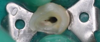
Endodontic treatment involves filling the root canals.
The materials used are divided into several groups:
- According to physical properties: plastic (pastes), hard (pins).
- According to the functions performed: for sealing (sealers), for filling (fillers).
- Depending on the duration of installation - for permanent or temporary filling.
Types of adhesion
In dentistry, there are two types of adhesion:
Mechanical – due to micromechanical adhesion of the material to the tooth tissues;
Chemical – due to the formation of a chemical bond between the material and dentin and enamel.
Only GIC has chemical adhesion. All other materials used in dentistry have mechanical and micromechanical adhesion.
Mechanical adhesion is the connection of materials with hard dental tissues due to mechanical retention with the participation of micromechanical pores and roughness on their surface.
In 1955, Buoncore discovered that the surface of tooth enamel becomes rough after etching with phosphoric acid and this increases the adhesion of methacrylic filling material to the tooth surface. The technique of etching enamel with acid, born in this way 40 years ago, is the basis of modern adhesive methods of dental restoration.
Failure to complete this stage of work leads to disruption of the adhesion of the composite material to the hard tissues of the tooth, which is manifested by the appearance of a marginal gap, microbial invasion, staining of the edges of the filling, post-operative sensitivity, etc.
It should be noted that the strength of adhesion to enamel and dentin is significantly different. Thus, the main problem of ensuring effective adhesion to hard tooth tissues lies in the different anatomical structure of enamel and dentin.
The most popular types
Despite the wide variety of restorative materials of this type, dentists choose compomers from certain manufacturers. They differ in their characteristics and can be used in different cases.
Dyract eXtra
This filling composition is used to eliminate defects that occur in incisors and molars.
The main advantage of this material is the constant release of fluoride ions, which helps prevent caries, preventing its re-development after filling.
The main indications for use are cases when there is a need for restoration of carious cavities in children and adult patients.
Contraindications include:
- situations when one of the ingredients becomes an allergen for the patient;
- if during the manipulation it is not possible to eliminate the effect of salivary fluid or blood;
- when, after the formation of the stump, the use of a ceramic crown is assumed.
Important! The color selection occurs after the surface enamel is moistened. The composition of Dyract eXtra has 6 tones, which allows you to choose the desired shade.
Glasiosite
The restoration composition is available in capsules with a volume of 0.25 g. It is used in the following cases:
- to eliminate carious cavities in baby teeth;
- when performing veneering work on teeth located in the smile zone;
- for long-term restoration of class I and II;
- if extended sealing of fissures (natural pits and depressions on the chewing surface of teeth) is necessary.
When using Glasiosite material, the following advantages are noted:
- high level of strength ensures resistance to abrasion;
- there is a good connection with surface enamel and dentin;
- stable and long-term fluoride production;
- good color rendition, which is why the filling is almost invisible in the smile area.
Twinky Star
German manufacturers have taken an original approach to solving dental problems that arise in childhood.
The bright and different colors of the filling composition can reduce the child’s feeling of fear when visiting the dentist.
The compomer curing process occurs in two stages:
- under the influence of a halogen lamp during the filling installation procedure;
- within two months, under the influence of moisture, the hardness coefficient increases.
When using this composition, high quality of the final result is ensured. In addition, it has a high degree of biocompatibility and prevents the development of caries.
Twinky Star does not apply:
- if allergic reactions occur to its components;
- in case of need of direct covering of dental pulp;
- when it is not possible to completely isolate the tooth;
- in combination with cements containing zinc oxide;
- if a heart rate pacemaker is installed.
Comp Natur
The presence of a pink tint in the Comp Natural material allows restoration work to be carried out in areas with damage to the neck of the tooth (especially on the incisors). According to Black's classification, the composition is used in the treatment of class V cavities.
This type of cement composition is characterized by high strength, good color transfer and low abrasion. For convenience, Comp Natur is available in capsules.
MagieFil
This type of cement composition is used to treat temporary teeth. Like all representatives of these materials, it has double curing.
Simultaneously with the therapeutic effect, it has a preventive effect, preventing the occurrence of carious cavities. Available in 4 variations of different shades.
Ionosit Seal
A type of light-curing radiopaque material that is used to fill fissures (most often on baby teeth).
It contains fluorine and zinc ions, which prevents the development of caries. When used, no adverse reactions are observed, except in cases where one of the components causes symptoms of an allergic process.
Ionosit-Baseliner
Glaciosite is used as an insulating gasket. When used, practitioners note the following advantages of the material:
- has a natural tooth color;
- well adapted to narrow fissures;
- fills even minor depressions and pits;
- releasing zinc and fluoride ions, prevents relapse of caries;
- The convenient applicator ensures comfortable work.
In rare cases, due to hypersensitivity to the main or additional components, allergic reactions may occur.
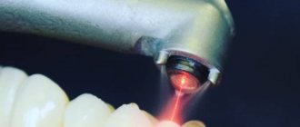
Composition and properties of the drug Kalasept and instructions for use in dentistry for dental treatment.
This post is all about using Proroot Mta Dentsply.
Here https://zubovv.ru/lechenie/zubyi/plombyi/irrigatsii-kornevyih-kanalov.html we will consider the requirements for solutions for root canal irrigation.
Mechanisms of adhesion of composites to the enamel surface
Enamel consists mainly of inorganic substances; in addition, the enamel contains a small amount of organic substances and water. Under the influence of acids, selective dissolution of the peripheral and central zones of enamel prisms occurs to a depth of 5-10 nm and the transformation of the enamel surface, which under an electron microscope becomes similar to a honeycomb or a horseshoe shape, or a combination of both forms.
As a result of mechanical beveling of enamel prisms and treatment of enamel with acid, the active adhesion surface with composite materials increases and the possibility of enveloping the surface layer of enamel with hydrophobic and viscous adhesives improves. Due to their high viscosity, they penetrate slowly to the entire depth of the etched enamel. After polymerization of the adhesive, processes are formed in the interprismatic areas, which mechanically adhere to the enamel surface and thus contribute to the microretention adhesion of the composite to the enamel surface.
Enamel etching
When etching enamel with acid, a layer 10 microns thick is removed from the surface and micropores 5-50 microns deep are formed.
The effect of acid etching of enamel depends on several factors:
type of acid used;
acid concentration;
forms of acid application (gel or liquid);
etching time;
time of rinsing with water;
ways in which etching is activated;
instrumental treatment of the enamel surface before etching;
chemical composition and condition of enamel;
enamel of baby or permanent teeth;
degree of enamel mineralization.
Most often in modern dentistry, phosphoric acid is used for acid etching of tooth tissue. The most optimal acid concentration is 30-40%. In some cases, the use of weak solutions of organic acids is recommended for etching dentin.
To prevent phosphoric acid from spreading to areas of the tooth where acid etching is undesirable, etchings are made in the form of colored gels.
The duration of acid etching of enamel is usually 30 seconds. Experimental studies using SEM showed that there were no differences in the degree of porosity of the enamel surface between exposures of 30 seconds and 60 seconds. In addition, it has been proven that exposure to acid for more than 60 seconds leads to the destruction of enamel prisms and deterioration of adhesion.
Depending on the resistance of the enamel, it is recommended to change the application time of the etching gel: with low enamel resistance it is reduced to 15 seconds, and with increased resistance it increases to 60 seconds.
The duration of removal of the etching with a stream of running water should be equal to the duration of exposure to the acid, i.e. 30 seconds.
To increase the adhesion force, it is recommended to create a bevel of the enamel, which allows increasing the area of contact of the composite with the enamel. The strength of this connection increases when it is formed along the cross section of enamel prisms, since in this case the interprismatic substance dissolves during etching, forming wider and deeper pores.
Classification and main components
The chemical composition of compomer materials includes:
- two types of acid (composite and polyacrylic);
- strontium fluorosilicon glass;
- strontium fluoride compounds;
- stabilizers giving the necessary consistency;
- substances that initiate the polymerization process.
Depending on the area of application of these restoration materials, they are either packable or fluid.
Packable
Allowed for use when the elimination of carious cavities does not occur on the frontal incisors of the front row due to the low level of aesthetic value.
The material is mainly used when installing fillings on molars or premolars, but only in that part of the tooth that experiences minimal pressure during the chewing process.
All materials in this group are characterized by high density and are highly filled. According to clinical studies, compomers do not differ in their characteristics from hybrid composites, and in some situations they are even inferior in strength.
The advantages of this type of material include:
- ease of use;
- high strength;
- radiopacity.
The disadvantages are the short working time (up to 1 minute) due to the rapid setting. Also, in some cases, problems arise with edge adaptation.
Liquid-flowing
Materials of this group are divided into high-, medium- and low-flow. Most often they are used as filling agents to eliminate carious cavities of classes III, V, when it becomes difficult to use conventional composites (narrow and hard-to-reach cavities, chips).
They can also be used to fix fixed dentures. In some cases, the combination of compomers with other materials allows them to be used as a base layer, which provides a good marginal seal.
Most practitioners note the following positive qualities of these materials:
- high degree of aesthetics;
- good for polishing.
The main disadvantage is high shrinkage and low strength.
Mechanisms of adhesion of composites to the dentin surface
The main problem in ensuring effective adhesion to dentin is its structure. The diacrylates that make up the composites have fairly high adhesiveness to tooth enamel, but in relation to dentin they behave as hydrophobic substances that do not adhere well to its surface, which is due to the peculiarities of the structure of dentin.
Dentin consists of 45% mineralized components, 30% organic structures, and 25% water. The nature of living dentin is such that its surface is always wet, and drying under clinical conditions is practically impossible. Due to the speed of fluid movement in the dentinal tubules, complete renewal of moisture repeatedly occurs on the surface of the dentin. In clinical conditions, even after drying the carious cavity, unnoticeable residual moisture is observed, which can affect the strength of the bond between dentin and composite. In this regard, dentinal adhesive systems must be hydrophilic, i.e. water compatible.
Another problem in the mechanism of adhesion of the composite to dentin is the smear layer, which is formed as a result of instrumental processing of dentin and consists of hydroxyapatite particles, destroyed remains of odontoblasts and denatured collagen fibers. Depending on the type of preparation, this layer reaches a thickness of up to 5 nm; it clogs the dentinal tubules and covers the intertubular dentin like a gasket. If at first it was considered as an insulator that prevents the penetration of microorganisms into the dentinal tubules, now there is no doubt that it interferes with the adhesion of the composite to the dentin surface and, accordingly, the formation of a strong adhesive connection.
In connection with the above, there was a need to search for completely new mechanisms of adhesion of composites to dentin, different from the mechanisms of adhesion to enamel.
Analyzing various adhesive systems for dentin and their adhesion mechanisms, two approaches are fundamentally distinguished. In the first case, the smear layer is completely preserved on the surface of the dentin and is impregnated with hydrophilic low-viscosity monomers and is directly used as a connecting layer between the dentin and the composite.
In the second approach, by dissolving the smear layer and superficial decalcification of dentin. This approach is the most common currently.
Dentin etching (conditioning)
In 1979, the Japanese physician Fuzayama first used dentin etching, and since then this procedure has been widely introduced into practice in Japan. In Europe, this happened later, when it was proven that pulpitis after etching is not associated with the damaging effect of acid on the pulp, but with the phenomenon of microleakage and depressurization of fillings. In addition, older generation adhesive systems were incompatible with dentin.
Modern dentin adhesive systems include mandatory pre-treatment of the dentin surface with so-called dentin conditioners, which promote the penetration of hydrophilic monomers into the surface layer of dentin and their chemical adhesion to the hydrophobic monomers of the composite.
Dentin conditioning is a chemical modification of the dentin surface using acids such as citric, polyacrylic, lactic, etc. In this case, the smear layer is removed completely or partially, and the dentinal tubules are also fully or partially opened. In addition, demineralization of the surface layer of dentin occurs, exposure of collagen fibers of the organic matrix and activation of dentin ions and apatites.
Conditioners in some adhesive systems must be removed using a stream of running water. The dentin surface must then be slightly dried. One of the main conditions for high-quality adhesion is the degree of moisture of the dentin after removing the etching solution. This is primarily due to the hyphrophilicity of the primer. Thus, the adhesion force decreases sharply when dentin overdries. In this case, collapse and loss of collagen fibers are noted, which impairs the penetration of the primer between them to form a strong bond. Dentin that is too wet also does not provide sufficient adhesion. The main criterion for the degree of dentin moisture is “sparkling” dentin, on which there are no “wet puddles”.
Subsequent application of a dentin adhesive system (primer) ensures the penetration of hydrophilic monomers into the open dentinal tubules, saturating the demineralized surface layer of dentin and adhesion to its exposed collagen fibers. With the formation of a hybrid zone. The hydrophilic resins that make up the dentinal adhesive penetrate into the dentinal tubules; the spaces previously occupied by hydroxyapatite encapsulate collagen fibers. After polymerization of the adhesive, a thin layer of new substance is formed, consisting of adhesive components and dentin collagen fibers. This is called the hybrid layer.
The hybrid layer not only provides reliable fixation of the composite to dentin, but also is an effective protective barrier against the invasion of microorganisms and chemicals into the dentinal tubules and tooth cavity. In addition, it blocks the movement of cerebrospinal fluid in the dentinal tubules and prevents postoperative sensitivity.
This mechanism of action is used, for example, in dentinal adhesives: Gluma (Bayer), Denthesive (Kulzer) and Scotchbond Multi Purpose (3M).
This adhesion mechanism can also be achieved by treating dentin with so-called self-conditioning primers, which contain, along with hydrophilic monomers, one or another organic acid. Under the influence of these primers, the smear layer of dentin is partially dissolved, and the dentinal tubules are also partially opened. The surface layer of intertubular dentin is demineralized and simultaneously impregnated with hydrophilic monomers. The smear layer is not washed off, but is sprayed, and its sediment falls on the surface of the dentin. The adhesion of the composite to dentin is achieved due to the penetration of polymers into the dentinal tubules and the formation of polymer processes and due to the impregnation of the surface layer of dentin with monomers. This mechanism underlies the following adhesive systems: ART – Bond (Coltene), Scotchbond (3 M) and Syntac (Vivadent).
Thus, an adhesive system for enamel and dentin should have the following properties:
provide good initial and long-term bond strength to enamel and dentin;
have good biocompatibility;
minimize marginal permeability;
prevent secondary caries and marginal staining (as a consequence of marginal permeability);
be convenient and easy to use;
have a long shelf life;
be compatible with a wide range of restorative materials;
should not be toxic and cause sensitization to staff and patients;
should isolate the tooth surface from oral fluids.
For dentin adhesive, we add three more requirements:
penetrate into etched dentin;
have hydrophilic properties;
If possible, remove the smear layer.
What to do in case of destruction of tartar and caries
If the destruction is minor and there is no pain, the doctor drills into the damaged darkened areas, cleans them of plaque, disinfects them and closes the resulting hole with a special compound. If a tooth hurts badly, it means that caries has affected the pulp - the loose, fibrous connective tissue that fills the tooth cavity. In this case, you will have to come for treatment more than once.
First, the doctor will clean the walls of the resulting cavity, then fill it with an antiseptic, fix it with a temporary filling and isolate the damaged surface from external influences.
If the pain does not go away after a certain time, it is necessary to remove the nerve, since the destruction has reached it. The doctor will cement the root canals and restore the tooth crown, selecting the necessary filling materials.
Materials for dental restoration
At the moment, there is no impeccable material in dentistry that is similar to natural teeth in all respects. But you can select impressions that match the natural shade of tartar as closely as possible.
For this purpose, the composition is pre-selected. Sealants are used to prevent caries. When cracks, grooves or other small flaws form on the enamel, they are sealed with special substances that form a film to stop further tooth decay. Fluoride ions inside the sealant protect against erosion.
Adhesives (substances that connect materials by surface adhesion) are needed to improve the adhesion of the filling to the walls of the tooth.
Some mixtures have anti-inflammatory and restorative properties. They are used as therapeutic pads under fillings.
There are compounds that are compatible with soft tissues and fill root canals well, but are not strong enough.
Temporary restorative pastes either quickly wear out or break down. Therefore, they are suitable only for short-term sealing of hollows during treatment and diagnosis.
To install long-term fillings, compositions that most closely match the characteristics of your own dental tissues are suitable. It is with their help that the exact shape of the cast is reproduced.
Research on restorative compounds
The search for a universal dental composition for dental reconstruction does not stop and every year filling materials continue to be improved. But before new products are widely used, they must undergo a series of studies to ensure compliance with existing standards. To do this, newly created samples are carefully studied in accredited laboratories in 3 areas:
The physical and mechanical direction makes it possible to find out:
• Consistency of the material; • change in temperature of the mass during hardening; • volume of the substance during the solidification process; • color stability; • the optimal amount of time required for curing the mass; • water absorption of the substance; • solubility coefficient in water and other media; • hardness level; • transparency of the substrate; • percentage of abrasion; • adhesion ability; • presence of radiopacity; • thermal conductivity value.
Tracking biological properties allows us to understand:
• how toxic the substances are if swallowed; • degree of cumulative toxicity of the material; • local irritant effect; • ability to cause allergic reactions.
Research into the biological compatibility of restoration materials provides a guarantee of additional safety.
Clinical trials
At this stage, the compositions are mastered in dental clinics at research institutes. Based on the experience gained, recommendations on the use of the compositions are given.
A final understanding of the qualities and properties of improved materials for reconstruction is provided only by long-term observations of their practical application.
Basic requirements for temporary filling materials:
1. They interact well with body tissues, do not irritate and do not enter into putrefactive or oxidative reactions in the oral cavity; 2. They have a strong and tight adhesion to hard tissues; 3.Do not differ in appearance from a natural tooth; 4. Correspond to the quality and properties of natural dental tissues; 5. Dentin is regenerated (a type of bone tissue covered with enamel in the crown and cement in the root).
Formulations for temporary use
Cement paste was first used for filling teeth in 1832.
The material consisted of a solution of phosphoric acid combined with calcium oxide powder. Later, to increase the strength of the substance, glass powder was added to the composition. Until the mid-20th century, cements, along with metals, were the main filling agents. In modern dentistry, various modifications of cements are used to solve many problems.
To close the hollow for a short time, use the following materials.
Zinc sulphate cements
A powder from a mixture of zinc oxide with the addition of kaolin or dextrin and zinc sulfate is mixed on a glass plate. Distilled water is used for preparation. The resulting paste is placed into the treated hole with a trowel or spatula. Seal with a cotton swab.
Zinc oxide provides good adhesion to hard tissues, and zinc sulfate with kaolin is responsible for the strength and ductility of the resulting substance. The mixture hardens within 2-3 minutes. This composition is inexpensive and easy to work with.
Minus: it wears out quickly, which is why it can last no longer than 2-3 days.
Zinc-eugenol cements
Dentine paste is available in ready-made form. There is also a mixture of zinc oxide and peach or clove oils, containing up to 70% eugenol, which form a paste that hardens in 2-3 hours.
Eugenol is an essential oil of natural origin. Has an anti-inflammatory and calming effect on the pulp.
It is used as therapeutic pads and for crown restoration. When interacting with composites, eugenol distorts the course of polymerization, which reduces the use of such compositions for treatment. The advantages of cements of this group include ease of use, high biological compatibility and healing properties that regenerate the pulp.
Zinc phosphate (zinc oxide and phosphoric acid)
To improve the properties, oxides of metals such as bismuth, silver, magnesium, and silicon are added in small quantities to zinc oxide. Then the powder is combined with orthophosphoric acid, diluted in water with small admixtures of zinc and aluminum phosphates. The addition of silver and bismuth gives the composition a bactericidal effect.
Excellent compatibility with soft tissues predetermines its primary use as insulating pads for permanent restoration. Root canals are also filled. If sealing is required for a period longer than 2-3 weeks, then it is used on a temporary basis.
Cements of this group are very plastic and adhere well to the walls of the tooth. The composition is characterized by low thermal conductivity and radiopacity. It is worth considering that the materials are not strong enough, decrease in volume when cured, are porous, and do not match the color of tooth enamel. They interact chemically with saliva and can irritate the pulp due to the acid reaction of the cement paste.
Polycarboxylate cements
Polyacrylic acid dissolved in water is combined with zinc oxide and magnesium oxide. The result is a plastic mass with excellent adhesion. It does not irritate the pulp and has low thermal conductivity.
Unlike phosphate cements, it is less susceptible to chemical interaction with saliva, but is prone to destruction from mechanical stress.
Disadvantage: too long time for complete hardening - 10-12 hours. Used for fastening orthodontic products, as insulating pads and as temporary fillings.
Adhesive systems for enamel
Enamel adhesive systems (adhesives) consist of hydrophobic liquid monomers of composite materials, which, due to micromechanical adhesion, ensure adhesion to tooth enamel. It should be noted that these adhesives do not provide adhesion to dentin, so it is necessary to either isolate the dentin from toxic effects with an insulating gasket or use a dentin adhesive system (primer).
Chemical Cure Composite Kits contain only enamel adhesives and are chemically cured.
Stages of working with adhesive systems for enamel:
etching the enamel surface for 30 seconds using 37% phosphoric acid, which is part of the etching gels;
removing the etching gel with a stream of running water for 30 seconds;
drying the enamel and controlling the quality of the etching (etched enamel has a matte tint);
mixing the components of the adhesive system in a 1:1 ratio;
introducing the adhesive system into the carious cavity using an applicator (applied to prepared enamel and an insulating lining);
distribution of the enamel adhesive system using a weak air stream;
introduction of composite material.
Adhesive systems for enamel included in sets of light-curing composite materials are single-component. In addition to them, the set includes an adhesive system for dentin (primer). The stages of working with these systems will be discussed a little later.
Features of use
When performing restoration work using compomers, the process of installing a filling is practically no different from using conventional composite materials.
Therefore, the following algorithm of actions is observed:
- Initially, professional cleaning of the sector in which the damaged tooth is located is carried out.
- To prevent relapse of caries development, preventive preparation is performed. This occurs with minimal cavity formation, as a larger area will increase the volume of the filling, which will reduce its strength.
- When dentin is deeply damaged, it becomes necessary to spot cover this area. For this purpose, calcium-containing pads are used. They are subsequently insulated with a glass ionomer cement composition.
- The adhesive system is applied.
- Layer-by-layer application of compomers is carried out by analogy with the use of composite compositions. The thickness of a single applied layer should not exceed 2.5 millimeters.
- Polishing and grinding of the tooth surface occurs at the end of the process.
You should know! Curing time for one layer is approximately 40 seconds. The use of an ultraviolet lamp should be directed to the location where the defect is being eliminated.
Dentin adhesive systems (primers)
In a fairly short period of time, several generations of adhesive systems for dentin have changed, with development going in two directions - simplifying the procedure for use and improving the adhesion itself. The term “generation” does not, by and large, have any scientific basis; nevertheless, it allows us to structure in a certain way the whole variety of adhesive systems present on the market today. Belonging to a particular generation is determined by the chemical composition, mechanical adhesion indicators and ease of use.
First generation
The first generation of adhesives appeared in the late 70s of the last century. They are characterized by high rates of adhesion to enamel, but adhesion to dentin is extremely low - as a rule, no more than 2 MPa. Adhesion was achieved through the interaction of the bond and calcium contained in the dentin. Naturally, the problem of debonding was extremely acute - difficulties arose within a few months. Therefore, this generation of adhesive systems were recommended for use only in Class III and V cavities. When used in posterior areas, significant postoperative sensitivity was often observed.
Second generation
In the early 80s of the last century, the second generation of adhesives appeared. Here, an attempt was made to utilize the smear layer to obtain higher adhesion rates to dentin. The result was an increase in this indicator to 2-8 MPa, which, of course, is absolutely insufficient for reliable fixation. In addition, microleakage was often observed with these systems, and the problem of postoperative sensitivity was also not resolved. The long-term stability of representatives of this generation was also problematic - after a year, up to 30% of restorations turned out to be unsuccessful precisely because of a significant deterioration in the adhesion index.
Third generation
At the end of the 1980s, two-component adhesive systems appeared, consisting of a primer and an adhesive. This, as well as a significant improvement in mechanical adhesion indicators (8-15 MPa), allows us to distinguish them into a separate generation of adhesive systems - the third in a row. Their appearance made it possible in some clinical situations to minimize tooth preparation, thus opening the era of ultra-conservative dentistry. In addition, with their use, a significant decrease in postoperative sensitivity was observed. For the first time, adhesives of this generation provided adhesion not only to teeth, but also to metals and ceramics.
The main problem was the fragility of bonding agents. Some studies have demonstrated a significant decrease in adhesion rates as early as 3 years after restoration. However, it was from this generation that the routine use of adhesives in restorations of posterior posterior teeth began.
Fourth generation
The advent of fourth generation adhesives in the early 90s transformed dentistry. The index of adhesion to dentin has reached “modern” values – 17-25 MPa, and postoperative sensitivity when using adhesives of this generation has decreased even more.
was revolutionary : after etching, the applied adhesive interacts with the collagen matrix of dentin, forming an intermediate layer that is neither dentin nor adhesive, which is called hybrid. It is the presence of this layer that is responsible for the high strength indicators.
The main success of fourth-generation adhesives owes to the emerging technique of total etching and the concept of wet dentin bonding.
The disadvantages of materials in this group include the presence of two or more components that must be mixed in precise proportions. This seems easy in a “laboratory” setting, but can be a problem in real life. It was precisely due to inaccuracies during mixing that problems arose when using fourth-generation adhesives.
These adhesive systems contain 3 components:
1) conditioner (phosphoric acid in the form of a gel for etching enamel and dentin);
2) primer (a mixture of hydrophilic low-molecular compounds that penetrate wet dentin, saturate it and form a hybrid layer);
3) enamel adhesive system (unfilled resin that ensures the connection of the composite with the hybrid layer and tooth enamel).
Types of 4th generation adhesive systems:
Provides for partial dissolution of the smear layer and partial opening of the dentinal tubules using weak solutions of organic acids included in the primer composition.
Stages of working with 4th generation adhesive systems Type 1:
1. etching the enamel surface for 30 seconds using 37% phosphoric acid, which is part of the etching gels;
2. removing the etching gel with a stream of running water for 30 seconds;
3. drying the enamel and quality control of the etching (etched enamel has a matte tint);
4. applying primer to the dentin of the carious cavity using an applicator (exposure 10 seconds);
5. primer distribution using a weak air stream;
6. introducing the enamel adhesive system into the carious cavity using an applicator (applied to prepared enamel and dentin);
7. distribution of the enamel adhesive system using a weak air stream;
8. photopolymerization of adhesive and primer;
9. introduction of composite material.
Type 2:
Provides for complete dissolution of the smear layer by etching dentin with phosphoric acid.
Stages of working with 4th generation adhesive systems Type 2:
1. etching the enamel surface for 15 seconds using 37% phosphoric acid, which is part of the etching gels, adding the gel to the dentin for 15 seconds;
2. removing the etching gel with a stream of running water for 30 seconds;
3. drying of enamel and dentin (quality control of etching - etched enamel has a matte tint, dentin should not be overdried - moist, shiny);
4. applying primer to the dentin of the carious cavity using an applicator (exposure 10 seconds);
5. primer distribution using a weak air stream;
6. introducing the enamel adhesive system into the carious cavity using an applicator (applied to prepared enamel and dentin);
7. distribution of the enamel adhesive system using a weak air stream;
8. photopolymerization of adhesive and primer;
9. introduction of composite material.
Fifth generation
In the fifth generation adhesives, the mixing problem was eliminated - the “one bottle” concept was implemented, i.e. the adhesive and primer were placed in one container (they became one-component).
The use of one-component systems also involves total etching of enamel and dentin. The mechanism of their connection is similar to the mechanism of adhesion of 4th generation systems. These materials have good adhesion rates to enamel, dentin, ceramics and metal (at the level of 20-25 MPa), but their most important advantage is the absence of the component mixing stage, the poor performance of which led to a decrease in adhesion rates in fourth-generation systems.
Fifth generation adhesive systems are still the most popular because they are easy to use and give predictable results. Postoperative sensitivity when using them is also low.
Stages of working with 5th generation adhesive systems:
1. etching the enamel surface for 15 seconds using 37% phosphoric acid, which is part of the etching gels, adding the gel to the dentin for 15 seconds;
2. removing the etching gel with a stream of running water for 30 seconds;
3. drying of enamel and dentin (quality control of the etching - etched enamel has a matte tint, dentin should not be overdried - moist and shiny);
4. applying the adhesive system to the enamel and dentin of the carious cavity using an applicator (exposure 15 seconds);
5. distribution of the adhesive system using a weak air stream;
6. photopolymerization of the adhesive system;
7. introduction of composite material.
Sixth generation
The next task of developers when improving adhesive systems was the need to remove the etching stage from the list of procedures performed. In sixth generation systems this problem has been solved.
Generation 6 adhesive systems are one-step, self-etching systems that come in 2 bottles and require mixing immediately before use. The system is then applied to the enamel and dentin. At the same time, etching, diffusion into the tooth tissue and the formation of a hybrid zone are ensured.
Compared to adhesive systems of the 4th and 5th generations, they are easier to use, working with them requires less time due to the reduction in the number of steps, and the risk of technical errors is reduced.
However, adhesion to dentin (18-23 MPa) remains virtually unchanged over time, while adhesion to enamel deteriorates.
Stages of working with 6th generation adhesive systems:
1. The components of the adhesive system are mixed outside the oral cavity (inside a disposable package or in a special cell);
2. applying the adhesive system to the enamel and dentin of the carious cavity using an applicator (exposure 15 seconds);
3. distribution of the adhesive system using a weak air stream;
4. photopolymerization of the adhesive system;
5. introduction of composite material.
Text of the book “Therapeutic Dentistry. Lecture notes"
Polymer filling materials
Represented by the following groups of filling materials.
The first group consists of unfilled polymer filling materials, the second group includes filled (or composite) materials. The third group is represented by compomers, the fourth - by metal filling materials. What is common to all polymer filling materials is that their hardening and formation of a certain structure is based on the phenomenon of polymerization. It represents the formation of a spatial structure of a material by grouping a large number of simpler particles into a more complex and larger one.
Unfilled polymer filling materials
presented in acrylic and epoxy materials.
Acrylic filling materials are a system of powder and liquid. The powder contains various components. Firstly, these are polymer particles, which are represented by polymethyl methacrylate, secondly, the initiator of the polymerization reaction is benzoyl peroxide, and thirdly, various pigment coloring particles that determine the color characteristics of the filling material. These components are mandatory for polymer plastics. In addition, the powder may contain various fillers. They are necessary to ensure certain strength characteristics, impart the ability to retain its volume after hardening, and withstand various factors acting on the filling material during its hardening and subsequently. It is possible to include plasticizers in the powder composition, which determine the plasticity of the material. The liquid of acrylic plastics contains particles of a monomer, which is the methyl ester of methacrylic acid, and an inhibitor of the polymerization process, which is represented by various substances belonging to the group of quinones (hydroquinone is most often used). The initiator of the polymerization reaction causes the beginning of the polymerization reaction, the inhibitor determines its controlled nature. In addition, a polymerization inhibitor is necessary to prevent the polymerization reaction from occurring under storage conditions or during transportation.
The group of unfilled polymer filling materials also includes materials based on epoxy resins.
Materials based on epoxy resins have two components. These components - resin and hardener - have a paste-like consistency.
Acrylic and epoxy filling materials are used very rarely.
Polymerization process
. The polymerization process can be diagrammed as follows. The first phase of the process is determined by the beginning of polymerization. This occurs due to the formation of active centers under the action of the polymerization initiator. Active centers contain groups represented by free radicals. The chemical bond between individual monomers during the formation of a polymer particle occurs due to the interaction of the monomer molecule with such a center. The formation of such chemical bonds between monomer molecules occurs in the second phase of the polymerization process. This phase is called polymer chain growth. The end of polymerization occurs in the third phase, called the chain termination stage.
Some unfilled polymer filling materials go through a number of stages during their maturation. Immediately after mixing the material components, the sand stage begins. During this stage, the material is not used for filling carious cavities. The second stage is characterized by the appearance of stretching threads of plastic. The name of this stage is appropriate - the stage of stretching threads. During this stage, the material must be used for filling. The third stage is doughy. At this stage it is necessary to form a filling. The final stage is rubber-like. Material at this stage of maturation is unsuitable for insertion into a carious cavity and formation of a filling.
It should be noted that the polymerization reaction is exothermic, that is, it occurs with the release of heat. This makes it possible to irritate the dental pulp due to excessive thermal effects. In addition, the irritant effect on the pulp may be due to the effect of a chemical agent on it in the form of a monomer that does not remain unchanged after the completion of the polymerization process. This unreacted monomer will naturally have an adverse effect on the odontoblast cells of the pulp. The disadvantages of polymer plastics will also be their rather low strength characteristics, the ability to absorb water, and become fiber-free. The effect of temperatures will also lead to changes in the volumetric characteristics of these materials. The aesthetic properties of polymer plastics are also quite questionable; moreover, under the influence of food dyes, their color changes.
Examples of unfilled polymer filling materials are Acrylic Oxide and Carbondent. To mix these materials, you need to mix the powder and liquid in a 1:1 ratio for 40 seconds. After this, the material must be introduced into the carious cavity, carefully rubbing it against the walls.
Composite filling materials,
or, as they are also called, composites.
Composites in their structure contain a number of structural components: matrix, filler and surface-active components. The polymer matrix - the basis of the composite - consists of monomer molecules connected to each other through chemical bonds. The chemical structure of the polymer matrix is bisphenol glycidyl methacrylate. In addition, this phase includes a polymerization catalyst (due to its presence, the polymerization process begins), an additional catalyst (used in composites with a chemical type of curing to ensure a more complete polymerization process). Instead of an additional catalyst, light-curing composite materials use a polymerization activator. In order to ensure a controlled nature of polymerization, optimal conditions for storage and transportation of composites, a polymerization inhibitor is also included in the composition of this phase.
The second phase - the composite material (filler) - is an inorganic substance from a chemical point of view. Filler particles can differ in size, shape, and the material that forms them. Depending on the characteristics of the filler particles, various composite materials will be considered, which therefore have different properties.
The third phase is surfactants, which provide adhesion between the composite matrix and filler particles.
There are various classifications of composite materials.
According to the curing mechanism, composites are usually divided into materials of chemical curing, light curing, and dual curing mechanism. In addition, there are composites in which the polymerization process occurs under the influence of heat; at present they are rarely used.
Depending on the size of the filler particles and the degree of filling, it is customary to distinguish the following groups. Macro-filled composites have a particle size from 8 to 12 microns, micro-filled composites have a particle size of 0.04-0.1 microns; mini-filled, particles of which have a size of 1–5 microns, and hybrid. Hybrid composites, in turn, can be divided into macrohybrid (contain filler particles 8–12 and 0.04–0.1 µm in size), microhybrid (contain particles 1–5 and 0.04–0.1 µm in size) and totally made (particles of which have a size of 5–8; 1–5; 0.01–0.1 microns).
In addition, they use a classification based on the purpose of composite materials, according to which it is customary to distinguish classes A and B. The first of them includes filling materials for filling carious cavities, which, according to Black’s classification, belong to classes 1 and 2. Class B includes materials for filling carious cavities according to the Black classification, belonging to classes 3, 4 and 5.
Composite materials, classified by the type of curing as those cured as a result of a chemical reaction, consist of two components. They can be produced in the form of a two-paste system or in the form of a powder and liquid. Composite materials in the form of powder and liquid are currently used quite rarely. The polymerization reaction in chemically cured composites is initiated by a catalyst that is present in one of the components and an additional catalyst that is present in the other of them. The formation of active centers occurs, which contain active groups represented by free radicals. These radicals interact with monomer molecules. The formation of a polymer matrix structure occurs. A positive property of composite materials with a chemical curing method is the implementation of polymerization in all layers of the filling. A negative property of these materials is the change in color of the filling due to the retention of part of the catalyst in its composition, which has not reacted. In addition, chemical composites have a short period of time during which it is possible to simulate a filling; they are characterized by a change in viscosity during the polymerization reaction. Composite materials with a light curing method are characterized by the fact that the initiation of the polymerization process is carried out under the influence of light from a special lamp. They are available in paste form. A positive property of light-curing materials is that after introducing the material into a carious cavity, it can be used for modeling for a long time. Light-curing composite materials are more convenient to use. It is possible to use various combinations of materials when placing one filling, thereby ensuring the best aesthetic effect. The negative properties of light-curing composites are their high cost, the need for a large amount of time, and the possibility of harmful effects on the cornea of the eyes from the light that the lamp emits.
Light-curing composite materials should be applied in thin layers. The first portion of the composite material is applied so that the layer thickness is 0.5 mm, the thickness of subsequent portions should be no more than 2 mm. The layers of the composite should be shaped like the letter U. When polymerizing the material, it is necessary to position the lamp so that there is an indirect light source.
Macro-filled composite materials are characterized by particle sizes from 8 to 12 microns. In addition, the presence of particles up to 100 microns in size is possible.
Inorganic filler particles provide fairly high strength characteristics of macrofilled composites, which is especially important when filling carious cavities located on chewing teeth, or in cases where strength characteristics are fundamentally important. At the same time, the particles of inorganic filler, which are large in size and also located at different distances from each other, form an uneven surface structure. This provides conditions favorable for plaque accumulation in the area of these irregularities. It is simply impossible to process such a surface to such an extent that it becomes smooth. From the point of view of aesthetic issues, the use of macro-filled composites is undesirable, at least without their combination with aesthetic micro-filled composites. This determines the fact that macrofilled composites are indicated for filling carious cavities of anterior teeth only in cases where aesthetic characteristics are not fundamental. For example, in class 1 according to the Black classification, when carious cavities are located in the area of the blind pits of the incisors, it is possible to install a permanent filling using macrofilled composites. In other cases, the use of these materials for filling teeth in the anterior group is impractical. In addition, macrofilled composites are used to restore teeth, which are supposed to be covered with artificial crowns in the future, or veneers will be made using a microfilled composite. Representatives of macro-filled composites are materials such as Evicrol, Adaptic, Profile.
Microfilled composites differ in particle size ranging from 0.04 to 0.1 microns. The reflection of light rays will be specular, so the surface of the filling will not look matte, but glossy. Microfilled composites have much less irregularities on their surface and are much easier to polish. All this determines the high aesthetic performance of these composite materials. At the same time, a decrease in the filler content in microfilled composites causes their lower strength characteristics. These features determine the scope of application of these filling materials. Indications for the use of microfilled filling materials will be those situations where there are high aesthetic requirements, i.e. filling carious cavities of the anterior teeth, according to Black’s classification, belonging to classes 3 and 5. It is possible to use microfilled composites together with macrofilled or hybrid ones for class 4 carious cavities. Representatives of microfilled composites are materials such as Evicrol Anterior, Isopast, Durafill.
Minifilled composites are characterized by the fact that, unlike other composite materials, they contain filler particles, the size of which ranges from 1 to 5 microns. From the point of view of the characteristics of strength and aesthetic properties, mini-filled composite materials represent average values between macro- and micro-filled ones.
Hybrid composites
characterized by particle sizes ranging from 0.04 to 5 microns. Due to the presence of filler particles of different sizes, the possibility of changing the number of both, and their ratio, it is possible to slightly change the characteristics of hybrid composite materials. With an increase in the content of particles with a larger size, the strength characteristics of materials also increase proportionally; with an increase in the number of filler particles with a small size, the aesthetic characteristics proportionally change for the better.
Hybrid composites have improved aesthetic properties compared to macrofilled composite materials, since they contain filler particles up to 0.76 microns in size. Filler particles up to 0.76 microns in size ensure the formation of specular and mixed reflections of a light beam. This determines the characteristics of the surface of the filling – a shiny appearance. In addition, by reducing the distance between particles, the surface is less rough and has a polished appearance. At the same time, the content of larger filler particles provides certain strength characteristics of filling materials. Representatives of hybrid composite materials are materials such as “Prizma”, “Prismafil”, Evicrol Posterior.
Microhybrid composite materials
characterized by filler particle sizes ranging from 0.04 to 1 microns. On average, particles are less than 0.76 microns in size. In this regard, the aesthetic properties of microhybrid materials are quite high. The fairly high strength characteristics of these materials are due to a certain change in the structure of their matrix. The positive properties of microhybrid composite filling materials determine their widespread use at present. Placement of fillings using these filling materials is indicated for filling carious cavities belonging to all six classes according to the Black classification, when making veneers. Representatives of microhybrid composite materials are Charisma, Te-Econom, Filtek Supreme, Definite.
Fluid-flowing composite materials
slightly different in composition from other composites. The polymer matrix of these materials is represented by resins that have the property of fluidity.
Flowable composites can be classified according to the following characteristics. According to the curing mechanism, they can be divided into chemical and light curing materials. Based on the degree of fluidity, fluid-flowing composites can be divided into medium-flow and high-flow. In terms of the size of the filler particles, they are often microhybrid.
The positive properties of fluid-flowing composites are satisfactory strength and aesthetic indicators, fairly low elastic modulus values, due to which they can, to one degree or another, compensate for the decrease in their volumetric characteristics after hardening, and sufficient ease of use. A negative property is the possibility of a decrease in volumetric characteristics after the material hardens.
Indications for the use of fluid composite filling materials are the following:
1) carious and non-carious lesions of teeth when they are localized in the cervical region;
2) filling of carious cavities classified as class 2 according to Black’s classification, using the “tunnel” preparation method;
3) filling of carious cavities, classified according to Black’s classification as classes 3 and 4;
4) fissure sealing;
5) splinting of mobile teeth for periodontal diseases;
6) use of inlays, veneers;
7) the presence of chips of hard dental tissues or chips of metal-ceramic structures when they are small.
Representatives of this group of filling materials are Filtek Flow, Flow-it.
Condensable composite materials
are another, slightly different group of composites. They contain filler particles of a fairly large size (more than 3.5 microns) and a slightly modified polymer matrix.
The positive properties of this group of filling materials are high strength properties, low rates of decrease in the volumetric characteristics of the material after it hardens, and sufficient ease of use.
At the same time, from an aesthetic point of view, these materials are undesirable for use when filling carious cavities in the frontal group of teeth.
Indications for the use of condensed filling materials are carious cavities, which, according to Black’s classification, belong to classes 1 and 2, and class 5 in the area of chewing teeth. The use of these materials in filling carious cavities in primary teeth in children is shown. When preparing a tooth for orthopedic treatment, the use of these materials is also very convenient. They can be used for splinting mobile teeth in the treatment of periodontal diseases.
Representatives of this group of filling materials are Filtek P-60, SureFil.
Lecture No. 9 Periodontal diseases
Periodontal diseases can be classified into the following groups.
1. Gingivitis
– is an inflammatory process in the gum tissue, which is not accompanied by a violation of the structure of the periodontal junction.
2. Periodontitis
– is an inflammatory process in the gum tissue, which is characterized by a disruption of the structure of the periodontal junction due to the destruction of periodontal tissue and bone.
3. Periodontal disease
– a disease in which dystrophic processes develop in the periodontium of teeth.
4. Idiopathic periodontal diseases
.
5. Periodontomas
– periodontal lesions that are of a tumor nature.
Etiology
Periodontal diseases occur in response to local and general factors.
Local factors include a small vestibule of the oral cavity, anomalies in the attachment of the frenulum, unformed contact points during the treatment of caries, the presence of carious cavities or failed fillings in the cervical area of the teeth, failed orthopedic structures, anomalies in the position of the teeth, etc.
General factors that lead to the development of periodontal diseases can be designated as general somatic diseases, which result in a disruption of the blood supply to periodontal tissues. Such factors include pathology of the cardiovascular, endocrine systems, gastrointestinal tract, etc.
Pathogenesis
Periodontal diseases are associated with the occurrence of inflammation under the influence of dental plaque microorganisms on periodontal tissues. In response to a damaging agent, blood cells exit the vascular bed and move, absorb the agent, and release inflammatory mediators. A complex of vascular reactions develops. The processes of cellular respiration and energy exchange are disrupted. Depending on the phase of inflammation that will predominate, three forms of gingivitis are distinguished: catarrhal - with pronounced phenomena of exudation, ulcerative-necrotic - with pronounced phenomena of alteration, hypertrophic - with pronounced phenomena of proliferation.