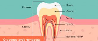
Periodontal disease
(
parodontosis
; Greek para about + odus, odontos tooth + -osis; synonym:
alveolar pyorrhea, Fauchard's disease, paradentosis, paradentitis, paradontopathia inflammata profunda
) is a periodontal disease characterized by damage to all its elements, destruction of the dentogingival junction and progressive destruction of the alveolar processes, leading to tooth loss without appropriate treatment.
Periodontal disease was first described in 1746 by P. Fauchard, who called it “false scurvy.” Then Toirak (A. Toirac) proposed the term “alveolar pyorrhea”. After the term “periodontium” became widespread among dentists to refer to periodontal tissues, O. Weski proposed calling pathological processes in periodontal tissues periodontal disease (this term was adopted in the USSR). In accordance with the International Classification of Diseases (1980), gum and periodontal diseases include acute and chronic periodontitis and periodontal disease.
Numerous epidemiological studies conducted in our country and abroad indicate a significant prevalence of periodontal disease, which closely correlates with age. Thus, according to the materials of G. I. Vishnyak (1962), M. G. Lunev (1967), A. I. Rybakov and G. V. Baziyan (1973), at the age of 12-13 years, periodontal disease was found in 2-4% , at 14-16 years old - 6-12%, at 17-18 years old - 19%. According to A.I. Evdokimov (1937), at the age of 20 years P. is observed in 14-29%, at the age of 30 years - in 50%, and after 40 years - from 60 to 90%. According to WHO (1978), the prevalence of P. depends on climatic and geographical conditions, and at the age of 35-44 years P. is found in 40-75%.
Etiology
The etiology of periodontal disease has not been fully elucidated. Its occurrence is associated with the influence of exo- and endogenous factors, changes in the reactivity of the body. Exogenous factors include nutritional disorders: deficiency, often relative, of proteins, vitamins, especially A, B, G, D and E, mineral salts (especially calcium and phosphorus), microelements (fluorine, iodine, etc.), excess consumption of carbohydrates and fat Of great importance in the occurrence of P. is the microflora of the oral cavity (see Mouth, oral cavity), enzymes and physiologically active substances of dental plaque (see Teeth) and fluid of the periodontal sulcus, as well as tartar. The emergence and development of P. is largely facilitated by malocclusion (see), especially accompanied by overload or underload of the periodontium, reducing its reserve functionality. Endogenous factors include genetic disorders, shifts in interstitial metabolism and its neurohumoral regulation, disturbances in the body's immunological reactivity, blood circulation, respiration, functional and organic changes in internal organs and other factors that cause a decrease in the resistance of periodontal tissues.
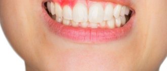
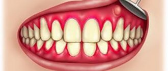
Symptoms of periodontal abscess
The pathological process is most often characterized by fairly vivid manifestations of symptoms. Among the characteristic signs of the problem, dental experts highlight the following points:
- mechanical irritation of the affected area provokes severe pain, which, however, is short-term. As a result, the patient cannot chew food calmly and experiences constant discomfort, including during a conversation,
Pain occurs while eating
- acute reactions to temperature contrasts in food and drinks appear,
- after some time, the gums in the affected area swell, a purulent lump appears, which gradually increases in size,
- at a more advanced stage, mobility of adjacent teeth is noted,
- a gap may appear between the tooth and the affected area of the mucosa, from which purulent exudate is released,
- symptoms of intoxication are reduced to fever, severe headache, loss of strength, development of nausea, diarrhea, and loss of appetite.
Important! This disease is especially dangerous for children and women during pregnancy; treatment should not be delayed under any circumstances. In representatives of this category of patients, even a minor infection can develop into a serious problem in the shortest possible time.
If you experience obvious discomfort or even severe pain in your gums, be sure to see a dentist - this should be done as soon as possible. Otherwise, complications cannot be avoided.
Pathogenesis
The pathogenesis of periodontal disease is determined by the interaction of general and local factors. At the same time, the above-mentioned exo- and endogenous etiological factors that induce P. (for example, functional disorders of internal organs and systems, oral microflora, dental plaque, etc.) are involved in the mechanism of its development and become pathogenetic.
Most researchers consider immunol disorders, reactivity of periodontal tissues under the influence of dental plaque components, metabolic disorders, inflammation of periodontal tissues (see), microcirculation disorders (see), arising as a result of neurohumoral changes in the body, to be the main ones in P.'s pathogenesis (see). Inflammatory mediators (histamine, serotonin and heparin released from mast cells, kinins - bradykinin, kallidin, prostaglandins, etc.), as well as enzymes of plaque and gingival crevicular fluid and other factors cause irritation and subsequent vegetation of the gingival epithelium deep into the periodontal gap, As a result, the circular ligament of the tooth is destroyed with the formation of a periodontal pocket. The inflammatory process in periodontal tissues is accompanied by symptoms of acidosis and pronounced functional disorders of microcirculation, which leads to degeneration of the bone tissue of the dental alveoli, their resorption and atrophy, loosening and loss of teeth.
A significant role in the pathogenesis of P. is played by functional disorders of microcirculation in periodontal tissues, beginning with a short-term phase of vascular dilation caused by the influence of biologically active substances (kinins, histamine, lysosomal enzymes, prostaglandins, etc.) followed by a long-term spasm (neurogenic genesis) and a decrease in volume blood flow speed. At the same time, the permeability of the vascular wall is disrupted - the initial increase in permeability is replaced by its decrease. The rheological properties of blood are also disrupted with thrombosis of the microvasculature of periodontal tissues. The level of oxygen tension and redox processes in periodontal tissues decreases, which indicates their dystrophy, as a result of which functional changes in blood vessels become organic, having a sclerotic character.
Due to the loss of teeth, pain when chewing, and mobility of teeth, food is not crushed enough, injures the mucous membrane of the mouth, esophagus, and stomach, which, along with a decrease in the secretion of gastric and pancreatic juice, contributes to the development of pathological changes in the gastrointestinal tract. Periodontal pockets, being foci of infection, can lead to sensitization of the body, allergic damage to the heart, kidneys, joints, etc.
Causes of abscess formation
First, you should figure out why you might encounter such a problem. In fact, this is a purulent abscess, the main cause of which is the penetration of infection into the gum pockets with the subsequent development of inflammation. Let's look at the most common provoking factors in a little more detail.
Infectious infection
A prerequisite for the development of periodontal abscess may be oral disease or poor dental care. Abundant accumulations of plaque and deposits lead to carious processes, which, in turn, cause rapid development of inflammation. If the pathology spreads to the periodontal pocket, the risk of developing a periodontal abscess immediately increases.
The photo shows tartar
“If I were you, I definitely wouldn’t put off going to the dentist. I had a very unpleasant story about this. I also put off treatment for caries for a long time, and the pain didn’t particularly bother me. And then one fine morning my gums and cheek became swollen, and I almost fainted. At this point, acute pain appeared. It turned out to be an abscess! I had to cut the gum and remove the pus. Unfortunately, the tooth could not be saved. Therefore, learn from other people’s mistakes.”
Vika V., Ufa, from correspondence on the forum www.32top.ru
Medical errors
Often the cause of the problem is poor quality dental treatment. This happens when the doctor injures the gums during the procedure. Pathogenic microorganisms begin to actively multiply in the wound, which leads to the development of an infectious process. If, in addition, the patient has a weakened immune system, the pathological process will develop twice as fast, which will result in the formation of purulent masses in the thickness of the soft tissues.
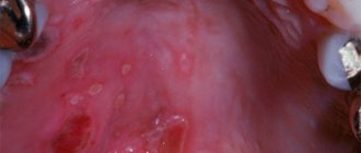
Foreign body in the oral mucosa
Another common cause of an abscess is the penetration of a foreign body into the mucous membrane or groove of the gums. This could be a bristle from a toothbrush, food debris, a fish bone, a piece of glass or a tooth. It is important to understand that only timely seeking medical help will solve the problem quickly and without consequences. Otherwise, in a short period of time, a small bump on the gum will transform into a full-fledged blister, which will have the most negative impact on the condition of neighboring teeth, and can also lead to deformation of the oval of the face.
Pathological anatomy
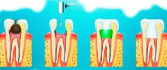
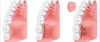
Rice.
1-3. Schematic representation of the formation of a periodontal pocket during periodontal disease (according to A. I. Evdokimov). Rice. 1. The relationship between tooth and periodontal tissue in a healthy person: 1—gingival groove (gap); 2 - interalveolar bone septum. Rice. 2. Initial stage of periodontal disease: 1 - periodontal pocket; 2—resorbable interalveolar bone septum; 3 - epithelium that has grown along the root of the tooth. Rice. 3. Severe stage of periodontal disease: 1 - periodontal and bone pocket formed as a result of resorption of the interalveolar bone septum; 2 - remains of the interalveolar bone septum. Periodontal disease usually begins with inflammation of the gums, occurring as chronic, catarrhal gingivitis (see). In the lumen of the gingival sulcus, abundant layers of loose basophilic masses are formed, including colonies of microbes and single epithelial cells. Later, on the basis of a dense supra- and subgingival plaque rich in microbial flora, tartar is formed (see), which aggravates the pathol, periodontal changes. Chronic gingivitis is accompanied by inflammatory infiltration of the gums with lymphoid, plasma cells, swelling of the gums, active proliferation, vegetation in the underlying connective tissue base of the gums, as well as increased desquamation of epithelial cells. The composition of the cellular infiltrate (lymphoid and plasma cells) is characteristic of a delayed-type hypersensitivity reaction. Already at this stage of the disease, mildly expressed lacunar resorption of the alveolar process bone is detected in the periodontal bone tissue (color fig. 1, 2).
The next stage of the pathological process is the formation of a periodontal pocket, characterized by the destruction of the epithelial cover in the area of the periodontal junction, the vegetation of epithelial layers deep into the periodontal gap. Electron microscopy, according to Carson and Sayegh (RE Carson, ES Sayegh, 1978), shows the disappearance of collagen fibrils of the periodontal ligament, which are normally woven into the cement. The contents of the periodontal pocket are necrotic detritus - structureless, basophilic and oxyphilic masses with colonies of microbes and destroyed leukocytes. The outer walls and bottom of the periodontal pocket are formed by granulation tissue, penetrated by branching layers of stratified squamous epithelium. In the bone structures of the alveolar process, pronounced resorptive changes are observed (tsvetn. Fig. 3), starting in the area of the apex of the alveolar process and leading to its complete resorption. Subsequently, resorbtin lacunae containing osteoclasts appear in the underlying sections of the walls of the dental alveoli, which leads to a gradual thinning of the interalveolar bone septa. At the same time, there is a thinning of the spongy bone in the thickness of the interalveolar septa. The resorption process can proceed as smooth resorption, lacunar resorption, and also as oncosis (as a result of bone cells acquiring the ability to osteoclase).
Against the background of patol, changes in the periodontium and as the periodontal pockets deepen in the adjacent areas of the periodontium, progression of hron and the inflammatory process is observed. Its morphological manifestations are the formation of dense lymphomacrophagic infiltrates with an admixture of plasma cells, the formation of fields of granulation tissue, and with exacerbation patol. process - solitary, and in some cases - multiple abscesses. With the progression of periodontal disease, periodontal pockets and the submersible growth of layers of stratified squamous epithelium, which are reactive in nature, reach the apexes of the roots of the teeth, and the periodontal ligament is completely destroyed. Further resorption of the bone tissue of the alveolar process leads to complete resorption of the interalveolar and interradicular bone septa (tsvetn. Fig. 3). With P., tooth tissue is involved in the pathological process quite early: cement resorption begins, which leads to the formation of deep cement and cemento-dentin niches. In parallel, the process of new formation of cement can be observed. If this process predominates, hypercementosis develops. In the pulp of teeth during P., dystrophic changes expressed to varying degrees are found, leading to its reticular atrophy, as well as to the formation of single and multiple petrificates and denticles in the pulp. Often with P., the development of retrograde pulpitis is noted, which can result in the death of the pulp.
Rice. 1. Histotopogram of the lower jaw with periodontal disease: 1 - teeth; 2 - Interalveolar bone septa with preserved contours of the apexes; 3 - areas of osteosclerosis; 4 - areas of osteoporosis; hematoxylin-eosin staining; x3. Rice. 2. Microspecimen of jaw bone tissue for periodontal disease: 1 - bone plates; 2 - artery with thickened walls and narrowed lumen; 3 — obliteration of the lumen of the vessel; hematoxylin-eosin staining; x 200.
Periodontal disease can occur with the predominant development of dystrophic changes in the periodontal tissues with characteristic atrophy of the bone trabeculae of the spongy substance, pronounced focal osteosclerosis, alternating with focal osteoporosis (Fig. 1). Patol, changes in bone tissue are combined with pronounced changes in its blood vessels: hyperplasia of the inner membrane, sclerosis and hyalinosis of the middle membrane, sharp narrowing or complete obliteration of the lumen of the vessels (Fig. 2).
In the connective tissue base of the gums, pronounced changes in blood vessels are also observed, manifested by proliferation of the endothelium, the formation of parietal thrombi, and sometimes the development of vasculitis; Against this background, swelling of collagen fibers, their fragmentation and even lysis are quite often observed. In general, the condition of the gums is characterized by the development of a sluggish inflammatory process in it; in the connective tissue stroma of the gums, an infiltrate of plasma and lymphoid cells is found, localized mainly in the area of the gingival sulcus.
Morfol, changes in the periodontium, similar to those with P. in humans, were obtained in experiments on animals.
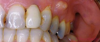
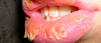
Deep damage to periodontal tissue
In the photo of gum disease, periodontal disease looks scary, especially in the last stage: the roots of the teeth are exposed and covered with hard deposits, and blood with purulent exudate leaks from the gum canals. Of course, such formidable changes are observed only when the disease is severely advanced. If the patient contacts the dentist in time, the development of the pathological process can be stopped.
Periodontal gum disease is characterized by the progressive atrophy of the tissues that surround the tooth and hold it in the jaw bones. The pathological process affects:
- gum;
- periodontal ligament;
- tooth root cement;
- alveolar processes.
With periodontal disease, the neck of the tooth becomes exposed. The gums swell and change color. Sometimes she becomes unnaturally pale or, conversely, turns red. After this, it begins to gradually retreat, exposing the root. It has a yellow tint, so it is very different in color from the crown. The patient can independently detect the disease at an early stage only if recession occurs on the front teeth. The changes will be noticeable when you smile.
Cosmetic defect is not the only problem of periodontal disease. Case histories of patients often indicate that patients experience loosening of teeth and the appearance of a putrid odor. Many patients have no pain at all, so they do not rush to the dentist for help. In some cases, tooth sensitivity increases. Pain occurs when eating sour or cold foods. And also while brushing your teeth.
The pathological process is dangerous because, starting with one tooth, it will gradually spread to neighboring areas. Without treatment, the situation will gradually worsen. She is in danger of losing her teeth. Therefore, dentists recommend carrying out preventive examinations as often as possible. Such measures allow you to identify the problem in time and take steps to stop the pathological process. Maintaining a medical history of moderate and initial periodontal disease is necessary for the dentist to monitor changes.
Clinical picture
The following stages of P. are distinguished: initial, developed and stage of stabilization (remission). P. usually does not appear immediately in the area of all teeth, but symmetry of the lesion is often observed. However, gradually the process becomes generalized and affects the entire periodontium.
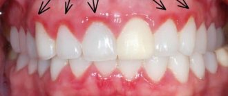
In the initial stage, P. occurs as gingivitis (usually catarrhal and hypertrophic, less often atrophic and desquamative). Bleeding gums, pain or a feeling of itching during eating and brushing teeth, anemia or hypertrophy of the gingival papillae of the tooth, burning or a feeling of numbness in the gums are noted, in most cases there is deposition of supra- and subgingival tartar.
Rice. 3. The appearance of teeth with periodontal disease: the teeth are displaced and rotated along the axis, their roots are exposed.
The developed stage of P., as a rule, is characterized by the presence of generalized gingivitis, pasty gums with symptoms of congestive hyperemia, their bleeding, the presence of periodontal pockets 5-6 mm deep or more, in most cases with the release of pus from them. There is an abundant deposition of dense subgingival calculus, which, unlike supragingival calculus, penetrates deep into the periodontal pockets in the form of thin granular layers, tightly attached to the roots of the teeth. The teeth become mobile, often shift, rotate along an axis (Fig. 3), and fall out. Free spaces form between teeth that were previously in close contact (see Diastema, Trema). Moving teeth injure surrounding tissues and increase inflammation. When the necks and roots of the teeth are exposed, their exposed areas are subject to prolonged irritation when eating, inhaling cooled air, talking, and become a source of pain. Displacement of teeth, impaired diction are noted, and some patients develop bad breath. The general condition, as a rule, suffers slightly, sometimes there is a low-grade fever. But in some cases the condition of patients worsens sharply.
The stage of stabilization (remission) occurs mainly after complex treatment. It is characterized by a subsidence of the inflammatory process and a decrease in tooth mobility, a decrease in the depth of periodontal pockets or their elimination, which occurs, for example, after surgical treatment of P.
In addition to P., which occurs in accordance with the described stages, P. occurs with a predominance of dystrophic changes, which proceeds slowly, without a tendency to exacerbation. At the same time, due to the uniform loss of bone and gum atrophy, periodontal pockets do not form, the teeth remain stable even in the case of significant atrophy of the bone of the alveolar processes. Patients complain of a feeling of itching and numbness in the gums, pain in the exposed necks of the teeth or in the area of wedge-shaped defects in the teeth. P. can be complicated by periodontitis (see), periostitis (see), inflammation of regional lymph nodes (see Lymphadenitis), ascending pulpitis (see), osteomyelitis of the jaws (see), phlegmon of the perimaxillary spaces and neck.
Periodontal disease: symptoms
The disease “periodontal disease” is characterized by several diagnostic signs, on the basis of which a preliminary diagnosis can be made:
- the gums and mucous membrane are pale, there is no inflammation;
- gradual receding gums: at the initial stage of the disease, the necks of the teeth are exposed, at the later stages - the roots;
- with mild severity, there are no periodontal (gum) pockets, and this is the main distinguishing symptom of periodontal disease from periodontitis and other periodontal diseases;
- There are practically no dental plaques, or they are weakly expressed;
- periodontal disease is combined with enamel erosion, wedge-shaped defect, and pathological abrasion of teeth;
- Even with significant loss of bone tissue, the teeth remain stable.
Such symptoms allow one to suspect periodontal disease at home, but to confirm the diagnosis, consultation with a specialist is necessary.
How does periodontal disease manifest in women?
Women often come to the dentist with complaints of tooth hypersensitivity, non-carious lesions of teeth and gum loss - the first possible symptoms of periodontal disease. Due to their nature and greater responsibility, periodontal disease in women can be diagnosed in the early stages of development.
During pregnancy or menopause, the symptoms of periodontal disease are expressed in significant loss of gums and rapid progression of the disease; the gums and interalveolar septa quickly reduce their height, which causes many inconveniences: dental hypersensitivity, aesthetic problems, nutritional disorders.
Symptoms of gum periodontal disease in men
In men, the disease "periodontal disease" in the vast majority of cases is diagnosed in the later stages of development. Men more often ignore preventive examinations and go to the dentist when the symptoms of periodontal disease are pronounced and interfere with normal life.
The main complaints of patients are reduced to exposure of the roots of the teeth, burning sensation in the mucous membrane and a decrease in its sensitivity. Sometimes you can see the addition of inflammation, which is why periodontitis is diagnosed, but the lack of positive dynamics in treatment prompts doctors to make a correct diagnosis and draw up a plan for effective treatment of periodontal disease.
Diagnosis
The diagnosis is established on the basis of patient complaints, wedges, pictures, data from radiological, laboratory and functional research methods.
Rice. 4. Panoramic radiograph of the lower jaw with periodontal disease: resorbing interalveolar bone septa, having a characteristic “eaten away” appearance (indicated by arrows).
Roentgenol, P.’s picture depends on the stage of the process. In the initial stage, osteoporosis (see) and destruction of the cortical substance of the apices of the interalveolar septa are detected. In the developed stage, truncation of the apices of the interalveolar septa is noted; along with horizontal resorption, vertical resorption also appears, often with the formation of bone pockets. The contours of the alveolar processes have a characteristic scalloped, “eaten” appearance (Fig. 4), and the deposition of subgingival tartar is determined. The increase in patol, the process is manifested by the appearance of foci of spotted osteoporosis and the formation of bone abscesses. The stabilization stage is radiographically manifested by compaction of the contours of the ridges of the interalveolar septa and the disappearance of foci of spotty osteoporosis.
Rice. 5. Panoramic radiograph of the lower jaw with periodontal disease: 1 - uniformly reduced interalveolar bone septa; 2 - foci of osteoporosis; 3 - focus of osteosclerosis; 4 - narrowed periodontal fissures; 5 - tartar.
The X-ray picture of periodontal disease with a predominance of dystrophic changes in the periodontium is characterized by a uniform decrease in the height of the interalveolar bone septa, sclerosis of the bone tissue of the alveolar process, and narrowing of the periodontal gap (Fig. 5).
Wedge, assessment of the periodontal condition is carried out on the basis of bone loss of the alveolar processes, the presence and intensity of inflammation in the tissues of the gingival margin and is expressed in the form of special periodontal indices. The degree of hygienic condition of the oral cavity is determined using colorimetric tests.
Differential diagnosis
carried out with eosinophilic granuloma, Lefebvre-Papillon syndrome, mobility of individual teeth against the background of their long-term trauma by antagonist teeth in case of occlusion. If there is an eosinophilic granuloma in the alveolar process (see) rentgenol, the picture is characterized by diffuse resolution of the bone, extending beyond the periodontal tissues. With Lefebvre-Papillon syndrome rentgenol, the picture is similar to that of the developed stage of P., however, palmoplantar keratosis is characteristic of this syndrome (see Lefebvre-Papillon syndrome). In the case of traumatic occlusion, the diagnosis is clarified by comparison with an x-ray of the teeth of the opposite side.
What is the essence of pathology
As the pathology develops, a cavity is formed in the thickness of the periodontal tissue, inside which purulent exudate begins to accumulate. The source of inflammation is usually localized in the immediate vicinity of the causative tooth. In fact, this is a small formation inside the gingival tissue, which gradually appears in the form of a lump on the surface of the mucosa, often with a fistula. The patient experiences obvious discomfort and acute pain.
Periodontal abscess, how does it manifest?
Typically, the cause of the problem is voluminous periodontal pockets and hard subgingival deposits. Most often, the acute form of the disease is diagnosed, while only 30% of patients who seek help suffer from chronic periodontal abscess1. This kind of trouble can occur in both adults and children; neither men nor women are equally immune from it. However, it is worth noting that the peak incidence occurs in the autumn-spring period, when most people have weakened immunity.
Treatment
P.'s treatment should be comprehensive, as individualized and systematic as possible. Complex to treat activities include local and general effects on patol, the process in periodontium.
Local treatment involves the elimination of local periodontal factors, removal of supra- and subgingival dental plaque, anti-inflammatory treatment and curettage of periodontal pockets, increasing the activity (or correction) of blood circulation in the periodontium and increasing the reactivity of its structures. Orthopedic treatment of P. is widely used—selective grinding of teeth, splinting, and clasp prosthetics. For the treatment of the initial stages of P., the use of ion-exchange membranes containing various microelements and biologically active substances is effective. In the treatment of periodontal pockets, antiseptics (hydrogen peroxide, thiobiargen solution, ethacridine lactate, iodinol, dimexide, etc.), enzymes (trypsin, chymotrypsin, hygrolithin, ribonuclease), glucocorticoids (hydrocortisone, prednisolone, dexazone, etc.), drugs are used. medicinal herbs (salvin, romazulan, carotolin, chlorophyllipt, etc.), antitrichomonas drugs (metronidazole, fasigin, etc.), protein anabolizers (methyluracil, pentoxyl). Treatment of periodontal pockets is carried out by rinsing with a syringe, instillations and medicinal gum bandages. When radically treating periodontal pockets in order to remove tartar, granulations, as well as deep-growing epithelium, deep curettage is performed - curettage according to Znamensky and Younger-Sachs using a periodontal set or special instruments. Sometimes cryo- and vacuum curettage are used using special devices. In some cases, they resort to gingivectomy or gingivotomy (excision or dissection of a section of the gum) according to the depth of the periodontal pockets or the so-called. patchwork operations. One of them is the Widmann-Neumann-Cieszynski operation, during which a mucoperiosteal flap is cut out from the vestibular side of the alveolar process in the area of four-six teeth and the affected periodontal tissue is exposed. This makes it possible to radically remove granulations and tartar. The mucoperiosteal flap is then placed in place and secured with sutures. However, after this operation there is significant exposure of the necks and roots of the teeth, which limits the indications for it. Favorable results are obtained from operations using allo- and explants. In order to eliminate hyperesthesia of the necks of teeth, fluoride varnish, applications of 10% calcium gluconate solution, 1 - 2% sodium fluoride solution, fluoride paste, etc. are used, and physiotherapeutic methods of treatment are also used.
General treatment is aimed at eliminating inflammatory changes in the periodontium, hyposensitization of the body, increasing the reactivity of the body and periodontium, preventing lesions (primarily microcirculatory) periodontal vessels, as well as treating common concomitant P. diseases. In order to reduce the intensity of inflammation in the periodontium in cases of severe process, broad-spectrum antibiotics (lincomycin, oleflavit, rondomycin), sulfonamides (biseptol) are used in the pre- and postoperative periods. To hyposensitize the body, diazolin, pipolfen, calcium chloride, and tavegil are used. Fluorine preparations, prodigiosan, ATP, trental, venoruton, antioxidants, adaitogens, etc. are used as agents for pathogenetic therapy. In the presence of diseases aggravating the course of P. (diabetes mellitus and other endocrine diseases, diseases of the gastrointestinal tract, cardiovascular systems), treatment of P. itself is combined with treatment of the disease aggravating the course of P.
Physiotherapeutic treatment is most effective in the initial stage of P. When using these methods, blood circulation and trophism of periodontal tissues improve. Physiotherapy is carried out using electro- and phonophoresis of heparin, antioxidants (dibunol, galascorbine, vitamin E), prostaglandin inhibitors (aspirin, indomethacin, ATP, trental). Hydro-, electro- and vibration therapy are used to treat periodontal disease, and hydrotherapy usually precedes other treatment methods. Electrophoresis of vitamins B1, C, etc. is carried out (see Electrophoresis). The use of pulse currents (see) - diadynamic, fluctuating and sinusoidal modulated currents - is important. Darsonvalization (see) and helium-neon laser are also used. Laser therapy (see Laser) reduces inflammation, normalizes blood circulation in periodontal tissues, and stimulates metabolic and regeneration processes. After curettage, local ultraviolet irradiation is indicated, which reduces tissue swelling and improves epithelization of the wound surface. Usually after 2 weeks. After surgical interventions, calcium or fluoride electrophoresis is prescribed. It is advisable to use resort factors (balneotherapy and peloid therapy).
In case of tooth mobility, secondary traumatic occlusion and purulent discharge from periodontal pockets, electrotherapy and massage are contraindicated; Hydrotherapy with the addition of medications is recommended.
Diagnostics
Diagnosis of periodontal disease begins with a visual examination, and the first signs of the problem are excessive formation of tartar with atypical exposure of the upper peri-cervical part of the teeth. It is recommended to go to the doctor for an emergency examination in the following cases:
- exposure of the neck or root of the teeth with the absence of severe inflammation;
- gum retraction;
- the presence of periodontal (gum) pockets;
- violation of the inclination of the teeth, the appearance of gaps;
- diagnosing metabolic disorders, problems with the endocrine or cardiovascular system.
Prognosis and Prevention
Forecast.
Treatment carried out according to indications slows down or stops the process. As a result of rational comprehensive treatment of periodontal disease, recovery with residual effects (relief) and stable recovery (stabilization of the process) can occur. Untimely treatment and irrational use of pathogenetic therapy lead to the gradual loss of several or all teeth.
P.'s prevention is carried out with mandatory consideration of professional characteristics, climatic and geographical conditions and concomitant diseases. Complex prevention includes clinical examination of patients, which is aimed at identifying the initial stages of P.; carrying out preventive measures, organizing specialized periodontal centers, departments and offices.
Primary preventive measures are aimed at developing hygienic skills, timely treatment of inflammatory changes in the tissues of the gingival margin, regular removal of dental plaque, treatment of dentition anomalies, and rational orthopedic treatment.
The complex of secondary preventive measures includes the elimination of early signs of pathol, changes in the oral cavity - monitoring its hygienic condition, eliminating factors that contribute to the retention of food accumulations in the interdental spaces, and preventing dental caries. Therapeutic, surgical and orthopedic measures aimed at eliminating pathol, the process, preventing complications and restoring the function of the masticatory apparatus are carried out both for therapeutic purposes and to prevent the progression of the process and its complications. They use means and methods aimed at stimulating microcirculation and trophism of periodontal tissues using physiotherapeutic methods, finger massage, rinsing the mouth with water at a contrasting temperature, as well as removing dental plaque. According to indications, surgical relocation of the frenulum and formation of the vestibule, elimination of primary traumatic contacts between teeth using selective functional grinding of the cusps, elimination and replacement of incorrectly made fillings and dentures, treatment of bruxism (see) and dentoalveolar anomalies are carried out.
The scheme of preventive recommendations should include a fractional balanced diet with a sufficient amount of fresh vegetables and fruits, vegetable oils, lactic acid products and others.
Bibliography:
Evdokimov A.I. On the issue of pathological anatomy, etiology and pathogenesis of periodontal disease (periodontal disease), Dentistry, No. 6, p. 10, 1939; Evdokimov A.I. and Nikitina T.V. Criteria for the curability of periodontal disease, ibid., t. 56, No. 5, p. 14, 1977; Shizhina N. A. and Prokhonchukov A. A. The role of disorders of the functions of internal organs and body systems as common background factors in the pathogenesis of periodontopathies, M., 1971; Ivanov V. S. Periodontal diseases, M., 1981; Kabakov V.D. and Belchikov E.V. Issues of immunology of periodontal disease, L., 1972; Kodola N.A., Khomutovsky O.A. and Tsentilo T.D. Periodontal disease, ultrastructure of the gums and pulp, Kyiv, 1980; Kopeikin V. N. Orthopedic treatment of periodontal diseases, M., 1977; Lemetskaya T. I. Periodontal diseases (periodontopathies), M., 1972; Novik I. O. Periodontal disease (pathogenesis, clinic, treatment), Kyiv, 1964; Platonov E. E. Tasks and ways of studying periodontopathies, Dentistry, No. 5, p. 3, 1964; Prokhonchukov A. A. and Zhizhina N. A. Pathogenesis of experimental periodontopathies, M., 1971; Guide to therapeutic dentistry, ed. A. I. Evdokimova and A. I. Rybakova, M., 1967; Rybakov A.I. and Ivanov V.S. Clinic of therapeutic dentistry, p. 192, M., 1980; Proceedings of the 6th All-Union Congress of Dentists, M., 1976; Experimental and clinical dentistry, ed. V. V. Panikarovsky, M., 1978; Experimental and clinical dentistry, ed. A. I. Rybakova, vol. 4, p. 89, 98, M., 1973; Epidemiology, etiology and prevention of periodontal diseases, Report of the WHO scientific group, M., 1980; Bernard R. D. La ionoforesi, Dent. Cadm. (Milano), v. 46, no. 10, p. 49, 1978; Carson RE, Sayegh FS a. Fedi PF Osteoclastic resorption of alveolar bone affected by periodontitis - correlation of light microscopic and scanning electron microscopic observations, J. Periodont., v. 49, p. 406, 1978; Glickmanl. Clinical periodontology, Philadelphia, 1979; Goldman H. M. a. Cohe n DW Periodontal therapy, St Louis, 1973; K 6-tzschkeH. Leitfaden der Parodontologie, Lpz., 1969; Kostlan J. a. Skash M., Onemocneni parodontu, Praha, 1967; Page RCa SchroederH.E. Pathogenesis of inflammatory periodontal diseases, Lab. Invest., v. 34, p. 235, 1976; Physikalische Therapie in der Stomatologie, hrsg. v. K. Jarmer, B., 1960; Waernaug J. Subgingival plague and loss of attachment in periodontosis as observed in autopsy material, J. Periodont., v. 47, p. 636, 1976.
V. S. Ivanov, T. I. Lemetskaya, T. V. Nikitina; R. I. Mikhailova (physiot.), V. V. Panikarovsky (pathological an.), A. A. Prokhonchukov (etiol., pathology).
Features of nutrition for periodontal disease
Periodontal disease imposes its own characteristics on the choice of products and menu planning. The main thing is to load the maxillofacial apparatus and give it as much chewing load as possible. Thus, it is possible to stimulate metabolic processes and give the body everything it needs to restore bone tissue. When chewing solid food, you must try to give an even load on the periodontal tissue - do not chew on one side.
The daily diet should include fresh vegetables and fruits, dairy products containing phosphorus and calcium. It is recommended to consume foods containing polyunsaturated fatty acids: vegetable oil, fish and seafood.
For the treatment and prevention of periodontal disease, it is recommended to include in the diet foods high in vitamins C, E, A, K, B6: vegetable oils, vegetables, fruits, yeast, legumes, berries. Due to vitamin B6 deficiency, mineral metabolism of bone tissue may be disrupted. The diet should include foods rich in minerals.
It is necessary to limit or completely eliminate: coffee, sweet soda, any drinks with dyes, sweets, foods with a lot of flavoring additives.