Device principle
The throat is a highly complex organ responsible for breathing, speaking, and moving food.
In short, its structure is based, as we said earlier, on the pharynx (pharynx) and larynx (larynx). Since this organ is a conducting channel, it is very important that all its muscles work harmoniously and correctly. Inconsistency in their activities will lead to the fact that food can enter the respiratory system and create a dangerous situation, even leading to death.
The structure of a child's throat is the same as that of adults. But children have narrower cavities and tubes. As a result, any disease in which swelling of these tissues occurs can be extremely dangerous. It is advisable for a person to know the structure of such an organ, as this can be useful in caring for it and during treatment. In the pharynx, the nasopharynx and oropharynx are distinguished.
Larynx
The larynx (Latin: larynx) is a tube reinforced with cartilage that passes air into the trachea and then into the lungs. The larynx is the first part of the lower respiratory tract. The structure of the larynx is represented by cartilages, paired and unpaired.
The paired ones include a pair of vocal cartilages, and the unpaired ones include the thyroid cartilage, the annular cartilage and the epiglottis. In addition, there are several small paired cartilages that are usually part of the ligaments. They are connected together by joints and links to form a moving device.
The structure of the throat is as follows:
- the larynx connects to the pharynx - a common part of the digestive and respiratory systems - and passes into the trachea;
- the entire larynx is movably attached under the jaw with the help of the hyoid bone, muscles and ligaments, its walls are sealed with cartilage;
- The largest cartilage of the thyroid gland creates a sexual difference, in males protruding forward and forming the Adam's apple.
Laryngeal muscles
The entire human throat is, first of all, muscles. The striated muscles are involved in the structure of the larynx. They control the movement of cartilage, determine the tension of the vocal cords and the width of the gap between them. The laryngeal muscles are divided into 3 groups.
Anterior muscles:
Musculus cricothyroideus - located from the anterior part of the annular cartilage to the lower edge of the thyroid cartilage. This muscle is divided into 2 parts: vertical and oblique. This muscular structure tilts the thyroid cartilage forward, simultaneously straining the vocal cords.
Lateral muscles
The lateral muscles include the following:
- cricoarytenoid lateralis;
- Thyroarytenoideus;
- thyroepiglotticus.
- M. cricoarytenoideus lateralis. It is located from the upper edge of the annular cartilage to the vocal protrusion. Performs internal rotation of the vocal cartilage and compresses the vocal cords.
- M. thyroarytenoideus. Located around the vocal cords, from the thyroid cartilage to the vocal cord. Promotes compression of the vocal cords.
- M. thyroepiglotticus. It is located from the cartilage of the thyroid gland to the edge of the epiglottis, extends beyond the epiglottis and expands the entrance to the larynx.
Posterior muscles
We refer to the posterior muscles as follows:
- cricoarytenoid posterior;
- arytenoideus.
- M. cricoarytenoideus posterior. It is located from the back of the annular to the prominence of the vocal cartilage. Plays a role in the inclination of the vocal cartilage and its rotation, at the same time opening the gap between the vocal muscles and tightening them. People with pathology of this muscle have a significant disturbance in sound and an obstacle to the free passage of air during breathing (they cannot take a “full” breath).
- M. arytenoideus. Connects the vocal cartilages at the back. The muscle has 2 sections - transverse and oblique. Its function is to reduce the gap between the vocal cords. This muscle includes a component that connects to the epiglottis, deflecting it back and narrowing the entrance to the larynx.
Anatomical structure of the throat
The structure of the throat consists of several parts: pharynx, larynx, trachea. To correctly diagnose the disease, it is necessary to carefully study the anatomy of the throat and analyze all its components in detail. Pathology can form in any area. Therefore, knowledge of the anatomy of the throat is one of the most important areas in otolaryngology.
If we talk about how the throat works, then in its structure it looks like an inverted cone, located near the 4th and 6th vertebrae. It originates from the hyoid bone, descends and passes into the trachea.
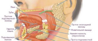
The diagram of the human throat is complex and is divided into several parts:
- The pharynx, which includes the nasopharynx, oropharynx, and swallowing department.
- The larynx, which is lined with tissue structures, blood and lymph vessels, nerves, glands, cartilage and muscles.
Detailed anatomy of the throat can be seen in the photo.
The pharynx, another name is the pharynx. It starts at the back of the mouth and continues down the neck. The wider part is located at the base of the skull for strength. The narrow lower part connects to the larynx. The outer part of the pharynx continues with the outer part of the mouth - it has quite a lot of glands that produce mucus and help moisten the throat during speech or eating.
When studying the anatomy of the pharynx, it is important to determine its type, structure, functions and risks of disease. As mentioned earlier, the pharynx is shaped like a cone. The narrowed part merges with the laryngopharynx, and the wide side continues the oral cavity. There are glands that produce mucus and help moisten the throat during communication and eating.
The larynx is located as follows:
- opposite 4 - 6 cervical vertebrae;
- behind - the laryngeal part of the pharynx;
- in front - formed due to the group of hyoid muscles;
- above - hyoid bone;
- lateral - adjacent to the thyroid gland with its lateral parts.
The structure of a child's pharynx has its own differences. Tonsils in newborns are underdeveloped and do not function at all. Their full development is achieved by two years.
The larynx includes in its structure a skeleton, which contains paired and unpaired cartilages connected by joints, ligaments and muscles:
- unpaired consist of: cricoid, epiglottis, thyroid.
- paired ones consist of: corniculate, arytenoid, wedge-shaped.
The muscles of the larynx are divided into three groups and consist of:
- thyroarytenoid, cricoarytenoid, oblique arytenoid and transverse muscles - those that narrow the glottis;
- posterior cricoarytenoid muscle - is paired and expands the glottis;
- vocal and cricothyroid - strain the vocal cords.
Entrance to the larynx:
- behind the entrance there are arytenoid cartilages, which consist of cornuform tubercles, and are located on the side of the mucous membrane;
- in front - epiglottis;
- on the sides there are aryepiglottic folds, which consist of wedge-shaped tubercles.
The laryngeal cavity is also divided into 3 parts:
- The vestibule tends to stretch from the vestibular folds to the epiglottis.
- Interventricular section - stretches from the inferior ligaments to the superior ligaments of the vestibule.
- Subglottic region - located at the bottom of the glottis, when it expands, the trachea begins.
The larynx has 3 membranes:
- mucous membrane - consists of multinucleated prismatic epithelium;
- fibrocartilaginous membrane - consists of elastic and hyaline cartilages;
- connective tissue - connects part of the larynx and other formations of the neck.
The larynx (larynx) is lined with various tissue structures, blood and lymphatic vessels, and nerves. The mucous membrane, covered from the inside, consists of multilayered epithelium. And underneath there is connective tissue, which in case of illness manifests itself as swelling. When studying the structure of the throat and larynx, we observe a large number of glands. They are absent only in the region of the edges of the vocal folds.
See the photo below for the structure of the human throat with a description.
The larynx is located in the throat in the shape of an hourglass. The structure of the larynx in a child differs from that of an adult. In infancy, she is two vertebrae higher than normal. If in adults the plates of the thyroid cartilage are connected at an acute angle, then in children they are at a right angle. The structure of the larynx in a child is also distinguished by a long glottis. In them it is shorter, and the vocal folds are of unequal size. The diagram of a child’s larynx can be seen in the photo below.
Another element in the structure of the human throat and larynx is the oropharynx.
This fragment is located behind the oral cavity. Its main function is to conduct air flow from the mouth to the respiratory organs. This part is more mobile compared to the nasopharynx. Thanks to this, when the muscle tissue of the oral cavity contracts, a person can speak.
We already know that the structure of the throat has certain components, but they will also include other, even smaller components. Among them is the tongue, which helps, by contracting the muscular systems, move food into the esophagus. There are also tonsils, which are very often involved in throat diseases.
The pharynx (Latin: pharynx) is the common area of the digestive and respiratory system through which food moves to the esophagus and stomach. The pharynx plays a very important role in both breathing and swallowing.
The pharynx is a funnel-shaped tube that connects the nasal cavity and mouth to the stomach. The pharynx is located at the back of the throat, its length is about 15 cm. Similarly, as in the case of the structure of the throat and larynx, the surface of the pharynx is lined with a mucous membrane, which protects it from the effects of gastric juices. The structure of the pharyngeal wall consists of ligaments and muscles.
The pharynx is located on the cranial base, and at the level of the annular cartilage of the larynx passes into the tubular esophagus. The border between the nasal and oral sections is formed by the soft palate. It serves as a “crossroads” between the respiratory and swallowing tracts.
The pharynx consists of 3 parts:
- nasopharynx,
- mouth part,
- laryngeal part.
The nasopharynx (nasopharynx, pars nasalis) balances the pressure between the nasopharynx cavity and the middle ear cavity, the posterior wall of which is lymphatic tissue.
The oropharynx (otopharynx - oropharynx, pars oralis) lies behind the oral cavity. In the oropharyngeal wall, lymphatic tissue extending from the root of the tongue is associated with the palatine tonsils. This part of the pharynx is a component of the body's defense system; it is a barrier located at the site of the most frequent penetration of infections.
The laryngeal part (larynopharynx, pars laryngea) is the third part of the pharynx. It continues down from the oropharynx to the C6 vertebra, where the pharynx passes through the esophagus. At the junction there is a narrowing between the laryngeal cartilages (anteriorly) and the spine (posteriorly).
The structure of the human larynx and throat with photos and descriptions: diseases and pathologies
The throat is a human organ that is classified as the upper respiratory tract.
Functions
The throat helps move air to the respiratory system and food through the digestive system. Also in one part of the throat there are ligaments and a protective system (prevents food from getting past its path).
Anatomical structure of the throat and pharynx
The throat contains a large number of nerves, important blood vessels and muscles. There are two parts of the throat - the pharynx and larynx. Their trachea continues. The functions between the parts of the throat are divided as follows:
- The pharynx moves food into the digestive system and air into the respiratory system.
- The vocal cords work thanks to the larynx.
The photo shows the ligaments during laryngoscopy
Swallowing department
The lowest section of the pharynx with a self-explanatory name. It has a complex of nerve plexuses that help maintain synchronous functioning of the pharynx. Thanks to this, air enters the lungs, and food enters the esophagus, and everything happens at the same time.
Diseases, pathologies and injuries
The following problems exist:
Related problems that cause sore throat:
- Smoking
- Smoke inhalation
- Inhaling dusty air
- acute respiratory infections
- Whooping cough
- Scarlet fever
- Flu
To determine the exact cause of your throat pain and irritation and to prescribe appropriate treatment, consult your doctor immediately.
Popular video on the structure and functions of the larynx:
Source: https://gidmed.com/otorinolarintologija/obshhie-svedeniya-ot/stroenie-gortani.html
What is the throat, larynx and pharynx
The pharynx (pharynx) is a cone-shaped structure turned upside down. It is located behind the mouth and goes down to the neck. The cone is wider at the top. It is located near the base of the skull, which gives it more strength. The lower part is united with the larynx. The layer of tissue covering the outside of the pharynx is a continuation of the layer of oral tissue lying outside. It has many glands that produce mucus, which is involved in the processes of moistening the throat when eating and talking.
A common misconception is to call the throat only the small area behind the tongue that turns red and hurts with sore throats. In this case, what is located below is often excluded. For example, there is a difference between the throat and larynx because it is part of this system below and is connected to the pharynx and trachea.
The throat is not an anatomy term; it is the common name for the part of the upper respiratory tract from the hyoid bone to the level of the clavicle or manubrium of the sternum. The throat contains:
- pharynx or oropharynx - begins in the visible part of the mouth, the entrance gates are the tonsils, they are also tonsils that do not allow infections below;
- nasopharynx - cavities located above the palate;
- swallowing department - a small area behind the epiglottis that pushes food and liquids into the esophagus;
- The larynx is a cartilaginous tube for air, lined with mucous membrane and blood vessels.
The epiglottis serves as a valve that prevents food and water from entering the larynx, at the top of which are the vocal folds (cords). Their closing and opening gives us the opportunity to make sounds. The larynx is protected in front by the thyroid cartilage, and behind it is the esophagus.
The pharynx, or as it is otherwise called “pharynx,” is located behind the oral cavity and extends down the neck. The shape of the pharynx is a cone turned upside down. The upper part of the cone, wider, is located at the base of the skull - this gives it strength. The lower part, narrower, is connected to the larynx.
Nasopharynx
The pharynx consists of three parts. These parts have their own location and perform certain functions. The uppermost part is the nasopharynx. From below, the nasopharynx is limited by the soft palate and when swallowing, the soft palate moves upward and covers the nasopharynx, thereby preventing food from entering the nose. The upper wall of the nasopharynx has adenoids.
Oropharynx
The oropharynx is the part of the pharynx that is located behind the oral cavity. The main function of the oropharynx is to promote air flow from the mouth to the respiratory organs. The nasopharynx is less mobile than the oropharynx. Therefore, as a result of contraction of the muscle mass of the oral cavity, speech is formed. In the oral cavity there is a tongue, which, with the help of the muscular system, helps move food into the esophagus and stomach. But the most important organs of the oropharynx are the tonsils, which are most often involved in throat diseases.
The lowest part of the throat performs the function of swallowing. The movements of the throat must be very clear and synchronized to simultaneously ensure the penetration of air into the lungs and food into the esophagus. This is achieved through a complex of nerve plexuses.
Anatomy of the pharynx
The pharynx is a funnel-shaped canal, which is the beginning of the digestive tube running from the oral cavity to the esophagus. There are 3 parts of the pharynx:
- nasopharynx;
- oropharynx;
- hypopharynx.
The nasopharynx is a cavity that connects the upper part of the pharynx with the nasal passages. The ear-nose-throat system is interconnected in every possible way. For example, two walls of the nasopharynx, located on the sides, are connected to the mouths of the auditory tubes.
A special accumulation of lymphoid tissue in which protective lymphocytes multiply is located on the posterior-superior wall of the nasopharynx and forms the nasopharyngeal tonsil
In case of serious diseases, the tissue can grow, filling the entire space of the nasopharynx. Such growths are called adenoids and require surgical removal.
In the nasopharynx, the inhaled air is warmed and purified. In addition, this part of the pharynx is designed as a resonator, thereby slightly modifying the voice. The nasopharynx is followed by the oropharynx or middle part of the pharynx. It is partially separated from the upper part by the hard palate.
The oropharynx is lined with mucous tissue, under which there are muscles. The muscles help push the bolus of food further into the esophagus. It is noteworthy that they are in constant motion, helping the pharynx to perform many imperceptible actions: swallowing saliva, inhaling air, etc.
From below, the oropharynx is limited by the root of the tongue, near which there is also a special spherical accumulation of lymphoid tissue - the lingual tonsil. The oropharynx smoothly passes into the laryngopharynx. The lower part of the pharynx begins immediately from the lingual tonsil and then passes into the esophagus. All 4 tonsils form the pharyngeal lymphadenoid ring.
In cases where the tonsils no longer cope with their main task - protection against germs and bacteria - and lead to complications in diseases, it is recommended to remove them.
The human pharynx connects the esophagus and the oral cavity
The pharynx is one of the resonators of the voice. It is where the respiratory and digestive tracts intersect. The physiology of the pharynx is such that food and air do not intersect, due to the fact that the corresponding channels reflexively open or close.
Swallowing is so synchronized with breathing that it only takes a couple of seconds for the pharynx to transform to accept a bolus of food. Swallowing itself is divided directly into several phases:
- Free Chewed and compacted food moves towards the pharynx, and then with the help of the tongue it is pressed against the hard palate, swallowing occurs, which cannot be interrupted naturally.
- Pharyngeal. On the back wall of the pharynx there are special receptors that are irritated by a bolus of food. A signal is sent to the brain that allows pharyngeal muscle contractions to occur.
- Esophageal. Food passes into the upper esophagus and then enters the stomach.
There are numerous taste buds on the soft palate and the root of the tongue, which help analyze taste and send the appropriate signal to the brain. It is designed so that when a foreign body enters the throat, the muscles of the pharynx reflexively contract, which is a protective function of the body.
The surface of the pharynx is lined with epithelial tissue. The mucous membrane has a large number of glands that secrete the necessary mucus. On the sides of the pharynx, near its walls, there are arteries and veins that provide the necessary blood supply.
Nasopharyngeal compound
The structure of the throat and larynx includes the structures that form them, such as the nasopharynx and oropharynx mentioned above. Let's consider one of them.
The nasopharynx is the part of the pharynx that occupies the upper position. It is limited from below by the soft palate, which begins to move upward during the process of swallowing. Thus, it covers the nasopharynx. This is necessary to protect it from food particles entering the respiratory tract. In the upper wall of the nasopharynx there are adenoids - tissue accumulations located behind its wall. This organ also has a tunnel connecting the throat to the middle ear. This formation is called the Eustachian tube.
How does the throat work?
In the most general terms, the structure of the throat is made up of two main parts: the pharynx (pharynx) and the larinx (larynx). The pharynx is responsible for moving food into the stomach and bringing air into the lungs. The main function of the larynx is to regulate the functioning of the vocal cords, that is, to provide us with the ability to speak.
The structure of the throat in a child and an adult is the same, but in children all the cavities and tubes located here are much narrower. Because of this, any disease accompanied by swelling of the tissues in this area can be dangerous in terms of blocking the airways.
Our throat
When we imagine the structure of the human throat, we first of all have in mind the pharynx, which is scientifically called the pharynx. It is located behind the oral cavity and goes down. In shape it resembles a cone with a narrowing downward. The upper expanded part of the pharynx provides the strength of this organ. The narrow lower part is connected to the larynx.
Since both food and air pass through the pharynx through different channels, the work of the muscles that block one of these channels is very important, otherwise food particles risk entering the respiratory system and leading to disastrous results.
Nasopharynx
The nasopharynx is the cavity that connects the inside of the nose and the top of the throat. Choanae, special holes, provide this connection. Next, the nasopharynx, descending down, passes into the middle pharynx. On the sides of the nasopharynx are the openings of the auditory tubes. The inside of the nasopharyngeal cavity is lined with a mucous membrane, which is penetrated by blood vessels, nerve endings and mucus-producing glands.
The structure of the nasopharynx allows it to perform several functions. It warms the air entering the body, moisturizes it, traps dust and germs, and also gives us the opportunity to smell.
Oropharynx
The structure of the mouth and throat is easy to see in the mirror. The oropharynx is the middle part of the throat. It is limited by the soft palate and the hyoid bone.
Main parts of the oropharynx
- Solid sky,
- Tongue,
- Tonsils.
The tonsils are one of the most important parts of the pharynx, providing our protection against infection. The oropharynx contains the palatine tonsils, also called tonsils. They are a collection of lymphoid tissue that produce substances that resist pathogenic microbes.
The oropharynx connects the oral cavity to the larynx. Its main function is to pass air flow to the bronchi and lungs.
Anatomy of cartilage
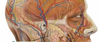
The epiglottis (Fig. 2, a, 4) consists of elastic cartilage, which enters the superior notch of the thyroid cartilage with the so-called stalk and is attached from the inside to the plates of this cartilage, forming the tubercle of the epiglottis (b, 1). The posterior surface of the epiglottis is covered with numerous pits in which the grape-shaped mucous glands are located. Inflammation often develops in these glands, ending in an abscess of the epiglottis.
Rice. 2. Rear view of the larynx: a - muscles of the larynx: 1 - uvula; 2 - palatine tonsil; 3 - root of tongue; 4 - epiglottis; 5 - aryepiglottic muscle; 6 - oblique arytenoid muscles; 7 - cricothyroid muscle; 8 - posterior cricoarytenoid muscle; 9 — plate of the cricoid cartilage; 10 - transverse arytenoid muscle;
11 - lateral lingual-epiglottic fold; b — laryngeal cavity: 1 — tubercle of the epiglottis; 2 - ventricular fold; 3 - vocal fold; 4 - external thyroarytenoid fold; 5 - cricoid cartilage; 6 - thyroid gland; 7 - cricothyroid muscle; 8 - vocal muscle; 9 - ventricles of the larynx; 10 - thyroid cartilage
The internal structure of the larynx is shown in Fig. 3. The anterior surface of the epiglottis is connected to the body and horns of the hyoid bone through the broad ligament (a, 7). In children and some adults, the epiglottis is presented in the form of a semi-folded sheet covering the entrance to the larynx. Such an epiglottis is a significant obstacle when examining the larynx using indirect laryngoscopy.
Rice. 3. Internal structure of the larynx with the right plate of the thyroid cartilage removed: a - elastic cone and quadrangular membrane: 1 - hypoglottic ligament; 2 - medial cricothyroid ligament; 3 - quadrangular membrane; 4 - thyroid cartilage; 5 - fold of the vestibule; 6 - vocal fold;
7 - elastic cone; 8 - cricoid cartilage; 9 - sublingual-thyroid membrane; 10 - lateral hyoid-thyroid ligament; b — muscles and ligaments of the larynx (right side; sagittal midline section): 1 — lateral hyoid-thyroid ligament; 2 - medial cricothyroid ligament; 3 - cricothyroid muscle; 4 - thyroarytenoid muscle; 5 - vocal fold; 6 - fold of the vestibule; 7 - thyroid epiglottis muscle; 8 - median hyoid-thyroid ligament
The thyroid cartilage is located on the cricoid cartilage. Its plates, connecting in front at an angle of 38°, protect the internal structures of the larynx from external mechanical influences. At the upper edge of the angle of the thyroid cartilage there is a superior notch (a, 10). Paired sternothyroid and thyrohyoid muscles are attached to the outer surface of the plates of the thyroid cartilage, the first of which lower the larynx, the second raise it.
The posterior edges of the plates of the thyroid cartilage pass into the superior and inferior horns. The superior horns (a, 1) are connected to the horns of the hyoid bone (a, 14) by means of the hyoid-thyroid ligaments (a, 13). From the anterior notch and the entire free edge of the thyroid cartilage, the median hyoid-thyroid ligament runs upward (a, 12). Anteriorly and laterally, the lower edge of the thyroid cartilage is connected to the arch of the cricoid cartilage through the broad cricothyroid ligament (a, 9).
The cricoid cartilage serves as the base of the larynx; from below it is firmly connected with the trachea, and above and in front - with the thyroid cartilage through the ligamentous apparatus and corresponding joints. These joints are formed by the articular surfaces of the cricoid cartilage and the lower horns of the thyroid cartilage (see Fig. 1, a, 4).
The arytenoid cartilages get their name from the shape of their movement, reminiscent of the counter-movement of oars during rowing. These cartilages have the shape of a triangular pyramid and are located on the superoposterior edge of the plate of the cricoid cartilage, to which they are connected by the cricoarytenoid joints. On each arytenoid cartilage there is a vocal process, to which a vocal fold is attached, which converges anteriorly at the angle of the thyroid cartilage with the vocal fold of the opposite side. A number of muscles of the larynx are attached to the vocal processes and cricoid cartilage (see Fig. 1, a, 5-8)
All cartilages of the larynx, consisting of hyaline cartilage (except for the epiglottis), begin to become saturated with calcium salts from the age of 25-30. The process of ossification of the cartilage of the larynx progresses steadily, and by the age of 65 the ossification of the larynx becomes complete. Partially, this process may also affect the ligamentous apparatus, as a result of which the cartilages of the larynx become inactive, its acoustic properties “fade”, the voice weakens, becomes muffled and rattling (senile voice)
When studying the structure of the larynx, special attention should be paid to the cartilage present.
They are presented as:
- Cricoid cartilage. This is a wide plate in the form of a ring, covering the back, front and sides. On the sides and edges, the cartilage has articular areas for connection with the thyroid and arytenoid cartilages.
- Thyroid cartilage, consisting of 2 plates that fuse in front at an angle. When studying the structure of a child’s larynx, these plates can be seen to converge in a rounded manner. This happens in women too, but in men it usually develops an angular protrusion.
- Arytenoid cartilages. They have the shape of pyramids, at the base of which there are 2 processes. The first, the anterior one, is the place for fastening the vocal cord, and the second, the lateral cartilage, is where the muscles are attached.
- Horn-shaped cartilages, which are located on the tops of the arytenoids.
- Epiglottic cartilage. It has a leaf-shaped form. The convex - concave surface is lined with mucous membrane, and it faces the larynx. The lower part of the cartilage extends into the laryngeal cavity. The front side faces the tongue.
The throat, like the pharynx and larynx, consists of osteomuscular and visceral parts. The osteomuscular apparatus provides movement of the head in three planes. Visceral consists of:
- swallowing, respiratory, thyroid and salivary glands,
- pairs of large nerve and vascular bundles,
- lymphatic centers.
The throat is cranially limited by the lower edge of the jaw and the external protrusion of the occipital bone. It is bounded caudally by the margin of the sternum, the clavicle, and the C7 protuberance.
The tissues of the throat are vertically and horizontally divided by three leaves of the cervical fascia. They provide plasticity and mobility of the cervical muscles and organs during movements of the head and neck without obstruction in the vascular, swallowing and respiratory tracts.
The superficial and pretracheal sheet of the cervical fascia is connected to the clavicle and manubrium of the sternum, which prevents the spread of inflammation caudally. The space between the pretracheal and vertebral sheets, which contains the cervical internal organs and the neural vascular bundle, is weakly connected caudally to the intercostal space. The blood supply to the throat is provided by the branches:
- carotis ext.,
- thyroidea inf.
- A. vertebralis.
The main venous outflow is directed to v. Jugularis int., the smaller one goes to the vertebral and inferior thyroid veins. Motorically, sensitively and vegetatively, the organs of the throat are moved by n. VII, IX, X, XI, XII (nerves 2–5 arterial arch), 8 laryngeal nerve and cervical sympathizer, slightly - with the help of n. V.
Internal structure of the larynx
The laryngeal cavity resembles an hourglass. The upper and lower parts of the larynx are expanded, its middle part is narrowed and during phonation it is almost completely blocked by the vocal folds. The narrowest part of the larynx is called the vocal or respiratory fissure, which is formed above by the folds of the vestibule, below by the vocal folds; The space above the glottis is called supraglottic, and below it is called subglottic.
The vocal folds (see Fig. 3, a, 6; b, 5) represent two muscular-ligamentous cords of whitish-pearl-colored color. They distinguish between upper and lower surfaces and a free edge. The vocal folds at the apex of the dihedral angle formed by the plates of the thyroid cartilage form a commissure. Posteriorly, the vocal folds diverge at an angle and with their posterior ends are attached to the vocal processes of the arytenoid cartilages, forming, together with the latter, the interarytenoid space.
The folds of the vestibule (see Fig. 3, a, 5; b, 6) are located above the vocal folds. Between them there are slit-like ventricles of the larynx (see Fig. 2, b, 9). Vestibular folds can be the site of occurrence of various tumor and inflammatory diseases, and in functional terms they can, to a certain extent, compensate for the phonatory function lost by the vocal folds.
The ventricles of the larynx (ventriculi laryngis; see Fig. 2, b, 9) have the form of two diverticula located between the folds of the vestibule and the vocal folds. They extend upward and outward towards the aryepiglottic folds and sometimes reach the level of the middle part of the thyrohyoid membrane. The clinical significance of the ventricles of the larynx lies in the fact that with tumors of the laryngeal folds, they lose their natural outlines earlier than other anatomical landmarks.
The vestibule of the larynx is bounded below by the folds of the vestibule, behind by the interarytenoid space, scoops and aryepiglottic folds, on the sides by the upper parts of the plates of the thyroid cartilage, in front by the epiglottis and the upper part of the angle of the thyroid cartilage. The main clinical significance of the vestibule of the larynx is that it is often in this place that a foreign body becomes fixed, and banal inflammatory processes and neoplasms arise.
The subglottic space is located below the vocal folds and has the appearance of a cone tapering downward, extending to the level of the first ring of the trachea. In early childhood, it contains a large amount of loose hydrophilic connective tissue, in which edema can quickly develop (false croup, subglottic laryngitis, etc.).
Introduction to the larynx
The structure of the throat has another important component fragment - the larynx.
This organ occupies space at the level of the 4th, 5th and 6th cervical vertebrae. The hyoid bone is located above the larynx, and a group of hyoid muscles is formed in front. The lateral areas rest against the thyroid gland. The area located behind contains the laryngeal fragment of the pharynx.
Cartilage forms the skeleton of this area, connecting to each other through ligaments, muscle groups and joints. Among them, paired and unpaired are distinguished.
Paired cartilages:
- arytenoid pair;
- horn-shaped pair;
- wedge-shaped pair.
Unpaired cartilages:
- cricoid;
- supraglottic;
- thyroid.
In the muscular system of the larynx, there are three main groups of muscle formations. Among them there are tissues responsible for reducing the lumen of the glottis, tissues intended to expand the vocal cords, and tissues that strain the vocal cords.
What is in a person's throat?
The throat is a human organ that is classified as the upper respiratory tract.
The throat helps move air to the respiratory system and food through the digestive system. Also in one of the parts of the throat are the vocal cords and a protective system (prevents food from getting past its path).
The throat contains a large number of nerves, important blood vessels and muscles. There are two parts of the throat - the pharynx and larynx. Their trachea continues. The functions between the parts of the throat are divided as follows:
- The pharynx moves food into the digestive system and air into the respiratory system.
- The vocal cords work thanks to the larynx.
Photo of vocal cords during laryngoscopy
Another name for the pharynx is pharynx. It starts at the back of the mouth and continues down the neck. The shape of the pharynx is an inverted cone.
The wider part is located at the base of the skull for strength. The narrow lower part connects to the larynx. The outer part of the pharynx continues with the outer part of the mouth - it has quite a lot of glands that produce mucus and help moisten the throat during speech or eating.
The pharynx has three parts - the nasopharynx, oropharynx and swallowing section.
The uppermost part of the pharynx. She has a soft palate, which limits her and, when swallowing, protects her nose from food getting into it. On the upper wall of the nasopharynx there are adenoids - a collection of tissue on the back wall of the organ. The nasopharynx is connected to the throat and middle ear by a special passage - the Eustachian tube. The nasopharynx is not as mobile as the oropharynx.
Middle part of the pharynx. Located at the back of the oral cavity. The main thing this organ is responsible for is the delivery of air to the respiratory organs. Human speech is possible due to contractions of the muscles of the mouth. The tongue is also located in the oral cavity, which facilitates the movement of food into the digestive system. The most important organs of the oropharynx are the tonsils; they are the ones most often involved in various throat diseases.
The lowest section of the pharynx with a self-explanatory name. It has a complex of nerve plexuses that help maintain synchronous functioning of the pharynx. Thanks to this, air enters the lungs, and food enters the esophagus, and everything happens at the same time.
The larynx is located in the body as follows:
- Opposite the cervical vertebrae (4-6 vertebrae).
- At the back is the immediate laryngeal part of the pharynx.
- In front - the larynx is formed thanks to a group of hyoid muscles.
- Above is the hyoid bone.
- Laterally, the larynx is adjacent with its lateral parts to the thyroid gland.
The larynx has a skeleton. The skeleton has unpaired and paired cartilages. Cartilage is connected by joints, ligaments and muscles.
Unpaired: cricoid, epiglottis, thyroid.
Paired: horn-shaped, aryten-shaped, wedge-shaped.
The muscles of the larynx, in turn, are also divided into three groups:
- Four muscles narrow the glottis: the thyroarytenoid, cricoarytenoid, oblique arytenoid and transverse muscles.
- Only one muscle widens the glottis - the posterior cricoarytenoid. She is a steam room.
- Two muscles tense the vocal cords: the vocal cord and the cricothyroid.
The larynx has an entrance.
- Behind this entrance are the arytenoid cartilages. They consist of horn-shaped tubercles that are located on the side of the mucous membrane.
- In front is the epiglottis.
- On the sides there are aryepiglottic folds. They consist of wedge-shaped tubercles.
The laryngeal cavity is divided into three parts:
- The vestibule stretches from the vestibular folds to the epiglottis, the folds are formed by the mucous membrane, and between these folds there is the vestibular fissure.
- The interventricular section is the narrowest. Stretches from the lower vocal cords to the upper ligaments of the vestibule. Its narrowest part is called the glottis, and it is created by intercartilaginous and membranous tissues.
- Subvocal area. Based on the name, it is clear that it is located below the glottis. The trachea expands and begins.
The larynx has three membranes:
- The mucous membrane, unlike the vocal cords (they are made of squamous non-keratinizing epithelium), consists of multinucleated prismatic epithelium.
- Fibrous-cartilaginous membrane - consists of elastic and hyaline cartilages, which are surrounded by fibrous connective tissue, and provides this entire structure with the framework of the larynx.
- Connective tissue is the connecting part of the larynx and other formations of the neck.
The larynx is responsible for three functions:
- Protective - the mucous membrane has ciliated epithelium, and it contains many glands. And if the food gets past, then the nerve endings carry out a reflex - a cough, which removes the food back from the larynx into the mouth.
- Respiratory - related to the previous function. The glottis can contract and expand, thereby directing air flow.
- Voice-forming - speech, voice. The characteristics of the voice depend on the individual anatomical structure. and the condition of the vocal cords.
The picture shows the structure of the larynx
The following problems exist:
- Laryngospasm
- Insufficient hydration of the vocal cords
- Tonsillitis
- Angina
- Laryngitis
- Laryngeal edema
- Pharyngitis
- Laryngeal stenosis
- Peritonsillitis
- Pharyngomycosis
- Retropharyngeal abscess
- Scleroma
- Parapharyngeal abscess
- Damaged throat
- Hypertrophied palatine tonsils
- Hypertrophied adenoids
- Injuries to mucous membranes
- Burns of mucous membranes
- Throat cancer
- Injury
- Cartilage fracture
- Injury to the junction of the larynx and trachea
- Suffocation
- Tuberculosis of the larynx
- Diphtheria
- Acid intoxication
- Alkali intoxication
- Phlegmon
Related problems that cause sore throat:
- Smoking
- Smoke inhalation
- Inhaling dusty air
- acute respiratory infections
- Whooping cough
- Scarlet fever
- Flu
To determine the exact cause of your throat pain and irritation and to prescribe appropriate treatment, consult your doctor immediately.
Popular video on the structure and functions of the larynx:
In this article, the reader will find information about the structure of the human throat, its components and functions. In addition, we will look at what the nasopharynx, oropharynx and larynx are. Let's get acquainted with the features of the anatomical structure of these structures.
The throat is one of the most important organs of the human body, belonging to the upper respiratory tract. Its structure facilitates the movement of air through the respiratory organs, and allows food to enter the digestive tract. In addition, the area includes a huge number of nerve tissues, blood vessels and pharyngeal muscles important for human life. In the structure of the throat, the main parts are represented by the pharynx and larynx.
By their continuation they form the trachea. The structure of the throat and larynx is designed in such a way that the first of these structures is responsible for the movement of air into the lungs and food into the stomach, and the second structure takes responsibility for the vocal cords.
The throat is a highly complex organ responsible for breathing, speaking, and moving food.
In short, its structure is based, as we said earlier, on the pharynx (pharynx) and larynx (larynx). Since this organ is a conducting channel, it is very important that all its muscles work harmoniously and correctly. Inconsistency in their activities will lead to the fact that food can enter the respiratory system and create a dangerous situation, even leading to death.
The structure of a child's throat is the same as that of adults. But children have narrower cavities and tubes. As a result, any disease in which swelling of these tissues occurs can be extremely dangerous. It is advisable for a person to know the structure of such an organ, as this can be useful in caring for it and during treatment. In the pharynx, the nasopharynx and oropharynx are distinguished.
The pharynx (pharynx) is a cone-shaped structure turned upside down. It is located behind the mouth and goes down to the neck. The cone is wider at the top. It is located near the base of the skull, which gives it more strength. The lower part is united with the larynx. The layer of tissue covering the outside of the pharynx is a continuation of the layer of oral tissue lying outside. It has many glands that produce mucus, which is involved in the processes of moistening the throat when eating and talking.
The structure of the throat and larynx includes the structures that form them, such as the nasopharynx and oropharynx mentioned above. Let's consider one of them.
The nasopharynx is the part of the pharynx that occupies the upper position. It is limited from below by the soft palate, which begins to move upward during the process of swallowing. Thus, it covers the nasopharynx. This is necessary to protect it from food particles entering the respiratory tract. In the upper wall of the nasopharynx there are adenoids - tissue accumulations located behind its wall. This organ also has a tunnel connecting the throat to the middle ear. This formation is called the Eustachian tube.
Another element in the structure of the human throat and larynx is the oropharynx.
This fragment is located behind the oral cavity. Its main function is to conduct air flow from the mouth to the respiratory organs. This part is more mobile compared to the nasopharynx. Thanks to this, when the muscle tissue of the oral cavity contracts, a person can speak.
We already know that the structure of the throat has certain components, but they will also include other, even smaller components. Among them is the tongue, which helps, by contracting the muscular systems, move food into the esophagus. There are also tonsils, which are very often involved in throat diseases.
The structure of the throat has another important component fragment - the larynx.
This organ occupies space at the level of the 4th, 5th and 6th cervical vertebrae. The hyoid bone is located above the larynx, and a group of hyoid muscles is formed in front. The lateral areas rest against the thyroid gland. The area located behind contains the laryngeal fragment of the pharynx.
Cartilage forms the skeleton of this area, connecting to each other through ligaments, muscle groups and joints. Among them, paired and unpaired are distinguished.
Paired cartilages:
- arytenoid pair;
- horn-shaped pair;
- wedge-shaped pair.
Unpaired cartilages:
- cricoid;
- supraglottic;
- thyroid.
In the muscular system of the larynx, there are three main groups of muscle formations. Among them there are tissues responsible for reducing the lumen of the glottis, tissues intended to expand the vocal cords, and tissues that strain the vocal cords.
The larynx has an entrance in front of which there is an epiglottis, and on the sides there are aryepiglottic folds, represented by a number of wedge-shaped tubercles. Behind the organ lie the arytenoid cartilages, represented by horn-shaped tubercles. These fragments are located on the mucous membrane, along its lateral parts. The laryngeal cavity includes the vestibule, subglottic region and interventricular region.
The first part originates in the area of the epiglottis and extends to the folds. Here, thanks to the mucous membrane, special folds are formed, between which lies a gap called the vestibule.
The subglottic region is the lower fragment of the larynx, which passes into the trachea below.
The interventricular region is a narrow area between the upper folds of the vestibule and the lower vocal cords.
There are a number of membranes in the larynx:
- mucous;
- fibrocartilaginous;
- connective tissue.
The main functions of the larynx are protective, voice-forming and respiratory. Each of them has a special meaning.
The functions of breathing and protection form a close connection with each other. This is due to the fact that air flows are delivered to the organs of the lungs, and at the same time the direction of the flows is regulated. Regulation of the air path is ensured by the activity of the glottis, which is capable of contraction and expansion. In addition, glands located in the ciliated epithelium perform a protective function.
Although the structure of the ear, throat and nose is different, the interconnection of these organs in the human body is extremely great. They unite with each other and are located approximately in the same areas. The activity of each component affects the operation of the other. Their role is to produce an irritant reaction followed by causing coughing when food enters the respiratory tract and organs. Using this mechanism, the larynx removes food into the oral cavity. This organ is also involved in the formation of the voice. The parameters of its pitch and sonority are determined by the anatomical structure of the larynx. For example, a hoarse voice appears due to insufficient hydration of the ligaments.
The throat is a part of our body that has a rather complex structure that corresponds to the functions it performs. Vessels, esophagus, and nerves pass through the throat. It facilitates the process of breathing and swallowing, as well as speaking. The throat is considered to be an area that begins at the oral and nasal cavities and ends at the trachea. Why does an ordinary person need to know the structure of the throat and larynx? When we know how our body works, we can better understand its work, protect ourselves from diseases, or approach their treatment more effectively and correctly.
In the most general terms, the structure of the throat is made up of two main parts: the pharynx (pharynx) and the larinx (larynx). The pharynx is responsible for moving food into the stomach and bringing air into the lungs. The main function of the larynx is to regulate the functioning of the vocal cords, that is, to provide us with the ability to speak.
The structure of the throat in a child and an adult is the same, but in children all the cavities and tubes located here are much narrower. Because of this, any disease accompanied by swelling of the tissues in this area can be dangerous in terms of blocking the airways.
Our throat
When we imagine the structure of the human throat, we first of all have in mind the pharynx, which is scientifically called the pharynx. It is located behind the oral cavity and goes down. In shape it resembles a cone with a narrowing downward. The upper expanded part of the pharynx provides the strength of this organ. The narrow lower part is connected to the larynx.
Since both food and air pass through the pharynx through different channels, the work of the muscles that block one of these channels is very important, otherwise food particles risk entering the respiratory system and leading to disastrous results.
Parts of the pharynx
- Nasopharynx,
- Oropharynx.
Nasopharynx
The nasopharynx is the cavity that connects the inside of the nose and the top of the throat. Choanae, special holes, provide this connection. Next, the nasopharynx, descending down, passes into the middle pharynx. On the sides of the nasopharynx are the openings of the auditory tubes. The inside of the nasopharyngeal cavity is lined with a mucous membrane, which is penetrated by blood vessels, nerve endings and mucus-producing glands.
The structure of the nasopharynx allows it to perform several functions. It warms the air entering the body, moisturizes it, traps dust and germs, and also gives us the opportunity to smell.
Oropharynx
The structure of the mouth and throat is easy to see in the mirror. The oropharynx is the middle part of the throat. It is limited by the soft palate and the hyoid bone.
Main parts of the oropharynx
- Solid sky,
- Tongue,
- Tonsils.
The tonsils are one of the most important parts of the pharynx, providing our protection against infection. The oropharynx contains the palatine tonsils, also called tonsils. They are a collection of lymphoid tissue that produce substances that resist pathogenic microbes.
The oropharynx connects the oral cavity to the larynx. Its main function is to pass air flow to the bronchi and lungs.
The structure of the human throat and larynx is such that we can eat, breathe and talk, that is, make articulate sounds. It is the formation of sounds that is the main function of the larynx, which it can perform thanks to its structure.
This organ is located at the level of the fourth to sixth cervical vertebrae, at the top it starts from the hyoid bone and descends, passing into the trachea. Large vessels run along its sides, behind the pharynx, with which it connects at the place where the oral cavity begins.
Structure of the larynx
- Epiglottis,
- Vocal cords,
- Cartilage.
The larynx is surrounded by nine cartilages, which are interconnected, and these joints are movable. The largest of them is the thyroid. It consists of two elements resembling quadrangular plates in shape. They are connected right at the front of the larynx to form the Adam's apple. In men, it protrudes noticeably on the surface of the neck, because the plates of the thyroid cartilage are connected almost at a right angle. In women, this angle is obtuse, so the Adam's apple is practically invisible, but can only be felt.
The epiglottis and epiglottic cartilage have an important function. Thanks to this element, at the moment of swallowing, the entrance to the larynx is covered, and food does not enter the respiratory tract and vocal cords.
The movement of the arytenoid cartilages is accompanied by the movement of the vocal cords. They either come closer or diverge, which ensures an increase or decrease in the glottis. Other cartilages provide tension on the vocal cords. The volume of the voice depends on the degree of openness of the glottis, and its timbre depends on the tension of the ligaments.
The glottis is limited by the vocal folds, which consist of mucous membranes. In children, these tissues are looser than in adults, so laryngitis (inflammation of the larynx) in them is fraught with the development of life-threatening edema.
So, the structure of the human throat helps it to perform all its functions. If we know what it consists of and what role its elements play, we will take our health more seriously. In the case of throat diseases, of course, the doctor prescribes treatment, but basic knowledge of anatomical features helps us understand the meaning of the doctor’s recommendations and motivates us to consciously implement them.
The larynx is a multifunctional human organ; it is involved in the breathing process and provides the ability to pronounce sounds. Located at the level of the fourth, fifth and sixth vertebrae of the cervical spine, being a connecting link between the pharynx and the entrance to the trachea, it belongs to the proximal part of the vocal tube. Its upper part is attached to the hyoid bone, and the lower part enters the trachea. The principle of the structure of the throat and larynx in humans is similar to all tubular organs; it has three membranes.
The structure of the human larynx is largely related to the organs and tissues adjacent to it:
- The pharynx is located behind the larynx; it is the final part of the oral cavity and enters the pharynx through a small hole. The pharyngeal ring is a transitional part of the system and is located in the proximal part of the larynx.
- The anterior part of the pharynx is bordered by the muscles of the neck, they are located distal to the hyoid bone.
- At the top of the organ, the lobes of the thyroid gland are attached to its sides, the surface of which borders on the large blood arteries and veins of the neck.
The cavity is covered by a chain of paired and single hyaline cartilages; they are attached to each other by muscles, joints, ligaments, all cartilage connections ensure the mobility of the system. The human larynx is a cavity between the nasopharynx and trachea.
The thyroid hyaline cartilage does not have a pair; it consists of two plates that have a quadrangular shape. Both plates fuse with each other in the front of the neck; in men, their connection forms an acute angle, and in women, a more rounded, unfolded configuration is formed. It is the angle of the connection that is called the “Adam’s apple,” which is clearly visible in the stronger half of humanity and is easily palpated.
The basis of the larynx is the arytenoid cartilage; it goes below the thyroid cartilage and is connected to it by joints. The lower part of the cricoid unpaired cartilage borders on a pair of arytenoids, the apices of which become carotid - cartilagines corniculatae - cartilages. The wedge-shaped tubercle-cartilage in humans is a vestigial organ. The epiglottic cartilage is responsible for covering the larynx, if a person swallows, it is located in its upper part. The connection of all cartilages with each other occurs thanks to the joints. The tone of the larynx is provided by a number of muscles and ligaments. The largest muscles of the larynx are the caudal and lateral cricoid-arytenoid ligaments, in addition to them there are large muscles - the vocal cords.
The larynx is a classic tubular organ; a cavity is created inside it; therefore, it consists of three membranes:
- Internal. Mucous.
- Average. Musculocartilaginous.
- External. Loose connective tissue membrane - adventitia.
The mucosa is conventionally divided into the following sections:
- The proximal section has a characteristic narrowed lower part; this part is the vestibule of the organ.
- The medial section is limited by the false vocal cords (paired muscle growths) of the upper part and the true ligaments below. On the sides of the middle section, the mucous membrane forms two pairs of folds, the gaps between which have special formations, they are called the laryngeal ventricles.
- The distal section is located below the vocal cords and functions as a connection between the larynx and trachea.
The muscular-cartilaginous membrane contains a pair of vocal cords, they join the pair of arytenoids and the thyroid cartilage, creating a narrow space between them, this cleft is called the vocal cord. During contracting movements of the muscles of the larynx, tension is observed in the vocal cords, which leads to a change in the size of the glottis. When exhaling, the vibrations of the vocal cords produce a characteristic sound associated with their vibration; it is vibration that makes it possible to pronounce vowels.
To form consonant sounds, the participation of the tongue, palate and lips is also necessary. The characteristic male low timbre is due to the peculiarity of the shape of the existing vocal cords, which are longer than those of women. The loose connective tissue part of the pharynx and larynx is equipped with capillaries responsible for feeding the organ, the largest of which can be called the upper and lower arteries.
The structure of the throat and larynx presupposes the presence of anatomically separate cavities that ensure the passage of air flow, the movement of food, includes vocal cords, many blood vessels, and nerve endings. The structural features of the organ associated with the contractile activity of the muscles, the work of the tongue and the muscles of the mouth make it possible to change the volume of the laryngeal cavity; as a result of muscle contractions, the vocal cords are stretched, which makes it possible to obtain certain sounds when exhaling.
A person can regulate these processes, gaining the ability to pronounce individual words and make musical sounds. The formation of sounds is controlled by 16 types of muscles. When the vocal cords are in a relaxed state, the exhalation of a stream of air does not lead to their vibrations, which explains the soundlessness of this process.
Pitch and timbre depend on the structural features of the larynx and its volume. But the main factor is the condition of the vocal cords, their length, tension, elasticity, firmness; the degree of hydration of the ligaments plays an important role. When the vocal cords become dry, they become less elastic, which is a consequence of hoarseness. Hoarseness may occur when the laryngeal muscles are weakened.
With age, the structure of the organ changes significantly. Newborn babies have a shorter and wider laryngeal cavity; it is located three vertebrae higher than that of an adult. The final formation of the larynx in children occurs at the age of about 13 years. Children do not have corniculate cartilages, thyrohyoid membranes or ligaments, and the laryngeal opening is much wider than in adolescents or adults. Anatomical formations are formed only by 7-8 years.
The developmental features of the female body determine the gradual development of all parts of the organ, but in boys the laryngeal complex begins to develop intensively in the age period from 13 to 15 years. A sharp increase in the length of the ligaments causes such a phenomenon as a change in voice in teenage boys.
Muscles, ligaments, membranes and joints are used to movably connect the cartilages of an organ with each other. The work of ligaments and membranes attaches cartilage to each other and unites the larynx with neighboring systems.
Three muscle groups are divided among themselves according to functional characteristics:
- Dilators are muscle formations that are responsible for expanding the volume of the organ and increasing the space between the vocal cords.
- Constrictors are muscle formations that control the narrowing of volume and reduction of the space between the vocal cords.
- The remaining muscles are responsible for the functioning of the vocal cords; they change their tension depending on the configuration of the organs of the larynx.
The larynx is built on the principle of a tube; a feature of this organ is the presence of fibrous fibrinous tissue in the submucosal layer.
The entire cavity is lined with a special ciliated epithelium, only the vocal folds and epiglottic cartilage are covered with a characteristic layer of multirow ciliated squamous epithelium. The basis of the mucous membrane consists of unformed connective tissue with little intercellular substance, which contains many reticular (elastic) fibers; the basal layer is attached to the membranes of these laryngeal fibers. The deep layer contains glands responsible for moisturizing the mucous membrane, and lymphoid follicles control the protective properties of vulnerable membranes.
The anatomy and physiology of the larynx and other ENT organs are quite complex. Therefore, for the effective treatment of many pathologies, issues of topography are developed in depth and the mechanisms of the occurrence of pathologies are studied. The larynx is a multifunctional organ, it is characterized by:
- Protective role.
- Respiratory role.
- Voice education.
The protective role is associated specifically with the respiratory organs. Here, the inhaled air is preheated and humidified before entering the trachea and lungs. The peculiarity of the ciliated epithelium is that it does not allow small particles of dust to pass through, settling on the pile and not further entering the respiratory system.
The presence of glandular goblet cells provides moisture to the larynx and prevents dust from entering the lungs. A large number of nerve endings in the initial section provokes an immediate coughing attack when particles of food or water enter it.
The structural features of the human larynx and throat allow the corresponding organs to perform many specific functions.
The voice-forming role of the laryngeal complex is associated with a person’s ability to pronounce certain sounds when exhaling. This process is ensured by the work of the muscles of the larynx and vocal cords. The timbre of the voice varies not only depending on the anatomical and physiological characteristics of the larynx, its functional state, but also on the ability to use it. A person can train the functioning of the vocal organs; learning to do this begins from childhood during everyday communication or vocal practice.
General diseases, pathologies and injuries
The larynx has an entrance in front of which there is an epiglottis, and on the sides there are aryepiglottic folds, represented by a number of wedge-shaped tubercles. Behind the organ lie the arytenoid cartilages, represented by horn-shaped tubercles. These fragments are located on the mucous membrane, along its lateral parts. The laryngeal cavity includes the vestibule, subglottic region and interventricular region.
The first part originates in the area of the epiglottis and extends to the folds. Here, thanks to the mucous membrane, special folds are formed, between which lies a gap called the vestibule.
The subglottic region is the lower fragment of the larynx, which passes into the trachea below.
The interventricular region is a narrow area between the upper folds of the vestibule and the lower vocal cords.
There are a number of membranes in the larynx:
- mucous;
- fibrocartilaginous;
- connective tissue.
The main functions of the larynx are protective, voice-forming and respiratory. Each of them has a special meaning.
The functions of breathing and protection form a close connection with each other. This is due to the fact that air flows are delivered to the organs of the lungs, and at the same time the direction of the flows is regulated. Regulation of the air path is ensured by the activity of the glottis, which is capable of contraction and expansion. In addition, glands located in the ciliated epithelium perform a protective function.
Although the structure of the ear, throat and nose is different, the interconnection of these organs in the human body is extremely great. They unite with each other and are located approximately in the same areas. The activity of each component affects the operation of the other. Their role is to produce an irritant reaction followed by causing coughing when food enters the respiratory tract and organs.
If you experience any warning signs from coughing to a characteristic sore throat, especially in children, you should consult a doctor. There is no need to make a diagnosis yourself, prescribe treatment, and especially not use strong remedies, especially antibiotics. It is important to remember that no infection occurs locally, in isolation. Inflammation of the larynx can develop into tracheitis, which in advanced form will develop into bronchitis and descend into the lungs.
Pay attention to life-threatening phenomena for children and adults that may seem frivolous and not frightening enough.
The disease manifests itself in the form of discomfort, sore throat, temperature rise to 37 - 37.5 C. After some time, severe pain in the throat appears, and the mucous membranes swell. In a severely advanced form, ulcers and follicles may appear, the discharge from which collects on the back wall of the pharynx.
The infection can be caused by staphylococci and streptococci. A favorable environment is created by free breathing in the cold, frequent exposure to dusty rooms, and smoking. In the chronic course, the pain may not be acute, but characteristic dryness, burning and discomfort when swallowing remain.
Treatment of pharyngitis is a complex of actions from rinsing to taking vitamin A. There is no need to prescribe it yourself, since it is necessary to exclude other diseases. The doctor focuses on examination data and a general blood test.
The main danger of this phenomenon is the absence of warning signs and alarming symptoms in the form of temperature. Suddenly there is a noisy inhalation with a characteristic whistle, the child may begin to choke. Convulsive twitching of the arms and legs appears. In a state of activity this is dangerous; at night the phenomena stop on their own.
First aid is to distract the child and splash cold water on his face. Consult a doctor to diagnose rickets, hydrocephalus and other systemic diseases.
Occurs as a consequence of syphilis, allergic phenomena (Quincke's edema), diphtheria and laryngitis. Manifests itself in the form of noisy breathing and hoarseness. In development, it can lead to complete closure of the respiratory passages and respiratory arrest. A very dangerous sign is the appearance of blueness on the skin.
First aid is to place the patient closer to a container with hot water and steam, ensure rest, and give a hot foot bath. In case of swelling, most often associated with allergies, call an ambulance for antihistamine injections.
These are the most dangerous infectious diseases that can lead to death or serious consequences. A rise in temperature, rash, the appearance of a characteristic white coating in the throat and “films” that make breathing difficult are signals that you need to immediately call a doctor. And before it appears, provide air access through the throat. Treatment of scarlet fever and similar diseases can only be carried out as directed by a doctor.
To summarize, we can say that the structure of the human throat is designed in such a way that when pathogenic microorganisms penetrate from the external environment, they retain them and prevent them from penetrating inside. Therefore, throat diseases are one of the most common pathologies.
The most common diseases of the pharynx and larynx are presented as:
- ARVI.
- Sore throats (tonsillitis of various forms and types).
- Pharyngitis.
- Laryngitis.
- Laryngospasm.
- Edema of the larynx.
- Laryngeal stenosis.
- Pharyngomycosis.
- Abscess of the throat, larynx, parapharyngeal abscess.
- Scleromas.
- Damaged throat.
- Hypertrophied palatine tonsils.
- Adenoids.
- Injuries and burns of mucous membranes.
- Throat cancer.
- Bruises and cartilage fractures.
- Choking.
- Tuberculosis of the larynx
- Diphtheria.
- Acid and alkali intoxication.
- Phlegmon.
To establish the exact cause of pain and irritation in the throat, to choose an effective and appropriate treatment, you must immediately consult a doctor.
During the cold season in countries with temperate climates, it is very easy to get a cold or sore throat. To avoid sore throat and viral diseases, you should:
- Clear your throat with gargles. For rinsing, you need to use warm water, gradually reducing its temperature. Instead of water, you can use a decoction of medicinal plants - calendula or sage, pine cones, eucalyptus.
- Change your toothbrush once a month and after illness, to avoid becoming re-infected by germs remaining on the brush, visit the dentist.
- Constantly strengthen your immune system with a varied and nutritious diet, drink not too hot tea with lemon or fruit juice made from wild berries and fruits. For preventive purposes, you can use rosehip decoction and syrup, propolis, and garlic.
- If possible, limit contact with sick people and use gauze bandages.
- Avoid hypothermia and getting your feet wet in cold weather.
- Periodically ventilate the room and carry out wet cleaning.
- At the first symptoms of a throat disease, protect it from cold and take antiviral drugs. The ideal medicine for the throat is honey - a natural antiseptic. Honey should be consumed not only during illness, but also for prevention every day.
- Seek medical help promptly. Only after consulting a doctor and on his recommendation can you take antibiotics. If the course of the disease is favorable, it is better to complete any course of treatment to avoid complications.
Do not forget that the throat and larynx must be carefully protected, since their diseases, especially in acute form, are fraught with serious consequences. If you cannot avoid the disease, you should visit a doctor, because self-medication and uncontrolled use of folk recipes can undermine your health.
The complex structure of the throat is due to many interacting and complementary elements that perform important functions for the human body. Knowledge in the field of throat anatomy will help you understand the functioning of the respiratory and digestive systems, prevent throat diseases and select effective treatment for emerging diseases.
Diseases affecting the throat and larynx
The ENT organs are more susceptible to diseases than others, since the mucous membrane is a fertile ground for reproduction for bacteria, viruses and fungi. The following main types of throat lesions are distinguished.
Epiglottitis
Inflammation of the epiglottis due to damage to its mucous membrane by bacteria of the genus pneumococcus, streptococcus and others. In addition, the inflammatory process can develop due to a burn or Candida fungus. The disease occurs in children between 2 and 12 years of age. It is dangerous because it can lead to suffocation.
As a rule, its symptoms are as follows: difficult wheezing, high fever, swelling of the larynx. Treatment boils down to antibacterial therapy and maintaining patency of the upper respiratory tract.
Rhinopharyngitis
Inflammation of the nasopharynx. The disease can be either viral or bacterial in nature. Main symptoms: nasal congestion, fever, acute sore throat. Rhinopharyngitis often spreads to the area of the auricle. As a rule, the disease does not last more than a week and with adequate treatment with antipyretic and antiviral drugs it quickly passes.
Laryngitis
A disease of the larynx that occurs due to overstrain of the vocal cords, hypothermia, smoking, and an allergic reaction. Characteristic symptoms of laryngitis are loss of voice or hoarseness, sore throat, pain when swallowing, and dry cough.
Acute laryngitis usually resolves within a week with complete vocal rest. If the disease has become chronic, the following treatment is recommended: warm drinks, hot foot baths, antihistamines, antitussives and immunostimulants.
Pharyngitis
A disease of the pharynx that develops against the background of damage to the upper respiratory tract by viruses or bacteria. Pharyngitis is characterized by the following symptoms: a feeling of rawness in the throat, pain when swallowing, radiating to the ear, enlarged tonsils, and possibly purulent discharge. Treatment is mainly represented by local agents: rinses, compresses, inhalations, lozenges.
Tonsillitis or sore throat
Inflammation of the tonsils is a disease that most often affects children. The symptoms of a sore throat are pronounced and quickly intensify: the tonsils are enlarged in volume, covered with purulent plugs, the body temperature rises to 39 degrees, the lymph nodes are painful when pressed and are enlarged, and severe cutting pain occurs when swallowing.
Treatment is carried out with the help of antibacterial and antihistamines, antibiotics if necessary, as well as local agents: gargles, throat lozenges, compresses.
Adenoids
Proliferation and inflammation of the nasopharyngeal tonsil. A similar phenomenon occurs more often in children, leads to deterioration of hearing and nasal breathing; if grade 2 or 3 of the disease is diagnosed out of three possible, surgical removal of the tonsil is required - adenotomy.
Cancer of the throat or larynx
Malignant tumors are the most serious pathology that must be diagnosed at an early stage, since delayed treatment can be fatal. Characteristic symptoms of a throat tumor:
- constant feeling of a foreign object in the throat;
- voice change;
- persistent cough;
- a sore throat;
- the appearance of blood when coughing;
- general weakness, loss of appetite;
- labored breathing.
Treatment occurs in the early stages and is mainly surgical, but chemotherapy is also often used. The structure of the human throat has been fully studied by doctors, making it easy to determine the cause and location of inflammation or damage. Modern medicine is able to correct the physiological structure of some parts of the larynx through surgery, which helps many people survive and lead a full life.
Organ structure
The most prominent part of the larynx, which can even be felt in the throat area, is its cartilaginous skeleton, which is covered on top with a connective tissue membrane with many elastic fibers (look at the photo).
Cartilages consist of hyaline tissue, some of them are paired (thyroid, epiglottis and cricoid), others are unpaired (sphenoid, corniculate, arytenoid). In order for the volume of the cavity and the position of the parts of the vocal apparatus inside to change, all elements of the cartilaginous skeleton are connected movably, through the articular surfaces, using ligaments and membranes.
The thyroid cartilage is the largest in this respiratory organ; it consists of two connected plates (left and right). It articulates with the cricoid cartilage, which forms the basis of the larynx and is connected to a pair of arytenoid cartilages. In the upper part of the latter there are horn-shaped cartilaginous elements of the skeleton.
The wedge-shaped cartilage is considered vestigial - during the process of human evolution, it decreased in size.
The epiglottis plays a very important role - it prevents food from entering the respiratory tract from the pharynx. When breathing, it moves back, closing the esophagus, while eating - forward, while the pharynx opens into the lumen of the digestive system.
The larynx contains a lot of muscle fibers in its walls. The largest muscles are the posterior and lateral cricoid-arytenoids (located between the same parts of the cartilaginous skeleton).
The most important muscles for the formation of the voice are the vocal muscles; they are located in the lumen of the organ and are capable of completely or partially blocking it when tense.
The vocal muscles are attached to the arytenoid and thyroid cartilaginous elements (diagram below). When air passes through the gap between them, they vibrate. In this way a person can make vowel sounds. What kind of sound it will be depends on the position of the lips, tongue, and soft palate.
Above these muscles there is a cavity - the vestibule, which is bordered by the pharynx. Above the laryngeal vocal muscles there is a supraglottic space, and under the vocal fold there is a subglottic space.