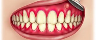
Costen's syndrome is a specific otoneurological disease. The disease appears as a consequence of dysfunction of the temporomandibular joint (TMJ). It occurs due to the absence of a large number of teeth, malocclusion, or the presence of deforming arthrosis of the jaw joints.
This syndrome was first described by the American otolaryngologist Kosten in 1934. In the process of studying the medical histories of his own patients, he discovered a certain relationship between ear pain and jaw pathologies.
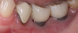
Often, the task of treating Costen syndrome falls on dentists involved in prosthetics of missing teeth or bite correction.
What is the TMJ and what are its functions?
Temporomandibular joint dysfunction (TMJ) is a fairly common disease.
According to statistics, up to 65% of patients of different ages have this problem. Diagnosing the pathology is difficult due to the many different symptoms that are difficult to associate. The disease can be confused with otitis media, dental diseases or other jaw pathologies. The specificity of the disease sometimes requires treatment from neurology and psychology. The temporomandibular joint is located in front of the ear and connects the lower jaw to the temporal bone of the skull.
Structure of the temporomandibular joint
It consists of the mandibular block, intra-articular disc, capsule, ligament and articular surface of the temporal bone.
The joint is very mobile, this allows us to move the jaw up and down, from side to side, talk and chew food. The movements of the joint are controlled by the muscles surrounding it. The TMJ bears a large load, which is associated with chewing, speech and facial expressions.
For various reasons, ANS dysfunction can develop, which causes diseases such as:
- myofacial syndrome;
- arthritis;
- arthrosis;
- chronic subluxation of the lower jaw;
- Costen's syndrome.
This leads to joint damage, inflammation and pain.
There are a number of different reasons that lead to the development of this syndrome. The main factors causing the disease include:
- incorrect structure of dentures;
- overstrain of the jaw muscles due to frequent eating of excessively solid foods;
- improper tooth filling;
- presence of bruxism;
- certain problems with the metabolism of minerals in the body;
- serious infectious diseases;
- problems with collagen production;
- incorrect jaw structure, which leads to incorrect load distribution;
- malocclusion.
Causes of the syndrome
There are many factors for the development of the syndrome. The main reasons are considered to be:
- absence of a large number of teeth;
- medical error by a dentist during prosthetics and fillings;
- overstrain of the masticatory muscles;
- untreated teeth grinding;
- disorders of mineral metabolism;
- infection;
- collagenoses;
- dental pathologies, due to which the load is distributed unevenly;
- malocclusion.
The syndrome can be caused by a combination of several of these factors, or by any one individually. In addition, there are risk factors that make the development of the disease more likely. These include:
- age over 50 years;
- menopause;
- jaw injuries;
- chronic inflammatory processes in the jaw area;
- osteoarthritis;
- hereditary tendency.
People at risk should be aware of the symptoms and treatment of Costen syndrome and its impact on the temporomandibular joint.
The syndrome is very similar in symptoms to acute otitis media and arthritis. But there is one main point for determining the syndrome - it is impossible to open your mouth wide. To prevent the development of this disease, it is necessary to regularly visit the dentist for dental treatment, prosthetics and bite correction.
Stages of development and consequences of Costen syndrome
It should be noted that quite often this syndrome develops without any symptoms. The main symptoms of this disease include:
- discomfort when chewing;
- crunching and clicking when opening the mouth;
- problems with jaw mobility;
- overstrain of the masticatory muscles;
- subtle facial asymmetry;
- pain in the jaw, eyes and ear.
With prolonged absence of treatment, in addition to deterioration of hearing and vision, complete immobilization of the jaw joints is observed.
Stages of disease development
There are four stages in the development of Costen syndrome:
- First . There is a certain reduction in the gap between the joints, as well as subtle looseness of the ligaments.
- Second . There is ossification of the condylar process, as well as a decrease in mobility in the joint.
- Third . The stage is accompanied by complete cartilaginous degeneration, extensive sclerosis of the surface of the joints, as well as limitation of jaw movements.
- Fourth . When diagnosing it, fibrous ankylosis can be detected, which can be treated exclusively with surgery. In addition, there is severe constant pain that cannot be pacified with the help of analgesics.
Complications and possible consequences
Without adequate treatment, the patient's condition will only worsen over time. The patient develops chronic pain caused by the following complications:
- Physiological changes in the jaw joint are observed. This leads to poor chewing of food and problems with the stomach and digestion. Patients with such problems can only eat soft foods due to the inability to chew.
- Subsequently, a significant weakening of the ligaments of the masticatory apparatus is observed.
- If treatment is not started promptly, this may lead to the inability to open the mouth.
When a joint is damaged, salts are deposited. Therefore, it is extremely important to promptly treat the disease. Taken together, this syndrome can seriously affect the deterioration of a person’s health through an indirect negative impact on various systems and organs.
Extra-articular pain syndrome (Hamstring syndrome, ARS syndrome)
Hamstring syndrome
It has been noted that in response to chronic trauma, changes occur not only in the joints.
Quite often, the localization of pain is associated with other tissues surrounding the joint, including synovial tissue. Frequent localizations of pain syndrome include enthesopathies at the sites of attachment of the tendons of the pelvic ring muscles. Clinical manifestations in this case are interpreted as Hamstring syndrome and ARS syndrome [1]. Hamstring syndrome is a traumatic injury to the muscles in the area of the ischial tuberosity (the posterior group of thigh muscles). Etiology. Hamstring syndrome is a fairly “young” disease. This syndrome was first discussed at the end of 1980 [2]. The cause of this syndrome is chronic
nic trauma to the posterior group of thigh muscles in the area of attachment to the ischial tuberosity. The cause of damage is too much contraction of the muscle, which can occur during sudden acceleration, a blow to a tense muscle, too much sports training, playing sports without proper warm-up and other situations. Hamstring syndrome is most often observed in athletes involved in athletics and, above all, sprinting and hurdles. However, Hamstring syndrome can also occur in non-athletes [4].
Chronic trauma to the muscles of the posterior thigh leads to inflammation and compression of the sciatic nerve. Inflamed tendons put pressure on the sciatic nerve, which in turn leads to a vicious circle. Pressure on the sciatic nerve leads to pain along the back of the thigh. Eventually a scar may form in this area.
Clinical picture.
In the clinical picture, the first place is pain in the gluteal region with irradiation along the back of the thigh. The pain intensifies with muscle tension and prolonged sitting. In addition, pain occurs with pressure (palpation) of the ischial tuberosity, passive flexion of the hip and extension of the leg, as well as with active flexion of the leg at the knee joint against the doctor’s resistance. With a long course of Hamstring syndrome, complete separation of the muscles from the ischial tuberosity is possible. Hamstring syndrome must be differentiated from a number of other possible causes of pain in the gluteal region and lower extremities (for example, sciatica, piriformis syndrome).
The following types of damage are distinguished:
- muscle stretching, with the formation of micro-tears;
- partial ruptures;
- avulsions or complete tears, when the muscle, no longer attached to the bone, contracts and gathers into a ball, running away from its attachment to the bone;
- avulsion fractures, when a muscle tears off a piece of bone.
Diagnostics.
To diagnose Hamstring syndrome, a thorough medical examination with the additional use of MRI and ultrasound is very important. MRI is decisive in determining the degree of soft tissue damage (complete, partial rupture) [3]. CT scans show the location and extent of tendon or muscle rupture, as well as associated swelling and hemorrhage. Magnetic resonance imaging shows thickening of the Hamstring muscles. X-rays are performed to exclude fractures.
MRI Complete tendon rupture
Unfortunately, Hamstring syndrome is practically unknown to domestic doctors, and the erroneous diagnosis of “trochanteric bursitis” is often made, which is much less common. That is why an integrated approach is necessary for a more accurate diagnosis.
Treatment.
At the first stage, conservative treatment is carried out, consisting of the prescription of non-steroidal anti-inflammatory drugs, steroid injections, physiotherapy, and / or acupuncture. There is data in the literature on the use of hyaluronic acid preparations for injection into joints and into the synovial membranes of tendons to relieve pain in extra-articular localization [1].
If pain and weakness do not go away, then surgical treatment is indicated. Surgical treatment consists of tenomyolysis of the biceps femoris muscle and neurolysis of the sciatic nerve. Surgical treatment in most cases leads to complete restoration of the function of the affected area. Pain relief occurs in most patients. Please be aware that surgery may lead to complications such as wound infections or nerve damage. The sciatic nerve must be released very carefully. The posterior femoral cutaneous nerve should not be disrupted or injured in any way [2]. In case of complete separation of the Hamstring muscles, surgical fixation of the tendon to the ischium is indicated. After surgery, when the hamstring muscles are separated from the ischial tuberosity, a special waist belt is worn for three weeks, which holds the leg in a bent position, which reduces the tension of the hamstring muscles and facilitates their growth to the bone [4].
Forecast
The prognosis for recovery is good. Avulsions of the posterior thigh muscles and avulsion fractures of the ischial tuberosity heal slowly and are accompanied by a high risk of recurrence.
Complications
Complications include the appearance of calcifications in the area of the ischial tuberosity or when muscles are torn from it; in this case, calcifications begin to irritate the sciatic nerve or cause pain when sitting and serve as an indication for surgical intervention.
Prevention
It is possible that warming up, which prepares the muscle for high loads, to some extent protects the athlete from muscle avulsions and avulsion fractures, but there is no convincing evidence of this yet.
Resources used: 1. M.A. Strakhov, A.V. Skoroglyadov, I.M. Kostiv, N.V. Chizhikov, D.E. Sannikov, I.O. Tsukurova “The use of low-molecular preparations of bound hyaluronic acid in athletes with pain syndrome of extra-articular localization” Moscow, journal Polyclinic 2/2013 pp. 54-60 2. Ian J. Young. Surgery for proximal hamstring syndrome. American Journal of Sports Medicine. December 2008 Vol. 36. No. 12. pp.2372-2378 3. Cohen, Stephen B. and Bradley, James, “Acute Proximal Hamstring Tear” (2007). Journal of the American Academy of Orthopedic Surgeons, June 2007, Vol. 15, No. 6, 350-355 4. travmaorto.ru/147.html
Author:
Nikolaeva S.V.
Ass. department oncology, radiation diagnostics, radiation therapy of the VSMA named after N.N. Burdenko
ARS syndrome.
ARS syndrome (Adduktor-Rectus-Symphysis) is a pathological condition of the tendons of the mm muscle complex. adductor longus et (or) brevis, m. gracilis, the distal part of m.rectus abdominis, as well as the anterior part of m. adductor magnus at the places of their attachment to the pubic or ischial bones. This symptom complex, first described by the Bulgarian doctor M. Bankov in 1958, should be considered as a manifestation of chronic microinstability of the anterior part of the pelvic semiring.
Etiology:
overload of the musculoskeletal system as a result of a discrepancy between physical activity and the possibility of compensatory reactions of the body.
Pathogenesis:
long-term similar loads associated with asymmetric contraction of the adductor muscles of the thighs, rectus and oblique abdominal muscles lead to microtraumatization of the ligamentous apparatus (tendon overload syndrome TOS (Tendon Overuse Syndrome)) of the pubic symphysis with the occurrence of inflammatory and then degenerative changes. These causes lead to enthesopathy, tendonitis and tendomyositis of the above localization.
Clinical manifestations:
Pain of varying intensity in the lower abdomen and groin areas, radiating along the muscles; local pain is noted upon palpation and maximum abduction of the hips and flexion of the torso with resistance in the places of attachment of the adductor muscles of the thighs and the rectus abdominis muscle to the pubic bone; pain, as a rule, accompanies physical activity (running, hitting a ball) and significantly limits a person’s functional capabilities.
Instrumental examination methods:
Ultrasound examination sometimes visualizes areas of hyperechoic structure of muscle tissue at the sites of attachment to the pubic bone.
The X-ray picture of ARS syndrome in cases of its protracted course is characterized by the presence of signs of osteochodrosis and osteochondritis of the symphysis pubis.
The arrow indicates the area of degenerative rupture of the m.adductor longus tendon at the site of attachment to the pubic bone.
MRI diagnostics:
The above images show increased MR signal within the adductor longus muscle at its insertion into the right pubic bone, which may be consistent with tendinitis.
Differential diagnosis:
Carry out with stretching of the adductor muscles of the thigh, fractures of the pelvic bones, inguinal hernia, prostatitis, osteomyelitis, rheumatoid arthritis, osteoarthritis, primary and metastatic tumors, urolithiasis, disease of the lumbar spine.
Treatment:
Conservative treatment includes local injections of corticosteroids, non-steroidal anti-inflammatory drug therapy, various types of ERT treatment (electrophoresis with anesthetics, Bernard currents, laser therapy and is associated with a significant number of relapses of this disease (up to 80% of cases). More positive results when using the extracorporeal shock method wave therapy Surgical treatment of ARS syndrome, which consists of performing partial myotomy and myofascioplasty of the rectus abdominis muscle and partial myotomy of the adductor muscles of the thighs.
Resources used: sport-travma.ru/stati/ars-sindrom/ smk-ffu.at.ua/index/0-26 koleno.kiev.ua/index.php?option=com_content&view=category&layout=blog&id=41&itemid=7
Author:
Milovanov N.V.
Radiologist LLC "MRT Expert", Moscow
How is the syndrome diagnosed?
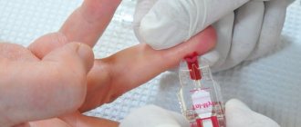
If the patient does not have joint damage, but has certain complaints, three-phalanx testing is used to diagnose the disease.
With the mouth open, the joints of the middle, ring and index fingers, placed between the phalanges, should pass clearly between the lower and upper incisors. If the masticatory muscles are damaged, this will be impossible to do.
Other diagnostic methods are used only to confirm the final diagnosis:
- conducting electromyography of the muscular masticatory apparatus;
- use of impedance examination of the middle ear;
- use of panoramic x-ray of the lower jaw;
- use of CT or MRI.
Classification
During the development process, pathological bite syndrome goes through four stages, each of which is characterized by the following symptoms:
- First stage. The size of the joint space narrows unevenly, and slight loosening of the ligament apparatus is observed. A person begins to experience the first symptoms of a pathological change in the cartilage disc.
- Second stage. The mobility of the temporomandibular joint decreases, ossification occurs in the area of the condylar process of the mandible.
- Third stage. Degeneration of the cartilage disc continues, and sclerosis of the articular surfaces develops. Movement of the lower jaw is severely limited.
- Fourth stage. The patient experiences a strong proliferation of fibrous tissue, and the mobility of the lower jaw is completely absent.
A person experiences persistent pain in the jaw area, radiating to the ear or cheek. At this stage, treatment is carried out exclusively by surgical methods.
Specifics of treatment
Treatment of the disease consists of the following measures:
- diet adjustments;
- use of laser, ultramagnetic and ultrasound therapy;
- Treatment of all problematic teeth is mandatory;
- the use of drug therapy to eliminate pain, improve tissue nutrition, reduce muscle tone, as well as the use of sedative herbal remedies.
Complex therapy methods are often used, which should eliminate inflammation and pain and restore the former mobility of the joint, namely:
- Cortisol injections are made into the joint cavity.
- Iontophoresis is used with the simultaneous use of novocaine and potassium iodide.
- Patients are often prescribed a special orthopedic device that limits jaw movement. This device must be worn for about 4 months. This method is aimed at reducing the load on the joint.
- If necessary, the bite is corrected using surgery or prosthetics.
If a set of such measures does not bring any visible results, the attending physician may suggest surgery to the patient.
Depending on the severity of the situation and associated problems, the cost of treating Costen syndrome can vary greatly. In some cases, you can get by with gentle treatment using only medications. In other situations, the patient can be cured with surgery and subsequent prosthetics.
Description of the problem
Costen's syndrome was discovered in the thirties of the 20th century. Otolaryngologist D. Kosten compared cases of patients complaining of ear pain due to pathologies in the structure of the jaw system, and came to the conclusion that there is a relationship between these factors.
Pathological occlusion syndrome, as Costen's syndrome is otherwise called, is a functional disorder of the temporomandibular joint.
This pathology affects the cartilage disc, which provides joint mobility in three directions, due to which complete chewing of food is carried out.
An unevenly distributed load between the jaws leads to inflammation of the cartilage disc, which can lead to its degeneration if not treated in a timely manner.
As a result of this phenomenon, there is a decrease or complete loss of mobility of the mandibular joint.
What do you need to know to avoid illness?
Doctors recommend avoiding frequent intake of very hard foods as a preventive measure for this syndrome. You should also completely abandon the habit of clenching your teeth when nervous.
At the first symptomatic manifestations and any discomfort when chewing, immediately consult a doctor to diagnose the cause of this condition.
Timely contact with a specialist and accurate diagnosis are a guarantee of successful treatment of Costen syndrome, which will make it possible to avoid more serious consequences and complications that accompany this disease.
Costen's syndrome is a rather serious pathology that affects various systems and organs of the human body. In order to minimize the risks associated with the occurrence of this disease and its consequences, you should be more attentive to your own body and, at the slightest problem, contact a doctor.
Symptoms
Costen's syndrome is characterized by the presence of a collection of symptoms that are rarely interrelated.
In addition, in the initial stages of the disease, symptoms may not appear, which delays the patient’s visit to the dentist and complicates timely diagnosis.
Most often, patients experience the following signs of disease development:
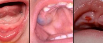
- pathologies in the functioning of the temporomandibular joint - difficulties during opening and closing of the jaw, the appearance of crunching and clicking, severe pain during chewing;
- the appearance of dryness and acute pain in the root of the tongue and pharynx;
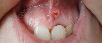
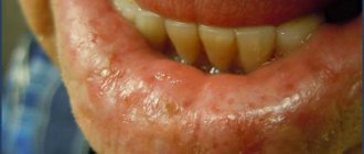
- the formation of a herpetic rash on the mucous membranes of the oral cavity and the outer edge of the ear canal;
- the occurrence of trigeminal neuralgia - severe pain of a paroxysmal nature that affects one side of the face - jaw, cheek, ear, eye;
- pain in the ears and head, which may worsen when sneezing or singing.
In some cases, patients may experience frequent dizziness, hearing loss, and pain in the cervical spine.