1716
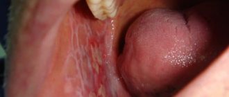
Superficial caries is a small roughness or cavity in the enamel, which is most often invisible to the patient. This is the next stage in the progression of destruction, after the stain stage. Typically, a person seeks medical help only when a tooth reacts to chemical irritants.
If caries at the spot stage does not manifest itself in any way, then the superficial one already begins to come into contact with the dentino-enamel border, the irritation of which is painful. But the dentin is not yet destroyed.
What does it represent?
The superficial form of caries is considered the initial stage . It is characterized by damage to the tooth enamel, without involving dentin in this process.
It is distinguished by smoothed symptoms and a long course. The pathological process can be detected by the appearance of yellowish spots on the enamel.
What is superficial caries
Superficial caries can be localized:
- on the front of the teeth,
- in the area of the contact surface,
- as well as near the gingival margin (cervical area).
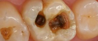
However, most often the defect is found on the fissures of the lateral teeth. In this case, fissure caries is diagnosed.
Fissures are natural depressions (grooves, pits) on the chewing surface of molars. This anatomical feature contributes to the rapid accumulation of plaque - food particles get into the deep fissures and are difficult to clean with a toothbrush.
As a result, the carious process begins in the central part of the tooth. The defect does not affect the cutting edges or the root zone, so it is quite difficult to diagnose this form, especially at the initial stage.
Fissure caries
Clinical symptoms
Despite the fact that the clinic of the superficial form is characterized by asymptomaticity, some manifestations still allow early diagnosis of caries :
- increased sensitivity of enamel. She begins to react not only to thermal stimuli, but also to the consumption of sweet or salty foods;
- while eating, a burning sensation is felt at the site of destruction, which is quickly relieved by rinsing with warm water;
- with strong mechanical impact, for example when cleaning with a brush, aching pain may occur;
- the area of destruction first becomes white and then yellow or brown;
- the structure of the affected area becomes heterogeneous and rough.
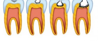
Stages of caries treatment:
1. Pain relief, anesthesia (if required)
2. Opening of the carious cavity.
Under the tooth enamel there is soft dentin, which, as a rule, is more easily destroyed. This is why the size of the entrance hole is deceptive, and the carious cavity is actually much wider than it appears from the outside. A drill is used to widen the inlet.
3. Cleaning the carious cavity.
At this stage, necrotic (dead) tissue will be removed using a drill or advanced instruments.
4. Treatment of the cavity before filling.
The shape of the carious cavity can be additionally processed with a drill for convenient installation of the filling.
5. Tooth filling.
The dentist fills the empty prepared space with filling material. In modern clinics, light fillings are used, which have a polymeric ability, i.e. the material hardens under the influence of ultraviolet light. Such equipment and material allow you to correctly and reliably fill a dental hole of any shape. And thanks to the features of light fillings, it is not difficult to choose a shade to match the color of the patient’s teeth, which means that the installed fillings in the teeth will not be noticeable.
The above stages of caries treatment are carried out in one visit to the dentist. However, treatment in a more advanced stage may require more time. If, when treating deep caries, your dentist will detect pulpitis. In this case, the pulp will need to be removed, and an additional visit to the dentist may be required.
How is the examination carried out?
At the diagnostic stage, it is necessary to correctly determine the form of the disease and identify the extent of enamel damage. This will help you select the optimal treatment technology, which will completely stop the further spread of caries.
Several effective research methods are used at once.
Condition assessment
Assessing the condition of the tooth makes it clear whether there is a need for filling it.
In addition to visual inspection, several other methods are used:
- Probing .
Justified only when diagnosing caries in adult patients, since it can be accompanied by painful sensations. To determine, the doctor uses a special probe, with which he taps the affected area and examines its structure for roughness. - Drying .
Recommended for diagnosing pathology in children. The dentist treats the surface of the enamel with an antiseptic and then dries it using a special lamp. Pathological areas stand out against the background of healthy tissue. The depth of damage to the dental layers is judged by the degree of dullness and change in shade.
Dyeing method
A method for more accurately determining the depth of damage to tooth tissue or for identifying caries in the absence of its external manifestation (stains).
The effectiveness is due to the use of special dyes that are able to penetrate into the narrowest pores of damaged enamel and firmly settle there.
The following substances are most often used for coloring:
- methylene red or blue;
- carmine;
- tropeolin;
- silver nitrate.
Our next publication will discuss the features of treatment of cervical caries of anterior teeth.
In a separate article we will talk about contraindications to local anesthesia in dentistry.
Using the link https://www.vash-dentist.ru/detskaya-stomatologia/d-zubi/karies-dz/lechenie-molochnyih.html we will discuss whether it is possible to treat caries of baby teeth without a drill.
The staining procedure is as follows:
- A retractor is installed.
- The dentition is isolated from saliva using a cofferdam or cotton wool rolls.
- The enamel is completely dried.
- The dye is applied using an applicator.
- After a few minutes, it is removed with a gauze swab, and the teeth are dried.
In those areas where enamel staining has occurred, superficial caries is diagnosed. The depth of the lesion is judged by the degree of its coloring.
Differential diagnosis
For comparison, the following symptoms of superficial margination are used : pain does not last long and appears only when exposed to chemical irritants, there is a small cavity on the surface of the enamel, dentin is not affected.
- Average caries. It also manifests itself as pain from chemical irritants. The dentin-enamel border is affected, there are areas of softened dentin, and sometimes the color of the entire tooth is changed.
- Chalky-mottled and erosive fluorosis. The appearance of the teeth is changed, the patient is bothered by pain from any irritant. Teeth are affected in groups, not individually. The entire enamel is covered with chalky-mottled defects and erosions.
- Erosion. Externally damaged teeth react to any impact. Localized on canines, premolars and incisors. Erosion forms from the convex part of the vestibular surface. The lesion is oval in shape with a smooth, glossy bottom.
- Wedge-shaped defect. It doesn't show up at all. Externally, the teeth look sharpened in the cervical region of the vestibular surfaces. The bottom of the wedge is dense and glossy. The dentin is affected.
Diagnostics
Due to the unexpressed symptoms of the initial stage of caries, patients can rarely suspect this disease on their own, attributing short-term pain to increased tooth sensitivity.
In this case, an enamel defect can only be determined in dentistry. Probing allows you to identify a chalky area, the surface of which is rough or has a shallow cavity filled with softened demineralized enamel of a yellow-gray hue. When probing the cavity, there is either no pain at all or it is insignificant.
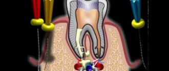
Often, in order to determine hidden enamel defects, transillumination is used, that is, the teeth are illuminated with a cold beam of light.
To determine the diagnosis, the hygiene index and the CPU index are taken into account. Electroodontodiagnosis does not reveal any deviation from the norm.
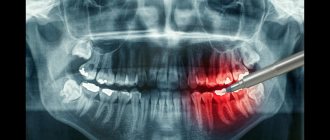
It is easier to recognize superficial caries when using special markers, that is, dyes that selectively stain carious defects. X-rays can also identify foci of the disease.
Primary caries should be recognized by conducting a differential diagnosis with such ailments as a wedge-shaped defect, erosion or hypoplasia of enamel, fluorosis, as well as intermediate caries.
Therapy
To treat superficial caries, 2 approaches are used: preparation and remineralization. Which one will be chosen depends on the condition of the enamel.
Roughness
If there is only roughness on the enamel, without the presence of a cavity, then the doctor grinds off the affected enamel and carries out remineralizing therapy to restore the surface layer.
The tooth is coated with a substance based on phosphorus, calcium and fluoride. The following drugs are used in modern dentistry:
- bifluoride 12.3% – colorless fluoride varnish for deep fluoridation. Contains calcium and sodium fluorides.
- Remodent 3% – powder from ground cattle jaws, made by vacuum drying. Contains: calcium, phosphorus, magnesium, potassium, sodium, chlorine, organic substances, manganese, iron, zinc, copper and other trace elements. Used for applications.
- 10% calcium gluconate solution, 10% calcium chloride, 2.4% calcium glycerophosphate are calcium-based preparations used for electrophoresis and applications.
- fluoride varnish is an alcohol solution with good penetrating ability. Designed to cover teeth to seal and prevent enamel damage by pathogenic microorganisms. The composition includes: sodium fluoride, fir balsam, chloroform, shellac.
- Belak is a remineralizing dental varnish. Apply with an applicator. Contains only fluoride.
How to treat root caries, read in the next review.
This material is devoted to the issues of conduction anesthesia in dentistry: what it is and in what cases it is effective.
Here you can watch a video of how the procedure for treating deep caries takes place.
Presence of cavity
If the roughness is not treated in a timely manner, a cavity will gradually form in its place. In this case, remineralization is no longer enough; preparation and full filling are required.
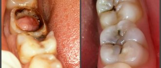
Photo: teeth after treatment
The doctor performs the following manipulations:
- Opening and expansion of the cavity. The overhanging edges are removed with a bur; the affected area should be clearly visible.
- Cleaning of necrotic dentin. It is done to healthy tissue to obtain a glossy bottom of the cavity.
- Formation of the working cavity. There are 2 options for preparation, the choice depends on the type of composite materials used.
A gentle method, the technology of which is aimed at preserving a healthy area of enamel, is used with light-curing composites.And Black preparation is suitable for installing a chemically cured composite filling.
- Finishing. Control cleaning of the enamel, after which all damaged areas should be removed, and the bottom of the cavity should be polished to a shine. This is necessary for better fixation of the filling. It is done with a diamond bur at low speed, with constant air-water cooling.
- Treatment. The doctor rinses the prepared cavity with 3% hydrogen peroxide (for chemical-curing fillings) or distilled water (for light-curing materials). As a result, the surface should be dry and sterile.
- Preparation and application of filling material. Additional mixing is done only when using a chemical seal; light seals are supplied ready-made by the manufacturer. The material is applied in such a way that it follows the shape of the tooth.
- Polymerization. In order for the light filling to harden, it is exposed to a halogen lamp. The chemical material itself becomes solid due to the interaction of the catalyst and the base.
- Finish: contouring and polishing. First, a correction is made to suit the specific occlusion (macrocontouring), then the filling is polished and ground until it is perfectly smooth.
The steps are clearly demonstrated in this video:
Icon
In modern clinics, the icon method is practiced, in which the cavity is cleaned and filled with a special gel, without the need for preparation. A detailed description of the principle of action and stages of icon caries treatment technology is in this article.
But for now you should not consider the method as one of the main ones, because it is available only in selected medical institutions.
Treatment in children
There are no special methods for treating children's teeth; when caries passes from the stained stage to roughness, the tooth already needs to be treated using remineralizing therapy, as described above. Treatment using the silvering method is no longer relevant, because... roughness and cavity formation is already destruction of tooth tissue.
The average price for regular treatment is about 1,100 rubles. According to the icon system - 3,500 rubles.
Treatment in the clinic
Let's consider the process of treating superficial caries in stages:
- Treatment of superficial caries consists of a series of stages performed by the dentist.
- The surface of the causative tooth is cleaned of plaque with special brushes and polishing paste.
- Preparation of a place of enamel that has been affected by caries. Anesthesia is usually used.
- The tooth is isolated from moisture so that secondary caries does not develop or the placed filling does not fall out. The best way to do this is to use a rubber dam.
- The enamel is etched with acid so that the filling can better adhere to the dental tissue. Upon completion of etching, the acid must be thoroughly washed off.
- A special glue (adhesive) is placed into the cavity, which will help secure the filling more firmly. After application, the adhesive is illuminated with a light-curing lamp.
- Filling. The filling material is introduced into the cavity, the desired shape is modeled from it, and it is illuminated with a special lamp.
- The surface is ground and polished.
Prevention
To avoid caries, you must adhere to preventive measures :
- Oral hygiene should be of the highest quality. To do this, in addition to a brush and paste, it is necessary to use additional cleaning products: floss, brushes, rinses, etc.
- You should avoid snacking on sweets and flour products, which are a breeding ground for bacteria.
- It is necessary to introduce foods rich in calcium and other minerals into the diet.
- One important point is regular professional cleaning at the dentist's office.
- A good prevention would be remineralization of the dentition.
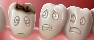
The appearance of stains on teeth is a signal of the development of a pathology that requires immediate treatment. The later you go to the dentist, the longer the recovery process will be and the greater the risk of losing a tooth.
If you find an error, please select a piece of text and press Ctrl+Enter.
Tags: caries treatment, caries prevention, tooth decay
Did you like the article? stay tuned
Previous article
Detailed description of the treatment of average caries
Next article
Empower braces: how do they stand out among analogues, what problems do they solve?
conclusions
For a patient, when asking how to treat dental caries, the most important factors are price, time, and lack of discomfort. To keep them to a minimum, it is necessary to promptly seek professional help, adhere to the doctor’s recommendations and monitor oral hygiene.
Calculator: calculate prices online
Light-curing fillings, caries treatment
The price is ALL INCLUSIVE except anesthesia, if necessary
Select the type of filling
0
Specify the number of fillings
Write a comment
Elena
September 27, 2020 at 06:08 pm
Caries is one of the most unpleasant problems with teeth, I experienced it myself. She began to notice that her teeth began to darken, some stains appeared, and when brushing, a nagging pain appeared, but she did nothing. When this started to get worse, I finally decided to see a dentist. They diagnosed caries and did staining. As a result, they did fissure sealing (the procedure is not very pleasant, but not terrible either). Now I constantly see my dentist and take basic preventive measures (I use teeth rinses, take a course of vitamins with calcium, etc.). If you notice something wrong with your teeth, I advise you to immediately run to the dentist to avoid making a mistake like me!
Igor
September 28, 2020 at 2:45 am
Caries, in general, is a family problem for us, for me, my father, and my brother. One grandfather ate apples every day and was distinguished by strong teeth, but that’s not about that now