Sometimes women come to the gynecologist with the question: “After an x-ray, when can you get pregnant?” After all, X-ray radiation is not harmless for the expectant mother. Many responsible women understand this and try to delay pregnancy after an x-ray. But there are situations when pregnancy occurs unexpectedly. The woman recalls that she recently (perhaps even in the current cycle) had an x-ray and is now worried about how the radiation will affect the baby’s health.
Let's talk to experts about how long it takes to get pregnant after an x-ray and whether there are reasons to panic.
Planning and x-ray
Any married couple wants to give birth to an absolutely healthy baby. And those future parents who take this issue seriously understand that they need to prepare for this important step in advance.
Planning a pregnancy is an important and responsible activity that involves a number of procedures aimed at identifying possible deviations from the norm in the bodies of future parents.
An X-ray examination may be prescribed by a doctor in the following cases:
- When diagnosing the cause when pregnancy does not occur for a long time;
- A routine fluorographic examination of the lungs, which every person must undergo once a year;
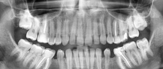
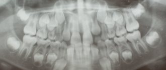
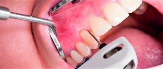
- Photograph of teeth during oral sanitation;
- For injuries;
- For some diseases, when ultrasound diagnostics are not enough.
And it often happens that a woman does not yet know that she is pregnant when she receives a certain dose of radiation.
When can you plan to conceive after an x-ray? Will pregnancy proceed with complications after an x-ray? Can irradiation affect the fetus; will it have developmental defects after this procedure? Let's look at these important issues.
To do or not
The question of whether it is possible to carry out this procedure without risk to the fetus can be definitively answered by considering the following factors:
- type of x-ray machine;
- expected radiation dose (hazard degree);
- indications for the procedure (emergency or life-threatening);
- the availability of an alternative (ultrasound can often help with the diagnosis without risk to the fetus, if it turns out to be sufficiently informative);
- consequences of failure (decompensation, complication of the disease due to lack of accurate diagnosis);
- duration of pregnancy (the first trimester is especially dangerous for negative consequences for the fetus, in the third trimester the negative impact is less);
- type of procedure (fluorography, computed tomography, high-rigidity studies, fluoroscopy are contraindicated for pregnant women).
The decision to take an image is made by the doctor together with the patient, so the woman must be informed of all the consequences of refusal or consent to the procedure. If there is a safer option for examination, then it should be preferred, except for conditions that threaten the life of the mother and child at the moment.
The question of choice is complex and requires consultation with a general practitioner, radiologist and obstetrician-gynecologist to make the most correct and safe decision.
Early dangers
X-rays in the early stages of pregnancy are more harmful to the health of the baby than in the last trimester. In the first months, the nervous system, organs of vision, and spine come under attack. Carrying out any x-ray examination before 6 weeks can cause congenital anomalies and deformities in the baby.
During this period, embryonic cells multiply most actively, the place of attachment of the fetus to the mother's womb is formed, and the main organs and systems are formed. In the fifth and sixth weeks, endocrine organs - the adrenal glands - begin to form. From the fourth to the eighth, the cardiovascular system actively develops and the heart is formed. At the sixth or seventh age, the thymus gland forms and the baby’s immune defense develops.
At this time, x-rays can cause the following abnormalities in the fetus:
- miscarriage;
- formation of extraembryonic organs;
- anemia;
- formation of a “cleft palate”, “cleft lip”, spinal cord cleft;
- defects in cardiac muscle tissue;
- valve defects;
- underdevelopment of the adrenal glands;
- pathologies of the thymus gland, severe congenital immunodeficiency;
- bone marrow suppression, aplastic processes;
- malformations of the lungs (agenesis and atresia of the pulmonary arteries, bronchi, absence of a lung, congenital bronchiectasis);
- intestinal malformations (intestinal obstruction, orifice atresia, absence of certain sections).
During this period, X-ray diagnostics are associated with a risk to life and the child and require careful decision-making. Most often, women are advised to choose an alternative and refuse.
Is it possible in the second trimester?
At this time, complex processes of differentiation of organs and tissues begin, and mitotic, that is, proliferative, activity decreases. This means that growth and development occur with less risk of X-ray damage.
In the second trimester, the brain begins to form, and the facial and cerebral parts of the skull are also formed: the baby’s face acquires recognizable features. The process of formation of hematopoiesis and the excretory system is actively underway. The digestive and respiratory systems are divided into more complex elements. At the end of the second trimester, almost all organs and tissues have already been formed, and the development of their functional component begins.
A photo taken after 12 weeks is less dangerous for the baby, but can cause:
- renal failure;
- diseases of the blood system;
- congenital anomalies and minor heart defects;
- underdevelopment of the respiratory system (non-opening of the lungs), excretory organs;
- congenital endocrine diseases (the sex glands, thyroid gland, and pineal gland are especially vulnerable);
- immunity defects.
Third trimester - features and dangers
At this time, the negative impact of x-rays on the fetus is minimal. After 24 weeks of development, the skeleton, muscle and adipose tissue complete their formation. Many processes in the baby’s body already occur without pronounced cell division. The child's DNA is less susceptible to external influence. But at this time there is a potential risk:
- delays in physical and mental development;
- immunity deficiency;
- hematopoietic pathologies;
- disorders of the reproductive system, thyroid function.
In the last months of pregnancy, babies are actively developing their brain and immunity, as the most complex structures, so you should be wary of the impact on these structures the most. During this period, it is permissible to conduct x-ray examination of almost all areas except the pelvis, abdominal cavity and spine. It is important to think about measures to protect the baby in the womb.
Is it possible to get pregnant immediately after hysterosalpingography?
When planning a pregnancy, the doctor will definitely prescribe an X-ray if the long-awaited conception does not occur for a long time. The test is called hysterosalpingography (HSG). It must be carried out in order to determine the quality of patency of the fallopian tubes. If adhesions are found in the tubes, fertilization is impossible.
To check whether the tubes are patent, an HSG is performed, during which a contrast agent is used. It is with its help that you can see in the pictures the condition of the pelvic organs. Thanks to this procedure, the doctor can also detect formations that are dangerous to the woman’s health.
An interesting fact is that after completing this study, a woman who has not been able to get pregnant for a long time may discover two cherished stripes.
There is a completely scientific explanation for this “miraculous” healing. The same liquid used for X-rays is injected under pressure. The result of this procedure is the divergence of small adhesions and restoration of the patency of the fallopian tubes.
This important point must be taken into account after the x-ray, and your doctor will tell you when you can become pregnant. But in any case, during the menstrual cycle when the woman was examined, she needs to use protection, since the egg received a serious dose of radiation.
What if pregnancy has already occurred?
Everyone knows that X-ray radiation in large doses can be harmful to health. But modern equipment reduces the harmful effects of the procedure to a minimum.
If the risk is minimal, is it possible to take x-rays immediately before conception and during pregnancy? Most gynecologists agree that it is undesirable for a pregnant woman to undergo this procedure. It is prescribed only in cases of extreme necessity.
With strong (and especially repeated) irradiation, living tissue cells can be harmed:
- Cells are damaged and are unable to perform their functions fully;
- Transform into forms of malignant formations;
- They die off.
The cells of the reproductive system are considered the most vulnerable to radiation. Irradiated sperm in men and damaged eggs in women are unable to conceive.
It is for this reason that the organs of the reproductive system must be protected using lead screens when undergoing an X-ray examination.
Since each organism has its own characteristics and reacts individually to any influence, the unborn child should be protected from the negative consequences of radiation.
It is worth planning conception two menstrual cycles after the body has received a dose of radiation - this is a sufficient period of time even for “reinsurers”.
However, many gynecologists are confident that it is possible to plan conception for the next cycle after the x-ray.
What are the advantages of x-rays?
Despite such serious consequences of x-ray, it remains the main method of diagnosing the condition of the body in case of dislocations, fractures and other types of research. The thing is that in order to get the side effects described above, you need to take pictures of the whole body at least 200 times in a row. In modern devices, the radiation dose received by the patient is relatively small and safe for his health. But it is still not recommended to do x-rays more than once a year, unless there is an urgent need for it.
Its main advantages:
- as a rule, no special preparation of the patient is required before the study;
- it can be done in almost any clinic or emergency room;
- relatively inexpensive cost of research;
- the quality of the images obtained during the study does not depend on the professionalism of the doctor performing the study (unlike ultrasound);
- the image is obtained immediately at the end of shooting;
- a high degree of detail in the image allows you to impartially assess the condition of the body;
- The method is not traumatic, no diagnostic incisions are required.
X-rays have a number of limitations:
- it is impossible to obtain a detailed image of soft tissues;
- the image is static, there is no way to evaluate the work of the organ in dynamics;
- Contrast is required for better visualization of the body parts being examined.
If other research methods are not suitable for detecting pathologies, the woman is sent for an x-ray.
Effect on the fetus: doctors' opinions
Sometimes a woman, after undergoing an x-ray examination, finds out that she was already pregnant at that time. And she is very concerned about whether this procedure affects the development of the fetus.
Doctors have different opinions about the safety of X-rays for expectant mothers.
Some doctors believe that modern equipment helps reduce risks to a minimum. Therefore, future parents have nothing to worry about.
Another part of the doctors claims that there are risks of complications. However, the consequences depend on what kind of examination was carried out and to what extent.
The risk of complications depends on how early the x-ray was taken.
If a woman was examined in the first half of the cycle, then there is no need to worry. After all, at this time the egg has not yet had time to mature and be released.
Having received irradiation in the second half of the menstrual cycle, when ovulation has already occurred, there is a high probability that the fetus may die or disturbances will be observed in its development. This applies to those who have had an X-ray of the pelvis or spine. Other studies, if done in compliance with all the rules, are practically safe.
So, it all depends on what kind of x-ray examination the woman underwent, how often she did it and what dose of radiation she received.
For example, if a pregnant woman took a photo of a tooth or hand, this will not affect the fetus in any way. But an X-ray of the pelvis, especially if it was done more than once, can cause many unpleasant consequences both for the subsequent pregnancy and for the planned one.
If a woman needs an x-ray for some reason while planning a pregnancy, it is better for her to choose the first third of the cycle, when the likelihood of getting pregnant is almost zero. Or use contraception for the entire cycle.
What are the dangers of x-rays during pregnancy?
The embryo is most susceptible to the harmful effects of ionizing radiation. In the first weeks of pregnancy, the formation of all internal organs, soft tissues, circulatory and nervous systems, and the brain begins. Now it is necessary to exclude any external influence on the expectant mother’s body so as not to interfere with this process. Moreover, it is impossible to predict the consequences of x-rays in the first weeks. The first trimester is the most dangerous and difficult time throughout pregnancy. An X-ray can interfere with the development of internal organs if it was taken at a time when the organ was being formed or actively developing.
At 5 weeks of pregnancy, most women find out about it. On an ultrasound, you can already notice a small dot measuring 2–4 mm. But at this point, ears and eyes are already forming, connective tissue is actively developing, and the rudiments of the lungs and intestines appear.
At week 6, the point stretches up to 7 mm in length, the embryo develops arms and legs, and the placenta is formed, which in the future will protect the fetus from external adverse influences.
When the fetus is 7 weeks old, its small heart beats quickly. While it is a pulsating vein. However, soon it will divide into chambers and will independently provide for the fetus.
When a tooth is photographed, the dose of x-ray radiation is minimal.
At 8 weeks, the embryo’s bones become stronger and the formation of the gastrointestinal tract, kidneys, heart, and bladder is completed. He is already floating freely in the space of the uterus.
When the embryo is 9 weeks old, it is able to make chaotic movements with its arms and legs and pushes off the walls of the uterus. The organs of the reproductive system are formed and the digestive system develops.
At 10 weeks, doctors begin to call the embryo an embryo. He is already almost like a baby, but much smaller. He weighs only 5 g, his internal organs are functioning. Marigolds appear on the fingers.
At week 11, the embryo’s tooth buds are formed, he can clench his hands into fists and unclench them, and hears loud, sharp sounds from outside.
Week 12 is an important stage in the formation of the nervous system. It is rapidly developing and improving.
At 13 weeks, the first trimester of pregnancy ends and the second begins - the most favorable time. The belly is still relatively small, the unpleasant sensations of the beginning of pregnancy have passed. Now you can enjoy your position.
The second half of pregnancy is a relatively safe time for various medical procedures that a pregnant woman requires. However, the third trimester is not always suitable for this, because the placenta is aging and may not be able to cope with external influences.
The first trimester of pregnancy is the most unfavorable for x-rays.