Ankylosis is a disease in which bones fuse together. In this case, the activity of the joints is partially or completely blocked. In dentistry, a distinction is made between ankylosis of the TMJ (temporomandibular joint) and dental ankylosis, when the tooth fuses with the bone. Most often men are affected by it. To eliminate the disease, you should contact an orthodontist and a surgeon: the first one works to bring the bite back to normal, and the second one performs surgical treatment of TMJ ankylosis, removing abnormalities in the joint and restoring its mobility.
Ankylosis of the TMJ appears:
- after a joint injury;
- after a long period of immobility, for example, being in a cast;
- due to inflammation or purulent process;
- as a consequence of arthritis.
When a joint fusions, as a result of the destruction of the articular cartilage, a bone growth appears near it, due to which the joints are connected and become immobile. Ankylosis is accompanied by breathing problems, tongue retraction, sleep apnea, snoring, and due to periodic lack of hygiene, tartar forms. This leads to tooth decay and other oral diseases. Ankylosis of the TMJ is eliminated with a surgeon’s scalpel.
The process of fusion of articular surfaces occurs gradually - as a result of violation of the integrity of the articular cartilage, the cavity around it is overgrown with connective bone tissue (growth), which ultimately leads to complete fusion and pathological changes in the joints, creating their immobility. Treatment of ankylosis of the temporomandibular joint is performed surgically.
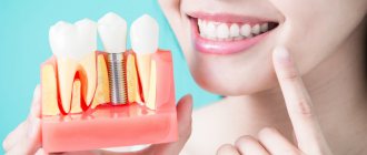
Advantages of implants Ankylosis
Ankylosis of the joint: how to recognize and treat the disease
All of the above features of Ankylos implants can be safely considered among the advantages, but the following advantages are worth highlighting separately:
- adequacy of pricing, which allows patients with different income levels to choose this manufacturer;
- the versatility of the product, which makes it possible to carry out fixation with minimal risk of infection of the patient’s body;
- installation of implants is carried out in the shortest possible time;
- the threat of implant rejection is minimal;
- possibility of combining designs;
- high levels of aesthetics;
- long-term service life of implants.
Bone ankylosis
With bony ankylosis of the TMJ, there is a connection between the condylar process and the zygomatic arch. It turns out that bone tissue fills the joint cavity. Bone ankylosis of the TMJ can be partial or complete, depending on whether there are intact areas on the surface of the joints. If the disease affects only one joint, the face becomes asymmetrical. When two joints are affected, the upper jaw moves forward, the chin region falls, which leads to malocclusion and problems with teeth, speech and breathing. A person with this condition will have difficulty eating solid food. The full form is characterized by the fact that the joint space is not visible on the x-ray, and the condylar process appears wider than it should.
Ankylosis of the tooth - what is it?
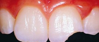
Dental ankylosis is a disorder in which the teeth become fused to the jaw. The oral cavity is a true miracle of nature. On the one hand, it contains teeth, considered the hardest structures in the body, and on the other, the soft but strong muscle of the tongue. The teeth are fixed in the jawbone. They are not attached directly to the jaw, but are held in place in depressions called alveoli by gingival and periodontal fibers. However, sometimes these gingival and periodontal fibers are missing, causing the teeth to fuse to the bone. This disorder is known as dental ankylosis. The causes of this disorder are unknown, although it is believed that trauma to the bone during tooth development may be a contributing factor.
Symptoms of dental ankylosis are not always obvious. If ankylosis of a baby tooth develops, there is a delay in its growth or incomplete eruption. In addition, a baby tooth that is fused to the jaw does not fall out as easily as regular baby teeth, or does not fall out at all, and therefore can prevent the eruption of permanent teeth. If the permanent tooth fuses with the bone, no symptoms may be observed and ankylosis is detected incidentally during an x-ray examination. The image reveals a lack of space and a visible boundary between the tooth root and the jaw.
One of the main problems associated with dental ankylosis is that sometimes the tooth does not grow back completely, and if it is a baby tooth, this can lead to a complete malocclusion. The fusion of a baby tooth with the bone also prevents the growth of the permanent tooth underneath. Moreover, since a tooth with ankylosis is attached directly to the bone and does not have any periodontal fibers, extraction of such a tooth is extremely difficult, since the bone plate may not withstand the removal process. Therefore, in such cases, there is a need for a minor surgical operation, which involves separating the tooth from the bone plate, and making it possible for its subsequent removal without damaging the bone plate. In addition to everything else, a significant problem faced by people with dental ankylosis is the inability to undergo orthodontic treatment due to the fact that tooth displacement is not feasible with this disorder.
If a fully erupted permanent tooth fuses with the bone, there is no need for treatment, since such a tooth does not cause any harm and does not cause any disturbances in the patient’s oral cavity. However, if ankylosis affects a primary tooth that has not erupted properly, surgery is required to allow the tooth to erupt. Similarly, if ankylosis affects a permanent tooth, causing oral problems such as crooked teeth, then removal of the fused tooth followed by orthodontic treatment is necessary.
In reality, dental ankylosis is not a very serious problem and most often remains asymptomatic. However, if a tooth becomes the cause of oral problems or interferes with the normal eruption and alignment of teeth, it requires evaluation, diagnosis and treatment as early as possible to avoid further complications.
source
The human tooth is a very complex organ that has a special attachment system in the socket. The periodontal ligament, which connects the root cement and the bone tissue of the alveoli, is responsible for its strong fixation in place. However, in some cases this link is missing. As a result, the root fuses with the bone, and the dental unit becomes attached directly to the jaw. This deviation from the norm is called “ankylosis of the tooth.”
You will learn about how this violation occurs, what it leads to, and whether it can be eliminated in today’s article.
Ankylosis is characteristic mainly of chewing teeth. In children, the second primary molar most often becomes problematic. The following symptoms may be a cause for concern:
- the tooth does not erupt completely or grows more slowly than the others,
- the tooth is visually lower than all the others in the row - it can even be called “integrated”,
- The baby molar does not fall out at all: this becomes an obstacle to the growth of a permanent tooth. For example, it can grow next to or even on top - due to the fact that there is already a rudiment or even a fully formed unit left in the bone, the tooth simply has nowhere to grow - there is no room left.
Look at the photo - it clearly demonstrates what ankylosis is.
A doctor can identify the disease by external signs. The problem unit is usually smaller in height than the others. Neighboring teeth are often tilted towards the ingrown segment. It is even possible that the antagonist tooth located on the upper jaw will be pushed out - this occurs due to the fact that it has no support left. True, such a consequence does not develop overnight - it takes time to “unscrew” the root system. Percussion (tapping) of the ankylosed unit produces a dull sound.
If we are talking about a permanent tooth, then ankylosis may not manifest itself in any way. As a rule, it is discovered accidentally during radiography. The picture does not show the border or gap between the root and the jaw bone.
The causes of the violation remained unexplored for a long time. However, they remain so to this day. But still, dentists identify several factors for the appearance of pathology:
- periodontal diseases: they can interfere with the normal development of the ligamentous apparatus or cause tissue necrosis, due to which there is no bridge between the root and the bone tissue and the tooth fuses with the cells of the jaw bone,
- dental injuries: lead to ankylosis if the repair process is disrupted - the formation of new bone tissue at the site of damage,
- hypodontia of permanent teeth – lack of units in a row,
- serious developmental pathologies: Down syndrome, cranial dysplasia, hypothyroidism in children, dwarfism and others.
It is also interesting that excessive calcium intake or poor absorption can lead to pathology. In particular, a mother during pregnancy or a baby in the first years of life. Of course, we are not talking about ordinary food - an excess of the mineral cannot occur, but you need to be careful with complex vitamins. Take only as directed and under the supervision of a physician.
Let's now see: if tooth ankylosis develops, what could this entail? It is customary to highlight several likely consequences:
- malocclusion: observed with pathology of mammary units and abnormal growth of permanent ones,
- loss of an antagonist tooth on the opposite jaw: it becomes loose due to the lack of full chewing load and then falls out,
- difficulty in removing the root: when it fuses with the jaw, there is a high risk of injuring the bone plate. Therefore, first you have to surgically separate the tooth from it,
- difficulty in correcting the bite: it is impossible to pull out ingrown units using braces - they fuse so tightly with the bone that it will not be possible to change their position.
Important! Often ankylosed units remain in place and function normally for decades. Sometimes patients do not pay attention to the pathology at all until they want to correct it. After an x-ray, it turns out that the tooth is shorter than the others for a reason - this is ankylosis.
Most dentists do not consider ankylosis of a permanent tooth to be a serious problem - of course, if it does not cause discomfort to the patient. However, if difficulties arise with normal eruption, you should seek surgical intervention to avoid unpleasant consequences in the future.
Before determining the treatment method, the doctor prescribes an examination. Modern clinics use computed tomography with 3D functionality. Panoramic and three-dimensional photographs will help identify the problem and ways to solve it.
If an ankylosed unit causes deformation of the entire row, it is advisable to remove it and then proceed to alignment using orthodontic appliances. For example, you can use braces, aligners or plates.
In cases where the problematic tooth does not interfere with its neighbors and the patient complains only about aesthetics, it is enough to install a crown made of metal-ceramics, pure ceramics, or zirconium dioxide. If it is not lowered too much, then veneers are also suitable. This allows you to correct your bite and restore aesthetics and functionality to the entire row.
“Since childhood, I have had uneven teeth, one on the lower jaw on the right is much lower than the rest, it looked very ugly. I decided to correct the situation with braces. But when I went to see the dentist and they took a picture, it turned out that this particular tooth had grown into the jaw bone. Therefore, braces will not help here. The doctor suggested putting a crown. They made it for me from metal ceramics, natural color. I can finally smile freely!”
Svetlana, patient at a dental clinic (Moscow).
We advise you to read: Causes of the first teeth in babies
It is worth remembering that ankylosis is easier to eliminate at the initial stage of its development. Therefore, you should visit the dentist regularly - this is especially true for children during the period of formation of a primary bite and subsequent change to a permanent one.
source
Is something bothering you? Do you want to know more detailed information about dental ankylosis, its causes, symptoms, methods of treatment and prevention, the course of the disease and diet after it? Or do you need an inspection? You can make an appointment with a doctor - the Euro lab clinic is always at your service! The best doctors will examine you, study external signs and help you identify the disease by symptoms, advise you and provide the necessary assistance and make a diagnosis. You can also call a doctor at home . The Euro lab clinic is open for you around the clock.
How to contact the clinic: Phone number of our clinic in Kiev: (+38 (multi-channel). The clinic secretary will select a convenient day and time for you to visit the doctor. Our coordinates and directions are listed here. Look in more detail about all the clinic’s services on its personal page.
If you have previously performed any tests, be sure to take their results to a consultation with your doctor. If the studies have not been performed, we will do everything necessary in our clinic or with our colleagues in other clinics.
You ? It is necessary to take a very careful approach to your overall health. People do not pay enough attention to the symptoms of diseases and do not realize that these diseases can be life-threatening. There are many diseases that at first do not manifest themselves in our body, but in the end it turns out that, unfortunately, it is too late to treat them. Each disease has its own specific signs, characteristic external manifestations - the so-called symptoms of the disease . Identifying symptoms is the first step in diagnosing diseases in general. be examined by a doctor several times a year in order not only to prevent a terrible disease, but also to maintain a healthy spirit in the body and the organism as a whole.
If you want to see a doctor, use the online consultation section, perhaps you will find answers to your questions there and read tips on caring for yourself . If you are interested in reviews about clinics and doctors, try to find the information you need in the All Medicine section. Also register on the Eurolab medical portal to be constantly aware of the latest news and information updates on the site, which will be automatically sent to you by email.
source
Tooth ankylosis is a disease in which the tooth root fuses with the alveolus (the depression in the jaw bone where the tooth is located). There is also ankylosis of the temporomandibular joint (TMJ) - fusion of its bones and a decrease in motor ability.
Usually the problem is typical for molars - chewing teeth. You should be alarmed by the fact that the child’s baby tooth does not fall out. Due to its fusion with the bone, it simply cannot be easily separated from the alveoli, as is normal, and remains in it.
Subsequently, the child’s jaw begins to develop incorrectly. The tooth is located noticeably lower than the others in the row, and the neighboring teeth are turned at an angle relative to it. This leads to significant bite problems and the inability to close teeth normally. Due to the lack of normal chewing load, the antagonist tooth on the opposite jaw gradually becomes loose and falls out.
Tooth ankylosis has other development options. The baby tooth may not fully erupt, as if it has slowed down in its development. Also, the growth of a baby tooth may be severely delayed, which is why a permanent tooth cannot develop normally in its place. As a result, the temporary tooth falls out, and the permanent tooth becomes fused to the jaw.
This problem occurs not only in children, but also in adults. The fusion of the temporomandibular joint occurs gradually. Ankylosis of the jaw is expressed in its limited mobility: the patient cannot fully open his mouth, feels that something is in the way in the joint, and complains of pain in it. It becomes more difficult to maintain oral hygiene, and caries forms on the last chewing teeth. Breathing disorders, snoring during sleep, and problems with diction often occur.
Ankylosis can be passed on genetically. The cause may be periodontal diseases that interfere with its normal development. Often the problem arises with hypodontia - a lack of teeth in the dentition.
Ankylosis of the joint can occur as a result of trauma, including prolonged immobility of the jaw due to being in a cast. It can also be caused by arthritis, an inflammatory process in the oral cavity complicated by suppuration.
It is impossible to treat ankylosis with traditional orthodontic methods (braces), since the tooth grows firmly into the bone and cannot be placed in the correct position. However, it is quite possible to restore functionality to teeth or a joint. In each specific case, experienced orthodontists at SM-Dentistry will select an individual solution, the result of which will be a correct, aesthetic dentition.
The patient undergoes X-ray diagnostics - dental computed tomography with 3D function on modern Galileos and Vatech devices. Detailed panoramic images enable the doctor to competently plan treatment.
If an incorrectly growing tooth does not interfere with its neighbors, the bite can be corrected using artificial crowns made of metal-ceramics, ceramics or zirconium dioxide. They are placed on a previously ground tooth and securely secured with dental cement. Crowns are made from individual impressions in certified laboratories with which SM-Dentistry directly cooperates. The problem of a tooth that has not fully erupted is solved by a dental surgeon.
Jaw ankylosis is most often treated surgically: the doctor will remove excess fibrous tissue and restore joint mobility. To consolidate the result, drug therapy and physical procedures will be needed.
The easiest way to get rid of ankylosis forever is at the initial stage of the disease. Therefore, at the first symptoms, you should seek qualified help from SM-Dentistry: through the online form on the website or by phone +7 (495) 777-48-06.
source
With ankylosis, the bones fuse together, which leads to blocking joint mobility. From a dental point of view, ankylosis of the TMJ (temporomandibular joint) and dental ankylosis can be observed, in which teeth fuse with the bone. Most often this disease is observed in men. Its treatment is carried out by orthodontists and surgeons: the former ensure the bite, bringing it to the correct state, and the latter surgically eliminate ankylosis of the TMJ, removing the disorders that have developed in the joint and ensuring its mobility.
- if the joint has been injured;
- if there has been prolonged immobility, for example, after applying plaster;
- as a result of inflammation or the onset of a purulent process;
- as a consequence of arthritis.
If the joint has fused after the destruction of the cartilage in the joint, a bone growth appears next to it. It connects the bones of the joint, which leads to its immobility. With ankylosis, breathing problems appear, the tongue sinks, apnea and snoring begin, and due to a decrease in the level of hygiene, tartar develops, which can lead to the development of caries and other diseases in the oral cavity. Ankylosis of the TMJ can be eliminated exclusively by surgery.
The articular surfaces grow together gradually - if the articular cartilage is damaged and destroyed, connective tissue begins to develop around it, forming a growth. Eventually, the joint acquires pathological changes, the bones fuse together, and immobility occurs. And without the intervention of a surgeon’s scalpel, the matter will no longer be possible.
We advise you to read: Signs of teeth cutting in infants
You often encounter ankylosis of teeth if their number is incomplete. This birth defect can cause teeth and bones to fuse together. There are other possible causes of the disease. For example, if a baby tooth does not fall out in a timely manner or a molar does not begin to form. In this case, the root of the tooth can grow together with the alveolar process, and the tooth has a reduced height. In such cases, the best solution is to install an artificial crown, which allows you to correct the teeth and correct the bite.
A patient who has developed this form of ankylosis is unable to fully open his mouth, and finds it increasingly difficult to do so. The x-ray shows that the fibrous tissue that has formed leaves a poorly visible joint space that appears discontinuous.
With bony ankylosis of the TMJ, the condylar process is connected to the zygomatic arch. In this case, the joint cavity is filled with bone tissue. This type of ankylosis can be complete or partial, in which intact areas remain on the articular surfaces. When only one of the two joints is affected, the face takes on an asymmetrical appearance. If both joints are affected, the upper jaw moves forward, the chin drops down, as a result of which the bite is disrupted, and problems arise not only with the teeth, but also with speech and breathing. Such a person eats solid food with great difficulty. In the full form of the disease, the joint gap is not visible during fluoroscopy; the condylar process appears wider than in the normal state.
The cause of the development of ankylosis of the TMJ in childhood may be the penetration of infection into the blood of a newborn, which is why ulcers appear in the bones and joints. Ankylosis can also be a consequence of injury, fracture of the condylar process and dislocation of the lower jaw. The joint cavity fills with blood, and hemarthrosis begins. In such a case, it is difficult for the child to eat, and a general delay in the development of the body may occur.
Orthodontists and oral and maxillofacial surgery specialists are involved in eliminating this disease. As a result of ankylosis, the lower jaw may remain underdeveloped and the bite may be disrupted, which doctors have to correct.
Doctors lengthen the damaged lower jaw and restore its mobility. At the same time, the bite is adjusted. After taking these measures, the facial skeleton will receive conditions for normal development and growth, full breathing will be restored, normal speech and the ability to chew food will return.
When the disease is just beginning to develop, it is possible to use therapeutic agents - ultrasound exposure and intra-articular injections of hydrocortisone. Under anesthesia, redressing is performed, fibrous adhesions in the joint are eliminated, and the jaws are moved apart with the help of applied force.
If ankylosis of the TMJ becomes persistent, surgical intervention is performed, after which orthodontic procedures are carried out. The surgeon's intervention eliminates the patient's cosmetic defects, and the lower jaw begins to function normally. Anesthesia for such an operation requires increased attention, since difficulties with intubation are possible. In some cases, it is necessary to use a tracheotomy to perform it.
To avoid secondary occurrence of ankylosis, after the operation the lower jaw is fixed with special splints, cosmetic procedures are organized, and the masticatory muscles are massaged. When everything returns to normal, the patient is treated by an orthodontist who completely corrects the bite.
source
Is something bothering you? Do you want to know more detailed information about dental ankylosis, its causes, symptoms, methods of treatment and prevention, the course of the disease and diet after it? Or do you need an inspection? You can make an appointment with a doctor - the Euro lab clinic is always at your service! The best doctors will examine you, study external signs and help you identify the disease by symptoms, advise you and provide the necessary assistance and make a diagnosis. You can also call a doctor at home . The Euro lab clinic is open for you around the clock.
How to contact the clinic: Phone number of our clinic in Kiev: (+38 (multi-channel). The clinic secretary will select a convenient day and time for you to visit the doctor. Our coordinates and directions are listed here. Look in more detail about all the clinic’s services on its personal page.
If you have previously performed any tests, be sure to take their results to a consultation with your doctor. If the studies have not been performed, we will do everything necessary in our clinic or with our colleagues in other clinics.
You ? It is necessary to take a very careful approach to your overall health. People do not pay enough attention to the symptoms of diseases and do not realize that these diseases can be life-threatening. There are many diseases that at first do not manifest themselves in our body, but in the end it turns out that, unfortunately, it is too late to treat them. Each disease has its own specific signs, characteristic external manifestations - the so-called symptoms of the disease . Identifying symptoms is the first step in diagnosing diseases in general. be examined by a doctor several times a year in order not only to prevent a terrible disease, but also to maintain a healthy spirit in the body and the organism as a whole.
If you want to see a doctor, use the online consultation section, perhaps you will find answers to your questions there and read tips on caring for yourself . If you are interested in reviews about clinics and doctors, try to find the information you need in the All Medicine section. Also register on the Eurolab medical portal to be constantly aware of the latest news and information updates on the site, which will be automatically sent to you by email.
source
Impaired regeneration of the periodontal membrane after various pathological processes (periodontitis, periodontitis, traumatic overloads) can lead to significant impairments in the supporting function of periodontal teeth, and in some cases to dental ankylosis - bone fusion of the tooth root with the alveolus [2, 3, 6].
In this case, the death of the periodontium occurs and the inclusion of root cement in osteoblastic and osteoclastic processes in the alveolar bone [7]. The causes of ankylosis of primary teeth are multimodal, and there are several theories of its occurrence:
- Genetic conditioning (family disease). Ankylosis is more common among siblings.
- Hypodentia of permanent teeth.
- Disturbance of the physiological balance between resorption and repair of bone tissue near the roots of the teeth.
- Tooth injury.
- Toxic effects on periodontium (improper use of the resorcinol-formalin method).
- Ideopathic ankylosis (the cause of the pathology cannot be identified).
The problem of normalizing occlusion in case of ankylosis of primary teeth deserves close attention of dentists [2]. After eruption, ankylosed milk teeth find themselves in a state of retention, while as neighboring teeth erupt, the growth of the alveolar process continues. In such cases, these teeth appear as “impacted” and are located below (on the lower jaw) and above (on the upper jaw) the occlusal plane [1, 5]. Also, bone ankylosis of baby teeth prevents their normal loss, and, consequently, the eruption of permanent teeth (Fig. 1).
Rice. 1. Orthopantomogram of a 32-year-old patient (ankylosis of 53, 65 teeth and retention of 13, 25 teeth).
Most often, deciduous molars, especially the lower ones, undergo ankylosis [2, 4, 6].
Purpose of the study: normalization of occlusion in ankylosis of primary teeth using orthopedic treatment.
In connection with this goal, from 2008 to 2012, 23 children aged 4 to 8 years with ankylosis of 28 primary molars were taken for orthopedic treatment. Mostly these were ankylosed primary molars on the lower jaw - 60.7% (17 teeth). To normalize occlusion, 6 children had 9 individual stamped thin-walled crowns made from metal sleeves 0.14 mm thick and fixed with glass ionomer cement.
In 15 children, 17 standard steel crowns for primary teeth produced by 3M ESPE were used for orthopedic treatment. For two children, 2 pin-stump inlays were made and then covered with two solid crowns (Fig. 2, 3).
We advise you to read: I had a toothache, I pulled it out, but the pain doesn’t go away
Rice. 3. X-ray of the 85th ankylosed tooth with hypodontia of the 45th tooth in a 4-year-old patient after endodontic and orthopedic treatment using a pin-stump inlay and an artificial crown.
All children were taken for clinical observation and regularly (2 times a year) invited for preventive examination of the oral cavity.
In all children, regardless of which orthopedic construction was used, in the first days supercontacts of teeth covered with artificial crowns were observed on occlusiograms. After 4-6 days, due to the functional restructuring of the periodontium of the primary non-ankylosed antagonist teeth, smooth occlusal contact was observed between all teeth.
In 3 children, at the stages of follow-up, the artificial crowns became uncemented, which did not present any difficulties in fixing them back. As the physiological change approached, all ankylosed milk teeth were removed so as not to interfere with the normal eruption of permanent teeth. Only in 4 children, for orthodontic reasons, due to hypodentia of permanent teeth, ankylosed primary molars were left for a longer period.
Timely orthopedic treatment of ankylosis of primary teeth with the use of artificial crowns makes it possible to normalize occlusion in children, and also helps prevent the occurrence of deformations and dental anomalies during the period of growth of the child’s dental system.
source
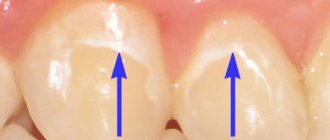
Problem: the patient is undergoing orthodontic treatment by O.A. Baranova. One tooth is short , but it is fused with the bone ( ankylosis of the tooth ), and it is impossible to move it with braces. It is necessary to resolve the issue of aesthetics during the period of orthodontic treatment.
Solution: for the period of orthodontic treatment, direct artistic restoration was performed using Spectrum light-curing material.
The patient contacted the Family Dental Orthodontist O.A. Baranova. The patient's complaints were uneven teeth, a short front tooth , which especially spoiled the appearance. After a comprehensive diagnosis, it turned out that the short tooth was an ankylosed tooth. In addition to the ankylosed tooth, the patient was diagnosed with crowded teeth, as well as a malocclusion (deep bite).
Ankylosis of teeth is the fusion of the cementum of the tooth with the jaw bone. The tooth sits firmly in the jaw, it is impossible to move it and difficult to remove without damaging the bone. With ankylosis, the tooth usually appears shorter and disrupts the aesthetics of the anterior teeth. The tooth opposite to the ankylosed one is pushed up, and the remaining teeth change their inclination towards the problem tooth. In case of ankylosis of the tooth, it is impossible to change the length with the help of braces, since there is no periodontal ligament, which is involved in the movement of teeth with braces (this is described in detail in the article by O.A. Baranova “How teeth move”). To change the aesthetics, correction with crowns is performed.
Orthodontist O.A. Baranova took part in the development of the treatment plan. and orthopedic dentist Tsukor S.V. It was decided to correct the orthodontic problems with the help of braces (the patient chose Incognito lingual braces) and the Herbst apparatus. a short tooth during the period of orthodontic treatment using artistic restoration, and after aligning all the teeth and correcting the bite, the issue of comprehensive aesthetic restoration will be resolved. It is worth noting that after orthodontic treatment, aesthetic correction is often required, since on teeth that were in an incorrect position, unevenly worn edges, asymmetrical angles, etc. become more noticeable after treatment. The patient in question already has noticeable problems with uneven edges of the lower teeth, unequal shape of the second incisors, and enamel defects. All this can be eliminated with the help of aesthetic correction after orthodontic treatment.
Before starting orthodontic treatment, hygienic preparation of teeth was carried out - removal of plaque and tartar. Professional dental plaque removal slightly changes the color of the teeth, making them lighter, and tooth restoration is carried out after cleaning, which ensures a more accurate color match. Teeth cleaning was carried out by hygienist E.P. Smirnova.
Orthodontist Baranova O.A. Incognito braces , individually made from impressions of the patient’s teeth . During aesthetic correction, the device is not fully installed to make the correction more convenient.
Orthopedic dentist Tsukor S.V. I built up a tooth in 1 hour using Spectrum, a material for direct artistic restoration of teeth. This method of restoring the aesthetics of the dentition by lengthening a short tooth with light-curing material is quite reliable, fast and inexpensive. To grow a tooth using direct artistic restoration costs 5,400 rubles. Spectrum material has excellent aesthetic properties and has the necessary strength.
Orthodontist Baranova O.A. finally installed all the necessary orthodontic equipment.
Having an orthodontist and a prosthodontist in the same building makes it much easier for patients to resolve complex situations. In this case, the installation of equipment and artistic restoration of the tooth were carried out at the same time, on the same day.
See other examples of artistic dental restoration performed by Dial-Dent specialists here.
source
- Doctors+
- 1,410 messages
- Gender: Male
- Yekaterinburg city
Question for the club of experts. How to get it now and with what? Who will recommend what from the toolkit? Because the length of the hogs is simply not enough here. The apex of the distal root is close to the canal, which can also add sensations)) The patient is 15 years old.
- Gender: Male
- City: Dp
Post edited by Romanson: June 17, 2020 - 09:38
- Gender: Male
- Moscow city
- Gender: Male
- Yekaterinburg city
Can you tell me more about what happened? The apex should not cause harm to the canal as it is not crooked, it should not break off, the shape protects against being pushed into the canal, as there is an extension towards the coronal part. Try to let the patient go and meet in 3 days - it should go easier. It seems to me that the best tools will be piezosurgical solutions.
Sent for removal 3.6. Along the way I found out that it was ankylosis. There is no play(((
- Gender: Male
- Yekaterinburg city
Stupidly, but carefully, you take the Lindeman cutter from the implant kit and drill it out.
This is where the life of the cutter will apparently end))
- Gender: Male
- Moscow city
- Gender: Male
- Moscow city
- Gender: Male
- City: Dp
- Gender: Male
- Yekaterinburg city
- Gender: Male
- City: Ukraine
- Gender: Male
- Moscow city
Post edited by Kolesnikov: June 17, 2020 - 11:56
- Gender: Male
- City: Moscow
- Gender: Male
- City: Tolyatti/St. Petersburg
- Gender: Male
- City: St. Petersburg
File away the vestibular area, there is only bone there. Root/two-way elevator. Under control of increase. Loosen medially and distally as Kolesnikov advises.
- Gender: Male
- City: St. Petersburg
Take a longer bur (nti has long surgical ones for turbines/raisers 31mm long)
- Gender: Male
- City: Kiev
So as not to create more topics. There was a recent case where the tooth fracture was 3.6 (long roots), I sawed it right along the bifurcation, I wanted to carefully remove it separately. When removing the medial and distal roots, I simply broke off pieces deeper and deeper, removed them, but quite traumatically. I couldn’t jam the elevator between the bone and the tooth. Question: what do you do in such cases, remove the bifurcation, saw the root around with a pick to pry it off, or remove part of the cortex approximally?
Ps such situations arise in multi-rooted teeth with fracture, when the roots are “stable”, fragments simply break off deeper and even more problems arise. What do you do when removing such teeth?
Post edited by Fatal: June 17, 2020 - 22:46
- Gender: Male
- City: Dp
source
Ankylosis of the TMJ in children: causes and treatment
The occurrence of temporomandibular joint ankylosis in children can be triggered by infection in the blood of newborns, as a result of which ulcers appear in the joints and bones. Also, ankylosis can manifest itself as a consequence of injury, in which the condylar process breaks and the lower jaw dislocates, blood collects in the joint cavity and hemarthrosis occurs. Since it is impossible to receive normal food in such a situation, there is a high probability that the child will develop a general developmental delay.
Eliminating this disease is the task of orthodontists and maxillofacial surgeons. Ankylosis causes the lower jaw to remain underdeveloped and doctors have to deal with malocclusion. Doctors must lengthen the injured jaw and restore its mobility. At the same time, it is necessary to correct the bite. These measures will become the basis for further normal growth and development of the facial skeleton, restoration of full breathing and the return of normal speech and chewing.
Dental ankylosis
You often encounter ankylosis of teeth if their number is incomplete. This birth defect can cause teeth and bones to fuse together. There are other possible causes of the disease. For example, if a baby tooth does not fall out in a timely manner or a molar does not begin to form. In this case, the root of the tooth can grow together with the alveolar process, and the tooth has a reduced height. In such cases, the best solution is to install an artificial crown, which allows you to correct the teeth and correct the bite.
Ankylosis of the TMJ: treatment
In the early stages of TMJ ankylosis, therapeutic procedures such as ultrasound and hydrocortisone injections are used into the joints. Also, under anesthesia, redressing is performed, eliminating fibrous adhesions in the joint with the jaws moving apart under the influence of external force.
Persistent ankylosis of the TMJ can only be eliminated during surgery. Orthodontic procedures are then carried out. Surgical intervention can relieve the patient of cosmetic defects and return the lower jaw to full functionality. During the operation, special attention is paid to anesthesia, since with this disease there may be difficulties with intubation, up to the need to perform it through a tracheotomy.
To prevent the recurrence of ankylosis, the lower jaw is fixed after surgery with splints and special means, cosmetic procedures and massage of the masticatory muscles are performed. Afterwards, the patient needs to visit an orthodontist to completely correct the bite.
Treatment methods
At the initial stage of development of ankylosis, it is possible to use conservative treatment methods: electrophoresis and injections of hyaluronidase and hydrocortisone; mechanotherapy; ultraphonophoresis. After several courses of physiotherapy, an attempt at redressing can be made - forced jaw extension (carried out under anesthesia).
Surgical treatment of TMJ ankylosis is indicated for persistent fibrous or osseous forms . The purpose of the operation is to restore normal functioning of the lower jaw and correct facial deformity. There are several options for operations for TMJ immobility:
- Osteotomy with atroplasty.
- Osteotomy of the lower jaw followed by skeletal traction.
- Bone grafting of the branches of the lower jaw with auto- or allograft.
The volume and nature of the intervention is determined by the degree of deformation of the jaw and facial skeleton.
In the postoperative period, the lower jaw is fixed with intraoral splints or orthodontic structures, the task of which is to prevent relapse. During the rehabilitation period, mechanotherapy, physiotherapy, and jaw muscle massage are used.
Removal steps
If the wisdom tooth has already erupted, the dentist may not carry out any preparatory procedures. If an unerupted or partially erupted “figure eight” is to be removed, the doctor will definitely refer the patient for an x-ray. And based on the results of the image, he draws up a plan for the operation. Its duration depends on the complexity of the procedure. Simple removal will take 5 - 10 minutes, complex removal will last for 20 - 160 minutes.
Easy removal
- Stage 1.
Selection of anesthetic. The doctor inquires about the patient’s allergies and asks him about chronic and past diseases. - Stage 2.
Conducting anesthesia. When removing an upper wisdom tooth, pain relief will take effect within 3-4 minutes. If you have to remove the bottom “eight”, you will have to wait 8-10 minutes. - Stage 3.
Actually, the removal or extraction of the “eight”. The doctor grabs the tooth with forceps or elevators and removes it from the socket.
If the removal was carried out against the background of inflammation, the dentist will wash the wound with an antiseptic solution and put an anti-inflammatory drug into the hole. In most cases, the operation ends with suturing the wound. This way it will heal faster and the risk of bleeding will be significantly reduced.
Simple wisdom tooth removal takes from 1 to 10 minutes. In this case, the gum is not cut, and the tooth is not drilled, but simply removed from the bone tissue.
Patient reviews
Review 1
Before describing the procedure, I want to talk about my pain threshold. I can NOT stand pain. At all! I LOVE IT!!! For example, before treating caries, I ask you to give me 2 painkiller injections. And then one day my wisdom tooth began to hurt. I endured it steadfastly! But patience has come to an end! I had to go to the clinic.
Sitting down in a chair, I, as usual, asked for 2 injections and mentally prepared myself for torture :) Instead of somehow supporting and encouraging me, the doctor said: - Nahhh... but the tooth is in an inaccessible place! You can’t even get close to him!!!
Then she asked me to open it wider, then even wider, then again and again... It seemed to me that the corners of my mouth were already torn, and the doctor kept asking me to make my “mouth wider”! At the same time, she said, “What can I do here...” and tried to pick up the tooth with pliers.. These attempts lasted about 5 minutes. There was no particular pain, it was just unpleasant that the doctor could not pick up the tooth and was scared from waiting for the outcome. In the end, I asked: - Maybe I’ll go already?!!! If she can’t pick up a tooth, then WHAT will happen next?! To my deep surprise, the doctor did not even try to persuade me to stay and complete the procedure. She simply said: “Go!”
Well, wow :) This has never happened to me before. Usually doctors persuade: - Well, be patient... If you came, then you need to be patient. And here on you - go... I ask: - And what to do now? The answer was: “Nothing!” Rinse! And don't eat anything for 2 hours. And don't drink anything hot all day. - Why??? - Because the wound should heal. - So you REMOVED my tooth??? - Yes!
Just like that! While I was preparing for “medieval torture” and many hours of torment, the specialist did her job! BRAVO!!! I was delighted! After a couple of hours, the freezing sensation began to subside and the pain returned. She was tolerable, rinsing helped. After a few days the pain went away, but the wound healed for probably a whole month! The pain was weak and dull... The gums ached a little and as if something was pulling. But everything ended well! Everything is overgrown evenly.
Review 2
One of the four teeth had grown into the gum and began to rot there. Therefore, when I came to my favorite dentist, there was only one diagnosis - the wisdom tooth needed to be removed. I honestly couldn’t even get scared when I was already given anesthesia and the fragile dentist started pulling my tooth.
From the sensations, I can’t say that it hurt, no, the anesthesia worked well, but there was a feeling, I felt the tooth being taken with forceps, how it was being pulled, and I was even surprised how my fragile doctor pulled out this chunky tooth. He fought hard to get out, but for professionals there are no problems.
After this, of course, my gums bled a little, I was prescribed mouth rinses so that everything would heal faster. My gums healed within a day. I did not feel any discomfort due to the missing wisdom tooth.
Therefore, I can say for sure that the devil is not as scary as he is painted. Yes, it was unpleasant, but not painful and it could be tolerated. And if I have to remove the rest of my wisdom teeth, I won’t be the least bit upset or scared. Since there is nothing scary in this procedure.
Types of temporomandibular joint diseases
The most common diseases of the temporomandibular joint are:
- Dislocation of the joint. It is the result of a violation of the relative position of the jaws due to muscle disorders. Accompanied by clicking, pain when opening the mouth, displacement of the components of the joint (head and disc). There are several types of displacement of the articular head: hypermobility, functional impairment (sprain), subluxation of the head. Displacement of the articular disc is called: disc dislocation (manifested by a click), disc subluxation (minor displacement, invisible to the patient), prolapse of the articular disc
- Arthritis. Acute arthritis is characterized by pain in the temporomandibular joint, limitation of movements of the lower jaw, swelling of the joint, increased body temperature
Temporomandibular joint
- Arthrosis. It is characterized by a disturbance in the movement of the jaw (when opening the mouth, a zigzag movement of the lower jaw), pain in the joint, ears, and chewing muscles. When moving, clicks and crunching sounds appear. Arthrosis is associated with injuries, inflammation, metabolic disorders
- Ankylosis. Caused by injuries and infectious diseases. The main symptom is limited movement of the lower jaw. In the case of purulent ankylosis in childhood, facial asymmetry, malocclusion, and numerous caries appear. With persistent changes in the joint, complete loss of movement may occur.
- Musculo-articular dysfunction. It is characterized by pain in the temporal region and ear, which increases during chewing, blocking of the lower jaw, clicking while moving the jaw, and facial asymmetry. Can be explained by endocrine and psycho-emotional factors. Leads to the development of arthrosis
Of great importance for diagnosing temporomandibular joint disease is radiography, which is used to determine the structure of the joint and its disorders.
Is it painful to remove a wisdom tooth from below?
Reviews about the removal of lower wisdom teeth are very contradictory. Some talk about the complete painlessness of the operation, others remember the minutes spent in the chair with a shudder. The fact is that when a wisdom tooth is removed from below, the patient’s sensations directly depend on the professionalism of the doctor.
Thus, infiltration anesthesia in the upper jaw is carried out in the gum in the area of the root of the wisdom tooth. And in the case of the lower “eight”, the dentist has to give an injection from the inside of the jaw. Carrying out such anesthesia is technically more difficult, and, alas, not all dentists are fluent in it.
If modern anesthetics are used, and the doctor is a professional in his field, pain during the removal of the lower wisdom tooth is completely eliminated.
It may hurt a little when the anesthetic is administered. Unpleasant sensations do not go away if an abscess has already formed in the area of the wisdom tooth. But in particularly difficult cases, the dentist may decide to use general anesthesia.
Symptoms
With this disease, patients complain of the inability to open their mouth wide enough, which makes it difficult to eat and causes difficulty in speaking clearly. Such patients are forced to eat liquid or semi-liquid food, which passes through the gap between the dentition or existing dental defects. Banal vomiting in such patients can cause death due to asphyxia.
If the disease develops in childhood, it may be accompanied by deformation of the facial skeleton, dental anomalies, malocclusion, and impaired teething. Unilateral temporomandibular joint ankylosis may be accompanied by a shift of the facial midline towards the affected side and the development of crossbite. With bilateral lesions, there is a posterior displacement of the chin, the formation of microgenia and underdevelopment of the lower third of the face.
Patients with this disease may experience breathing problems and difficulty or inability to perform proper oral hygiene.
What drugs are used for pain relief
Just a few decades ago, the only painkiller used by dentists was novocaine. Despite its low toxicity, it very often caused serious complications in patients: from an allergic reaction to anaphylactic shock.
The next step was the use of lidocaine. It is still used to this day in budget dental offices, and has an undeniable advantage: allergies to the drug occur extremely rarely. The problem is different: to prolong the effect of lidocaine, the dentist has to add a drop of adrenaline to it. Moreover, this is done “by eye”. And as soon as the doctor’s hand trembles a little, the patient is guaranteed such unpleasant symptoms as rapid heartbeat and dizziness.
It is much more comfortable for both the doctor and the patient of the dental clinic to deal with articaine anesthetics. These include:
- ubistezin
; - ultracaine;
- septanest
.
Firstly, such drugs are administered with a carpule syringe, which ensures that the injection itself is less painful. Secondly, the ratio of adrenaline and anesthetic in the drugs is adjusted to the nearest milliliter. This means that wisdom tooth removal will be painless, and the patient will not experience any side effects.
Important! If the patient is at risk, it is recommended to use an anesthetic that does not contain adrenaline.
Causes
In most cases, the causes of ankylosis of the TMJ lie in acute or purulent inflammation of the joint, as well as viral or bacterial damage to nearby tissues. Sometimes the disease develops due to the patient’s predisposition or due to inflammation of arthritis or arthrosis.
Other common causes of the disease include:
- viral or bacterial inflammation of the ENT organs;
- TMJ arthritis;
- osteomyelitis of the lower jaw;
- osteomyelitis of perimandibular phlegmons;
- purulent or acute otitis media of the middle ear;
- mastoiditis.
In some cases, the disease develops in connection with neonatal sepsis, which occurs with the formation of purulent inflammation in the joints or bone tissues.
In addition, bony ankylosis of the TMJ can form due to birth trauma or a blow. The following are recognized as provoking factors:
- falling from a great height;
- gunshot wounds;
- road accident;
- domestic injury.
In such situations, the disease may develop due to a fracture of the condylar part or due to dislocation of this area.
Any trauma to the lower area of the face sooner or later provokes deformation of the cartilage tissue. At the diagnostic stage, you can notice the proliferation of connective tissue, which subsequently becomes very dense. Then dense scar tissue forms in this area. Fusion becomes the cause of bone ankylosis of the TMJ.
Causes of temporomandibular joint ankylosis
The etiology of the disease can be primary and secondary. In the latter case, the occurrence of ankylosing changes is influenced by other primary conditions developing in this area: purulent inflammation of the joint, necrotic processes in the bone apparatus, purulent diseases of the inner ear.
Medical histories in children and adolescents most often indicate an infectious origin of TMJ ankylosis. The reason for this is the entry of infectious pathogens into the bloodstream, which become the source of the onset of purulent processes in the organs and joint apparatus.
Infection in the blood of adults occurs against the background of a concomitant infectious process, for example, scarlet fever, diphtheria, gonorrhea. Among the sources of pathology in adults is the development of primary pathology in nearby bones or joints (cervical, thoracic).
Less commonly, ankylosis occurs after a traumatic factor, which entails the fusion of its surfaces. For example, with a direct blow to the TMJ, a bone fracture occurs, bleeding occurs, and hemarthrosis develops.
The disease in newborns can occur after difficult labor, during which medical forceps were used. Often the development of the disease is influenced by other chronic diseases occurring in this department or in nearby areas.