Mechanical trauma is the most common type of damage to permanent and temporary teeth in children. It leads to a violation of the anatomical integrity, changes in the dentition or destruction of surrounding tissues. According to statistics, the front teeth, including the upper incisors, are most often susceptible to traumatic damage in children aged 6-10 years. The lack of proper treatment in this case often leads to serious complications, which can manifest as inflammatory-destructive processes, the appearance of cysts and other negative consequences.
Symptoms
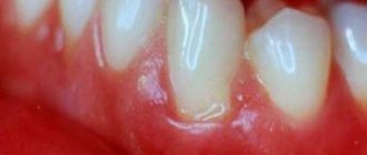
A strong blow to a tooth is accompanied by damage to periodontal tissue, and some fibers and small blood vessels rupture. There are no visible structural damage to the tooth; upon visual inspection, it appears intact. After a bruise, the tooth remains motionless, and minor mobility is rarely observed. The gums in the area of the injured tooth swell.
In the first hours after the impact, patients experience pain in the tooth, which intensifies when biting; the pain is aching in nature. The tooth feels high, and slight bleeding may occur from under the gum near it. When a bruise occurs, the neurovascular bundle of the tooth can be damaged, that is, the pulp is injured, and hemorrhage occurs in the pulp chamber, and the enamel becomes pink in color. A severe bruise can lead to the death of the pulp.
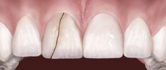
Often, when a bruise occurs, cracks appear on the tooth enamel, which can only be detected with a special examination.
Classification of dental injuries
There are acute and chronic dental injuries. Depending on the time of occurrence, they can be permanent or temporary. Primary teeth are characterized by dislocations and, less commonly, root fractures. Permanent injuries are considered: crown fractures, tooth root damage, as well as bruises and dislocations.
There are 8 known types of dental injuries:
- Tooth bruises – accompanied by violations of the integrity of the tooth structure (cracks, chips);
- Uncomplicated crown fractures;
- Complicated coronal fractures;
- Complete crown fractures;
- Fractures of crowns with violation of the integrity of the longitudinal root;
- Root fractures;
- Incomplete dislocations;
- Complete dislocations.
Regarding the completeness of the formation of the root system, the following types of dental injuries are distinguished:
- damage due to incomplete root growth;
- damage with incomplete formation of the root apex;
- damage with a fully formed root.
In addition to these injuries, there are also combined forms:
- incomplete dislocation and fracture of the crown (or root of the tooth);
- fracture of the dental crown and tooth root;
- dislocation with fracture of the root (or crown) of the tooth.
Bruised tooth
A tooth bruise is a closed mechanical injury to a tooth, in which its integrity, as a rule, is not compromised. The injury is accompanied by damage to periodontal tissue (tear or rupture of fibers). When the neurovascular bundle is completely ruptured, hemorrhage occurs into the pulp chamber and pulp necrosis.
The damage is characterized by severe aching pain, intensifying when biting or percussing the tooth. Swelling of the periodontal tissue can cause the tooth to appear as if it is coming out of its socket. Slight tooth mobility may also be felt. Hemorrhage into the pulp chamber leads to a change in the color of the tooth, which becomes pink.
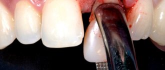
Diagnosis and treatment of tooth bruises
A tooth bruise is diagnosed after taking a history and based on clinical manifestations - darkening of the crown, pain. Instrumental data are also taken into account. An X-ray reveals the widening of the periodontal fissure and the likelihood of a tooth root fracture. A patient with a tooth bruise is expected to undergo monthly electroodontodiagnosis (EDD).
The main principle of treating injury is to minimize the load on the damaged tooth until the pain completely subsides. It is recommended to refrain from eating solid foods and chewing on the large side. In order to eliminate discomfort, the doctor can grind down the sharp edges of the tooth that cut the tongue and cheek. To stop inflammation, the dentist prescribes medication and physical therapy. If the doctor determines pulp necrosis, then cleaning is carried out with further filling.
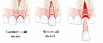
Tooth luxation
Tooth luxation is an injury in which a tooth partially or completely loses its connection with the socket. Incomplete tooth luxation (or extrusion) occurs due to tear (rupture) of periodontal fibers and due to damage to the walls of the alveoli. Such an injury is accompanied by pain, changes in the position of the tooth, its mobility, and the inability to chew. The displacement of the dental crown can be in the vestibular, distal, oral and other directions. The root of the tooth may deviate in the opposite direction. As periodontal tissues become damaged, bleeding “pockets” of teeth or gums occur. Incomplete dislocation is often accompanied by the formation of dental granulomas or root cysts, chronic periodontitis, disturbances in the formation of the tooth root, expansion of the dental canal, etc.
In case of complete dislocation (for example, traumatic extraction of a tooth), a rupture of periodontal tissues is observed with the capture of the circular ligament of the tooth, which is accompanied by its loss from the socket. The central incisors of the upper jaw are more susceptible to injury. During the examination of a patient with such a diagnosis, the tooth may no longer be in the row. In its place there will be only an empty hole with a blood clot.
Impacted tooth dislocation is accompanied by the penetration of the tooth root into the bone with the immersion of its coronal part into the cavity of the socket. With such injuries, the crown of the damaged tooth will be lower in level among others. The damage manifests itself as acute pain and alveolar bleeding.
Diagnosis and treatment of tooth dislocation
Diagnosis of tooth dislocation is made based on the clinical picture of the oral cavity and X-ray data. Studies make it clear what condition the alveolar bone is in and where the diseased tooth is located. The EDI procedure will help determine the viability of the pulp. In case of incomplete dislocation, treatment is aimed mainly at preserving the tooth. For this purpose, the dislocated tooth is returned to the socket and fixed with splints or mouthguards. A gentle regime and anti-inflammatory drugs are prescribed. Every month the patient must be observed by a dentist to monitor the process of tooth healing. If the pulp has died as a result of injury, the doctor performs endodontic treatment.
Treatment for complete dislocation involves prosthetics or dental implantation. If possible, replantation is performed and the tooth is returned to the socket, where it is fixed with splints. Impacted dislocations involve tooth repositioning under local anesthesia with further immobilization and orthodontic treatment. If the tooth cannot be restored, it is removed and replaced with a prosthesis.
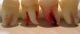
Tooth fracture
The following tooth fractures are distinguished: crown part within the enamel, crown detachment or root fracture. A crown fracture is noticeable to the naked eye: it is characterized by changes in the shape of the tooth, disrupting its aesthetics, and acute pain. The tooth remains motionless. The sharp edges of the broken coronal part injure the mucous membranes of the mouth, tongue and lips. Such an injury can provoke acute traumatic pulpitis (or periodontitis).
There are longitudinal, oblique, transverse and comminuted fractures of the roots of teeth, which are localized in the middle, lower or upper third, and can also be with or without displacement of the fragments. Fractures are characterized by severe pain that increases when biting or percussing the tooth. The crown can be movable to varying degrees.
Diagnosis and treatment of tooth fracture
A tooth crown fracture is diagnosed during a dental appointment. To confirm or exclude the diagnosis, targeted radiography is done. In order to assess the viability of the pulp, EDI is prescribed.
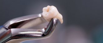
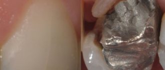
Treatment of damage to the crown is aimed at restoring its natural shape through the use of composite materials or using a stump inlay. If there are complications with a tooth fracture, treatment of pulpitis followed by tooth restoration may be required. Treatment for a root fracture involves tooth extraction or endodontic therapy followed by placement of a post.
Diagnostics
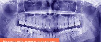
- Patients with a tooth injury are referred for x-ray diagnostics to exclude a root fracture. To obtain information using targeted radiography, it is sometimes necessary to take several images from different angles, which is undesirable for the patient.
The most accurate information about the condition of the roots is provided by computed tomography - an x-ray examination method that allows you to obtain a three-dimensional image of the tooth. The examination results are displayed on the computer screen and must be transferred to a CD or USB flash drive.
An X-ray examination of a tooth bruise reveals a slight widening of the periodontal fissure.
- The condition of the pulp after injury is monitored using EDI - electroodontodiagnosis. The method consists in determining the reaction of the nerve endings of the pulp to the influence of electric current. The level of electrical excitability of the pulp depends not only on its condition, but also on the degree of formation of the tooth root.
The examination is carried out 2 or 3 days after the injury, since on the first day the pulpal response may be reduced due to traumatic neuritis. Be sure to perform an electrical test on adjacent healthy teeth to compare sensitivity levels. 3-4 weeks after the injury, EDI is repeated.
- Another method of examination for a tooth bruise is transillumination, the essence of which is to pass a beam of cold light through the tooth and evaluate shadow formation. If there are cracks in the enamel after an impact, they will be clearly visible in the stream of light; The technique also helps to detect pulpitis. In modern clinics, all dental units are equipped with light guides for transillumination examination.
Treatment of dental injuries in children
The stages of therapy in children may vary in time. Sometimes this can be a few days, but more often it happens that treatment can take up to three years. Its duration depends on the severity of the injury, and in addition, on the degree of formation of the child’s dental system and the methods of treatment chosen. Children with dental injuries need rehabilitation measures. Typically, treatment is divided into the following three stages:
- The initial treatment stage lasts from the first visit to the doctor with a complaint directly to the provision of special medical care. Help can be provided to a child in any medical institution. If only the teeth are affected, and the soft tissue with the bones of the facial skeleton is intact and there is no concussion, then the child is referred to a dentist. It would be best to immediately contact a pediatric dentist. The doctor will take measures to assess the patient’s condition, make a diagnosis, provide pain relief or prescribe analgesics. As a rule, specialized therapy is delayed for several days.
- The second stage begins with collecting anamnesis. In this case, the cause of the injury is determined and special treatment is prescribed until complete recovery. This includes the preparation of medical documentation along with work with anamnesis, clinical examinations, x-rays, diagnosis and treatment.
- At the third stage, the functions of the injured incisors are restored, as well as follow-up treatment and clinical observation.
Dividing restorative post-traumatic therapy into these stages makes it possible to quickly assess how well the child is receiving help.
Complications after injury
With bruises, the prognosis is usually favorable, but in some cases the injury can lead to the following complications:
- Darkening of the enamel. After a bruise, the cause of darkening is hemorrhage into the pulp chamber: the pink color of the tooth darkens over time, the enamel acquires a brownish or gray tint. Depulped teeth tend to darken due to the fact that metabolic processes in them stop, the teeth become “dead”, and the enamel becomes dull.
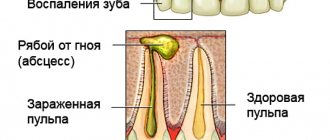
- The death of the pulp leads to the development of pulpitis: the pulp decomposes, inflammation develops in the tooth, which without treatment turns into periodontitis.
- Periodontitis is inflammation of periodontal tissue. It can be: post-traumatic, occurring a short time after the injury; chronic, develop as a consequence of pulpitis. When a purulent infection occurs, there is a high risk of tooth loss and purulent blood poisoning.
- The appearance of a post-traumatic odontogenic cyst at the root apex during the development of post-traumatic periodontitis.
- Stopping the development of roots of permanent teeth in children.
- In case of injury to milk teeth, the following are possible: disruption of the formation of the rudiments of permanent teeth, their death.
Treatment of dislocation
How to treat such acute dental trauma in children?
The symptoms of any dislocation are as follows: tooth mobility occurs, and in addition, complaints of pain in one or more incisors. During treatment, doctors decide whether to save the damaged tooth. In this case, it is advisable to assess the condition of the bone tissue. If up to half of the bone tissue at the root has survived, the tooth is preserved.
The damaged tooth is set under anesthesia, and then a state of rest is created. In addition, doctors determine how damaged the pulp is. If the nerve bundle is torn, the pulp may remain viable. Necrosis of the pulp often requires its removal, and in addition, filling of the canals. The condition of the pulp is checked by its reaction to electricity. If there is any doubt, the pulp is later re-checked and observed over time.
There are different types of dental injuries in children. When a tooth seems to be driven into the jaw, the child feels severe pain. In this case, you can see a reduced masticatory organ. In this case, it is advisable to fix the damaged tooth and remove the dead pulp as soon as possible so that the color of the tooth does not change. A complete luxation is the loss of an entire tooth. If the periodontal tissues have not been changed, the chewing organ undergoes replantation.
Treatment
- For mild bruises, treatment consists of resting the tooth for 3-4 weeks by reducing the load during chewing: the menu includes soft and semi-liquid foods, and a blender is used to grind hard foods.
- To ensure rest for baby teeth, temporary bite separation with the help of mouth guards is used; if a permanent tooth is bruised, splinting is performed. The splint allows you to immobilize an injured tooth and redistribute the load during chewing onto healthy teeth.
- If the pulp dies due to an impact, the tooth cavity is opened and the pulp is removed, after which the root canals are filled and a permanent filling is installed. If the crown of a tooth darkens, it can be whitened.
- When a baby tooth is bruised, grinding of the cutting edge of the crown of the antagonist tooth is used to prevent tooth contact and reduce pain. This method is not used for permanent teeth.
- To relieve pain, it is recommended to take an anesthetic tablet (ibuprofen, ketorolac, nimesulide) and apply an anesthetic gel (Dentol, Kamistad) to the gums around the tooth.
- When swelling of the soft tissues of the face accompanies a bruise, cold compresses are applied: a plastic bottle with cold water (not lower than +4°C) is wrapped in a cloth and held on the area of swelling for 15-20 minutes.
- A course of magnetic laser therapy is carried out - a combined effect on injured tissue of a magnetic field and low-intensity laser radiation. The method helps improve healing processes, relieves swelling and inflammation. The course consists of 10 daily procedures lasting 5 minutes.
- UHF therapy is indicated to accelerate tissue regeneration.
In dentistry, the following types are distinguished:
- Bruised tooth. It can be considered the mildest type of injury, which is provoked by blows, attempts to bite through a hard object or “taste” it, which is especially typical for children 1-3 years old.
- Tooth dislocation. Such an injury means that the tooth has moved from its usual place. Typically, this type of injury requires significant force, so it does not occur as often as bruises. There are cases of complete dislocation, when the entire tooth falls out of the socket. Modern pediatric dentistry can save even a lost tooth if there are favorable conditions for this, and parents brought their child in immediately after receiving an injury.
- Tooth fracture. Fractures are immediately visible - these are visible chippings of a corner, piece or most of a baby tooth. Most children break their front teeth; the most popular causes of such damage include fights.
- Fracture of the root or crown of a tooth. They are considered rarer species, because in normal everyday life and sports conditions it is extremely difficult to get such a deep fracture. Regardless of whether the child has acquired a simple tooth bruise or suffered a fracture, in any case it must be shown to a specialist so as not to endanger the health of the young body.
Prevention
To prevent dental injuries, you must:
- Observe safety precautions at work, arrange the workplace and carry out potentially hazardous work in accordance with established rules.
- Properly equip playrooms for children and supervise children while playing.
- Follow traffic rules and use seat belts when traveling, as even minor emergency braking of the car carries a high risk of injury.
- Avoid conflict situations that lead to fights.
- Ensure the safety of sports games using special equipment (helmets, masks, dental guards).
- Carefully select sports grounds: they must comply with safety requirements.
Tooth fracture in children
This type of tooth injury in children is very easy to diagnose because it is visible to the eye. Treatment in this case directly depends on the amount of tissue lost. Part of the crown can be restored using a filling composite material. It is important to cover the dentin with a special gasket immediately before filling. The crown can best be restored with a cap. Among other things, parapulp pins can also be used.
During the opening of the dental cavity in case of injury, anesthesia is performed, the pulp is removed and the canal is filled. In order to fix the filling as intensively as possible, the doctor installs a pin, which is fixed in the canal. It is possible to use composite fillings, caps, crowns or inlays. If a tooth is broken, it is necessary to restore it as quickly as possible, since the neighboring incisors will try to take up the free space and begin to tilt. In the future, with this development of events, prosthetics will be impossible without the intervention of an orthodontist.
The most common injury to a child's front teeth occurs.