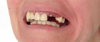
What is crack and fracture?
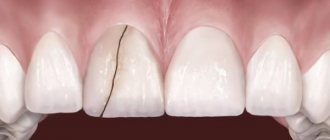
A crack is a violation of the integrity of the tooth enamel. (see photo)
It can occur as a result of injury or the abuse of very hot and very cold foods, especially when alternating (glaze lovers, think about whether it’s worth it!). It is easy to notice a crack if you shine a light on the tooth from the side. A crack in a tooth itself is not dangerous, but it can become a gateway for infection on the way to dentin or transform into fracture.
Fracture (literally fracture) is a chipping of part or the entire crown of a tooth. The most common causes of fracture are:
- Trauma . A car accident, playing sports without protection (hockey, boxing, etc. without a mouth guard), or simply hitting the tooth with a hard object can all lead to fracture;
- Large filling (especially on a pulpless tooth). The larger the filling, the greater the negative impact it can have on the surrounding hard tissues of the tooth (for example, when eating hot food, most fillings expand differently than the tooth tissues, especially for fillings made of a metal alloy-amalgam) and this is provided that the filling delivered with high quality. In the case when it is mobile, the probability of fracture increases sharply;
- Increased chewing load . I think any of you can remember a story told by a brother/friend/acquaintance about “how yesterday I ate crackers/pistachios/roach, and suddenly half a tooth chipped off!!!”. If the circumstances are unfortunate, solid food in combination with the strength of the chewing muscles developed can often lead to the chipping of a part of the tooth or a fracture of the wall.
Frakturs can be divided into:
- passing only through the enamel (the enamel chip may be insignificant in volume and does not always require treatment);
- including dentin (if left untreated, they can cause increased sensitivity of the tooth and accelerate the development of caries);
- affecting the pulp chamber (mandatory depulpation of the tooth is required due to severe pain and the possibility of developing inflammation in the periodontium ).
If the fracture line runs deep under the gum or a root fracture occurs, fracture can lead to tooth extraction.
If the doctor sees that the walls of the tooth are thinned for some reason and recommends that you cover the tooth with an artificial crown, listen to his advice more carefully, otherwise the tooth may be lost as a result of fracture!
Tooth extraction: what the patient needs to know
MCOH dentists believe that the tooth should be preserved whenever possible. However, there are often situations when its preservation threatens further complications, and removal cannot be avoided. In such cases, our specialists not only conduct a detailed examination to find the best option for dental care, but also justify each stage of the upcoming treatment in a form accessible to the patient.
The department has all the necessary equipment for diagnostics, which allows the examination to be carried out quickly and efficiently. Tooth extraction can hardly be called a simple procedure, since in each case it is necessary to take into account the individual characteristics of the tissue structure, predict the risks of complications, the patient’s age, concomitant diseases and much more. Tooth extraction is usually one of the stages of complex treatment, which will later be continued with prosthetics or other types of dental care.
The price is determined by the volume of medical care provided, as well as the type of materials used. For example, the cost of implants can vary significantly depending on the size, quality, manufacturer, etc.
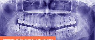
Fracture of tooth root
The most common causes of loss of tooth strength are root caries or root fracture.
Subject area
Dentists use various terms, including: "chipped tooth" or "cracked tooth." In order not to introduce misunderstandings into the conversation, they use a general classification of tooth cracks, which includes
- cracked tooth enamel;
- cracked tooth cusp;
- cracked tooth;
- chipped tooth;
- vertical root fracture.
A cracked tooth is a condition in which a crack has formed in the tooth, starting at the crown and continuing in an anteroposterior direction. This is a case of incomplete fracture. These types of fractures tend to involve the deeper apical areas of the tooth, causing root destruction and chipping of the entire tooth. In some cases, such a pathology is called a longitudinal root fracture; it must be differentiated from vertical root fracture.
A vertical root fracture is a crack that runs from the inner wall of the root canal to the surface of the tooth root.
Vertical fracture can occur in a healthy tooth in very rare cases; it usually develops on operated teeth as a result of improperly performed endodontic treatment. Most often, vertical root fractures affect the anterior roots of the lower molars and the second upper premolars.
Diagnosing vertical tooth root fractures is quite difficult, since similar symptoms, radiographic data and clinical picture are also characteristic of gingivitis, periodontal disease and other periodontal diseases.
Material and methods
The study was conducted on 25 intact single-rooted teeth removed for periodontal and orthodontic reasons. Previously, decoronation of the teeth and mechanical treatment of the root canals were performed (K-files, Dentsply; RaCe, FKG Dentaire SA) to the final size No. 25.06. The channels were subsequently unsealed by 2/3.
VTCs of teeth were obtained by applying a wedging force, in this case an anchor pin, which was screwed into the root canal of the tooth until a crack occurred. After removal of the pin, the presence of VTC of the teeth was confirmed using a stereomicroscope. 5 samples were not exposed and were selected as controls. Next, the tooth roots were fixed in self-hardening plastic (Rebaron, GC Corporation); then CBCT was performed.
CBCT was performed in the laboratory on a 3D Accuitomo 170 (3D Accuitomo; J. Morita Mfg. Corp., Kyoto, Japan) with a flat panel probe; field of study (FOV) - 4x4 cm, voxel size - 0.16 mm, radiation parameter - 80 kW, 4 mA, 30.8 s.
After obtaining X-ray data and analyzing the localization of cracks, transverse separation of the roots was carried out, dividing them into 3 parts. A total of 75 sections were obtained. For samples from the coronal and middle parts of the root, both cut surfaces were assessed, for samples from the apical part - 1. Only samples with a visually detectable crack were included in the study group. The control group (without crack) included 15 sections.
After preliminary preparation of the polished section, the crack size in the study group was measured using a Neophot 2 microscope at various magnifications. To record images of microobjects, a video eyepiece (Digital camera for microscope DCM-800) was used. Final image processing was performed using a photo editor (Arc Soft Photo Studio 5). A total of 40 thin sections were selected, forming three groups: control (without cracks), with cracks 50–150 µm in size (see figure).
VTK of the tooth is 50-150 microns. a - tooth root; b — axial section of CBCT; c — fragment of a microscopy data slice.
CBCT data corresponding to the acquired slices were isolated for subsequent evaluation. Images of these areas were presented in a PowerPoint presentation and assessed by 5 physicians not involved in sample preparation. A three-point scale was proposed for assessment: 0—no crack; 1—the presence of a crack is in doubt; 2 - there is a crack.
The research data are presented in the table.
Sensitivity, specificity and accuracy of CBCT for detecting root fractures
Sensitivity and accuracy when visualizing cracks >150 µm wide were significantly higher than when visualizing cracks <50 µm; specificity did not differ significantly ( p
=1.0). To determine reliability, the Wilkinson criterion was used, and to determine the agreement between researchers, the Cowan Kappa coefficient was used (on average 0.81).
crack at the root - remove?
Moderator: Lesya
Read also: Building up tooth enamel
crack at the root - remove?
Post by DmitryN » Wed Aug 15, 2012 15:39
In a private clinic, after a small chip, tooth 6 (46, an old problematic pulpless tooth) was prepared for prosthetics - an inlay and a metal-ceramic crown. The canals were partially filled, an impression was taken, and a temporary filling was inserted. The insertion of the inlay was scheduled after 9 days, but about a week later the prepared tooth began to sag a little. When the doctor removed the temporary filling to insert the tab, there was slight bleeding from the distal root and pain was felt. Examined by a therapist. She said that when the instrument is inserted, there is slight bleeding from two channels, therefore, most likely there is a crack. They sent it for deletion, which made us very upset, because... I wanted to preserve my roots. The picture shows a chipped tooth before filling and after filling, when there was bleeding. I wanted to clarify two questions: 1) do you really need to delete it, or are there any options with the tab? 2) what is better to place an implant (and which one) or a bridge with depulpation of the healthy five and a crown on the dead seven (doctors suggest)? The tooth hurts a little when pressed. Thanks in advance. Based on your answer, I will decide what to do next and where to get treatment.
Re: crack at the root - remove?
Post by Lesya » Wed Aug 15, 2012 21:49
Good evening. Yes, a root crack is a serious problem, especially when it comes to placing a tab on these roots. If there is indeed a crack or perforation in the root, the main problem is that blood and fluid will leak from this place, thereby soon this tab will become cemented and the crown will fall out. That's why the doctor started talking about removal. If there is no chance of sealing this crack and being sure that it will work, then it’s a pity to charge you money for an inlay and a crown. Now there are materials that make it possible to hermetically close perforations, but it all depends on the size of the crack or perforation.
I cannot tell you what to do, since I personally cannot check the tooth. If you are in doubt, it is better to show up to someone else. Let you have the opinions of at least two general practitioners. If the second one says that there is a perforation (or crack), then it is better to remove the tooth. Since any prosthetics for a cracked tooth will obviously be without a guarantee and most likely not successful. That is, at best, a year, then the root will crack and everything will collapse. If you are ready to take a risk, then you can try to seal the MTA perforations (if they are not large), do prosthetics without a guarantee and see what happens next. In this situation, the main thing is not to combine the 6th tooth with 7 crowns, let each one be on its own. So at least you don’t risk having to redo two teeth at once if the root cracks completely.
If the tooth is removed: what to do next? The 7th tooth is in such a condition that it also needs prosthetics. Most of the tooth is already a filling, and in the absence of the main chewing tooth (6), it will begin to crumble under the load. That is, it will have to be covered with a crown. And then it all comes down to finances and the condition of the 5th tooth. If the 5th tooth is without a filling, without caries, and you can afford to pay for an implant, then, of course, it is better to leave the 5th tooth alive and not make a bridge. If there is already caries on it, it is large and you understand that implantation is not yet possible for you, it is better to make a ceramic bridge, preparing the 5th tooth for this. Therefore, for now, I would advise having the 6th tooth checked by another doctor, listening to another opinion and only then deciding what to do. But you need to consult in person, since the decision to delete is never made solely on the basis of the image. You must see it with your own eyes and hands. how bad is everything there?
Fracture of the tooth crown
Fracture of the tooth crown.
The most common causes of loss of tooth strength are root caries or root fracture.
Subject area
Dentists use various terms, including: "chipped tooth" or "cracked tooth." In order not to introduce misunderstandings into the conversation, they use a general classification of tooth cracks, which includes
- cracked tooth enamel;
- cracked tooth cusp;
- cracked tooth;
- chipped tooth;
- vertical root fracture.
A cracked tooth is a condition in which a crack has formed in the tooth, starting at the crown and continuing in an anteroposterior direction. This is a case of incomplete fracture. These types of fractures tend to involve the deeper apical areas of the tooth, causing root destruction and chipping of the entire tooth. In some cases, such a pathology is called a longitudinal root fracture; it must be differentiated from vertical root fracture.
A vertical root fracture is a crack that runs from the inner wall of the root canal to the surface of the tooth root.
Vertical fracture can occur in a healthy tooth in very rare cases; it usually develops on operated teeth as a result of improperly performed endodontic treatment. Most often, vertical root fractures affect the anterior roots of the lower molars and the second upper premolars.
Diagnosing vertical tooth root fractures is quite difficult, since similar symptoms, radiographic data and clinical picture are also characteristic of gingivitis, periodontal disease and other periodontal diseases.
Etiology and pathogenesis
Can contribute to the development of vertical root fracture
- dental injuries of various origins;
- reduction in the thickness of the dentin layer during treatment or when preparing the root canal for an inlay (or pin);
- strong mechanical loads during endodontic treatment, during condensation of gutta-percha, during fixation in the root canal.
The thickness of the layer of preserved dentin plays a very important role. Interclinical studies showed that with a force equivalent to 3 kg applied to the spreader, no fractures occurred when the dentin layer was reduced by 20-30% of the natural thickness. When the thickness of the dentin layer was reduced by 40%, root fractures formed in five incisors; at 50%, in seven incisors.
Lateral condensation is part of the picture and should always be taken into account when assessing risks. The application of excessive force to the plugger or spreader when compacting gutta-percha is one of the factors in the possible formation of tooth root fractures.
There are many things to consider to ensure successful treatment. The type of spreader used is important. Steel tools require twice as much force as nickel-titanium ones, this is the disadvantage of the former. Experts generally recommend abandoning the use of hand spreaders, which create excessive stress when compacting gutta-percha, and preferring finger side condensers.
Practice shows that vertical root fractures are formed on the central incisor of the upper jaw when a force equivalent to 7 kg is applied, while smaller root sizes require the application of less mechanical force for the appearance of fractures.
What are cracks and fractures?
A crack is a violation of the integrity of the tooth enamel.
It can occur as a result of injury or the abuse of very hot and very cold foods, especially when alternating. It is easy to notice a crack if you shine a light on the tooth from the side. A crack in a tooth itself is not dangerous, but it can become a gateway for infection on the way to dentin or transform into fracture.
Fracture (literally fracture) is a chipping of part or the entire crown of a tooth. The most common causes of fracture are:
- Injury. A car accident, playing sports without protection (hockey, boxing, etc. without a mouth guard), or simply hitting the tooth with a hard object can all lead to fracture;
- Large filling (especially on a pulpless tooth). The larger the filling, the greater the negative impact it can have on the surrounding hard tissues of the tooth (for example, when eating hot food, most fillings expand differently than the tooth tissues, especially for fillings made of a metal alloy-amalgam) and this is provided that the filling delivered with high quality. In the case when it is mobile, the probability of fracture increases sharply;
- Increased chewing load. If the circumstances are unfortunate, solid food in combination with the strength of the chewing muscles developed can often lead to the chipping of a part of the tooth or a fracture of the wall.
Frakturs can be divided into:
- passing only through the enamel (the enamel chip may be insignificant in volume and does not always require treatment);
- including dentin (if left untreated, they can cause increased sensitivity of the tooth and accelerate the development of caries);
- affecting the pulp chamber (mandatory depulpation of the tooth is required due to severe pain and the possibility of developing inflammation in the periodontium).
If the fracture line runs deep under the gum or a root fracture occurs, fracture can lead to tooth extraction.
If the doctor sees that the walls of the tooth are thinned for some reason and recommends that you cover the tooth with an artificial crown, listen to his advice more carefully, otherwise the tooth may be lost as a result of fracture!
Fracture of the tooth crown.
A crack is a violation of the integrity of the tooth enamel. (see photo)
It can occur as a result of injury or the abuse of very hot and very cold foods, especially when alternating (glaze lovers, think about whether it’s worth it!). It is easy to notice a crack if you shine a light on the tooth from the side. A crack in a tooth itself is not dangerous, but it can become a gateway for infection on the way to dentin or transform into fracture.
Fracture (literally fracture) is a chipping of part or the entire crown of a tooth. The most common causes of fracture are:
- Injury. A car accident, playing sports without protection (hockey, boxing, etc. without a mouth guard), or simply hitting the tooth with a hard object can all lead to fracture;
- Large filling (especially on a pulpless tooth). The larger the filling, the greater the negative impact it can have on the surrounding hard tissues of the tooth (for example, when eating hot food, most fillings expand differently than the tooth tissues, especially for fillings made of a metal alloy-amalgam) and this is provided that the filling delivered with high quality. In the case when it is mobile, the probability of fracture increases sharply;
- Increased chewing load. I think any of you can remember a story told by a brother/friend/acquaintance about “how yesterday I was eating crackers/pistachios/roach, and suddenly half a tooth chipped off. ". If the circumstances are unfortunate, solid food in combination with the strength of the chewing muscles developed can often lead to the chipping of a part of the tooth or a fracture of the wall.
Frakturs can be divided into:
- passing only through the enamel (the enamel chip may be insignificant in volume and does not always require treatment);
- including dentin (if left untreated, they can cause increased sensitivity of the tooth and accelerate the development of caries);
- affecting the pulp chamber (mandatory depulpation of the tooth is required due to severe pain and the possibility of developing inflammation in the periodontium).
If the fracture line runs deep under the gum or a root fracture occurs, fracture can lead to tooth extraction.
If the doctor sees that the walls of the tooth are thinned for some reason and recommends that you cover the tooth with an artificial crown, listen to his advice more carefully, otherwise the tooth may be lost as a result of fracture!
Diagnostics
Diagnosis of transverse tooth root fractures is often difficult due to the fact that they are located in a hard-to-reach place. Usually the doctor makes an assumption about the nature of the pathology, based on the patient’s complaints and the presence of previous injuries. To clarify the diagnosis, specific methods are used:
- Sound percussion (tapping). Based on the sound produced when the tooth is tapped, the dentist determines whether there has been hemorrhage in the apex.
- Radiography. Allows you to determine the presence of large cracks and displacement of tooth parts relative to each other, the degree of injury to surrounding tissues. Subsequently, the tooth is probed to accurately determine the location of the damage. X-rays are taken several times during the entire treatment to adequately assess the correctness of the diagnosis, the execution of manipulations and the success of treatment.
- Electroodontometric study. Necessary for examining the condition of the pulp.
Read also: Tooth enamel restoration
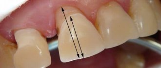
Symptoms of a cracked tooth
Symptoms of a cracked tooth: mild to very severe spontaneous pain consistent with irreversible pulpitis, pulp necrosis and apical periodontitis. If pulp necrosis occurs, an acute or chronic apical abscess may occur. In other words, once the crack has reached the pulp, significant pulpal or periapical pathology is likely to be present.
Cracks often form between the ages of 30 and 50, predominantly in women. The most commonly affected teeth are the mandibular first molars, followed by the upper premolars, upper first molars, and mandibular premolars. In restored teeth, cracks are more common in the case of non-adhesive type restorations and sharp internal corners of the cavity (amalgam and gold restorations).
It is assumed that microcracks form as a result of occlusal stress during chewing and repeated thermal expansion of the restorative material. On the other hand, the occlusal load can be distributed through the adhesive layer in adhesive-type restorations. Age is also a contributing factor to cracks. In fact, dentin's resistance to crack propagation decreases with age and dehydration.
The American Association of Endodontists, in its document "Cracking the cracked tooth code", defines 5 types of cracks, as shown in Figures 1, 2 and 3.
Fig. 1 – Crack and fracture of the tubercle
Fig. 2 – Tooth crack
In the tooth, a crack propagates from the occlusal surface of the tooth apically without separating the two fragments. The crack is predominantly located along the center of the tooth in a mesiodistal direction and may involve one or two marginal ridges.
Fig. 3 - Cracked tooth, view from the occlusal surface after amalgam removal.
Fig.4 Vertical root crack
In the event of a cracked tooth, the patient should be fully informed of the possibility of the crack spreading and the possibility of the tooth splitting. Although treatment will be successful in many cases, some cracked teeth may become fractured, requiring extraction.
Clinical case No. 1
A 47-year-old woman complained of spontaneous, continuous pain that began 1 week ago. The pain is caused by the right upper second molar and radiates to the right zygomatic bone. The patient had a history of bruxism. Visual inspection revealed a stained composite restoration and several structural cracks. The tooth is sensitive to touch and responds to cold testing. X-ray examination revealed no discernible changes in the structure of the tooth and surrounding bone.
Fig. 5 – X-ray and mesial view of the tooth
Rice. 6 – Occlusal surface, painted 10-year restoration
Rice. 7 - Probing revealed a 6mm loss of periodontal attachment at the level of the crack
Based on clinical examination, radiological assessment and medical history, a diagnosis of tooth fracture was suggested.
Rice. 8 — The old restoration is removed under anesthesia. The crack line was examined using a high-speed bur and traced until pulp involvement was detected.
Rice. 9 – The crack ends in the pulp chamber.
Rice. 10 — Treatment of the canals was carried out with the Protaper Universal system, the vestibular canals were passed to size F1, palatal canals to F2. All channels are sealed with the Thermafil system.
The prognosis of the tooth is considered relatively unfavorable. Instead of removal, the decision was made to treat the canals. In order to minimize the cost to the patient, a direct restoration with cuspal overlap was performed. After 30 days the symptoms disappeared. The patient continues to be monitored.
Rice. 11 — At a follow-up visit 5 years after endodontic treatment, the tooth is asymptomatic and functional. Unlike the first visit, the probing depth is less than 6mm.
Clinical case No. 2
A 53-year-old man presented with localized pain in the upper right second molar radiating to the right zygomatic bone. Visual inspection revealed an amalgam restoration and some structural cracks. The tooth is sensitive to touch and responds to cold testing. X-ray examination revealed no discernible changes in the structure of the tooth and surrounding bone. Based on clinical examination, radiological assessment and medical history, a diagnosis of tooth fracture was suggested. Under anesthesia, the old restoration was removed, the crack lines were opened using a high-speed bur and traced until communication with the pulp was found. The prognosis of the tooth is considered relatively unfavorable. Instead of removal, the decision was made to treat the canals.
Rice. 12 – Right: Rg after pulpotomy and Ca(OH)2 treatment lining. Left: completed endodontic treatment.
The canals were treated with the Protaper Universal system, the vestibular canals were treated to size F1, the palatal canal to size F2. All channels are sealed with the Thermafil system. A few days later, the symptoms disappeared, and the patient was called for a follow-up examination.
Rice. 13 - After 2 years of observation, the patient complained of pain, and a vertical root fracture was diagnosed. The tooth was removed and replaced with an implant.
Clinical case No. 3 The patient presented with symptoms of an abscess; pain and swelling in the area of the left lower molars. Percussion is positive for tooth 36, vitality test is negative.
Rice. 14 – Preliminary Rg image.
Endodontic access
Endodontic access was performed. During the work, a copious amount of purulent exudate was released from the tooth. The canal was treated at the same visit with irrigation with sodium hypochlorite and the EndoVac system. After 45 minutes, a paper pin inserted into the canal determined the continuation of exudation. A cotton ball is placed into the tooth cavity and a temporary filling is installed. The patient was scheduled for irrigation every 3 days for 30 days. The release of exudate from the canals did not stop. Severe inflammation developed, as in the first days of complaints. The decision was made to remove the tooth.
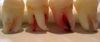
Rice. 15 – After removal, a fracture line was found on the distal root.
Rice. 16 – Close-up of the crack. The cemento-enamel junction and periodontal ligament are visualized.