Lichen planus is a chronic dermatological disease in which characteristic nodules, plaques and erosions appear on the mucous membranes. The disease can be localized on almost any part of the body, including the oral cavity. Before treating any dermatoses, differential diagnosis is necessary, since the choice of treatment strategy depends on it. General and local medications are usually used. From the article you will learn what lichen planus looks like in the oral cavity and how to treat this disease.
Determination of lichen planus on the oral mucosa (LP)
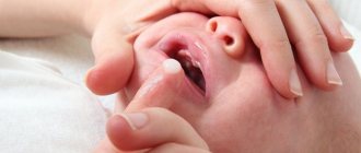
Lichen planus (LP) on the oral mucosa is a pathological condition of the mucous membranes, in which strong papules, erosions and plaques of a characteristic bright color appear. In addition, trophic processes are observed that complicate communication and food intake, and ulcers may appear on the palate. Foci of lichen can be located on the back of the cheeks, gums, tongue and mucous membrane of the palate. It is not a contagious disease; infection occurs against the background of malfunctions of the human immune system and some types of allergic stomatitis.
Young women are most susceptible to this pathology; in children and the elderly it is much less common.
This is one of the most common types of oral dermatoses. According to statistics, the disease occurs in 1-2% of all people. In 50-75% of cases it develops with the cutaneous form of lichen.
Lichen planus can manifest itself in acute or chronic form. There are often cases when the disease disappears on its own without any medical intervention.
Causes of lichen planus in the mouth
Lichen planus of the oral cavity (LP) is accompanied by inflammatory and degenerative processes affecting the mucous membrane. Granules (papules) up to 2 mm in size are formed on it, a change in color from pale pink to dark red with a purple tint, ulcers and erosion are noted.
It has not yet been established why LP, in the presence of the same provoking factors, occurs in some individuals and does not affect others. However, several main reasons for the development of the disease have been identified:
- Neurogenic. Stress, nervous exhaustion, depression and mental disorders provoke the appearance of LP.
- Toxic-allergenic. Lichen planus occurs as a result of an allergic reaction when taking certain drugs: antibiotics, beta blockers, analgesics, antihistamines, anti-inflammatory drugs and anti-malaria medications.
LLP is accompanied by inflammatory and dystrophic processes affecting the mucous membrane.
Important! A relationship has been established between the disease and patients who come into contact with the substance paraphenyldiamine at work. It is found in reagents for developing color films, hair dye, and is used to reinforce tires and plastic.
Provoking factors also include:
- Gastrointestinal diseases.
- Simultaneous occurrence of hyperemia and type 2 diabetes mellitus.
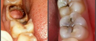
- Injuries to the oral cavity (domestic or during medical procedures), permanent damage from low-quality dentures and chipped enamel. This also includes allergic reactions to materials of fillings, crowns, preparations for sanitation and changes in the composition of saliva due to the presence of dental structures made of different metals in the mouth.
- Smoking.
- Regular consumption of large quantities of spicy, smoked, salted, pickled foods.
- Autoimmune diseases. LP is activated when the body's defense mechanisms are weakened.
Symptoms of forms: erosive-ulcerative, typical and others
Signs of lichen planus differ depending on the form of the disease. In modern medicine, there are six main types of pathology with distinct external manifestations:
- typical _ It occurs most often (in 45% of cases). During a visual examination, the patient has gray or white nodes of undefined shape and small diameter. Over time, they merge and form a characteristic pattern;
The typical form in rare cases leads to the onset of erosive processes.
- exudative-hyperemic. With this form, small gray-white papules appear on the mucous membrane, against which slight swelling and hyperemia of the oral mucosa occurs;
- hyperkeratotic . Gray plaques form, which expand and become rougher over time. You almost always feel severe dryness in the mouth, the mucous membrane of the tongue and the inner surface of the cheeks becomes rough. There is discomfort while talking and chewing food. If at the same time there is a sharp lack of B vitamins in the body, then the development of desquamative glossitis is possible;
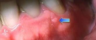
- erosive-ulcerative . The most severe form of lichen ruber is in the oral cavity. Occurs in 25% of all cases, more often it is a disease that has been neglected for years. Erosion and ulcers of various shapes form on the entire surface of the oral cavity. They are covered with a white or red coating, when removed, minor bleeding appears. This form is characterized by severe pain and burning;
- bullous _ A rare form of the disease in which dense blisters of small diameter appear on the surface of the oral mucosa. They contain pus and blood cells. Most often, they burst after 2-3 days, leaving behind erosions and ulcers covered with plaque;
- atypical _ It occurs in only 3-4% of all cases of lichen ruber. Usually localized on the inner surface of the upper lip and gums. The mouths of the salivary glands often expand. The worst to treat.
Lichen planus can transform from one form to another over time. There is a tendency towards remissions and relapses. Even after proper treatment, there is a risk of another outbreak of the disease several years later under the influence of provoking factors.
In 1% of cases, malignancy of lichen planus in the oral cavity is noted. Signs of malignant neoplasm in this disease are more often diagnosed against the background of the erosive-ulcerative type.
Symptoms
LLP is localized mainly on the inner surface of the cheeks. Less commonly, lichen appears on the tongue, lips, gums and palate.
Ringworm is located mainly on the inner surface of the cheeks.
Important! LLP is a non-viral disease. Therefore, it cannot be transmitted by contact, household or airborne droplets.
The disease is accompanied by the formation of papules. Their shape, number and size depend on the shape and degree of pathology. The following clinical signs are noted:
- Individual granules of round, oval or elongated shape are formed. Their size varies from 1 mm to 2 mm.
- The papules are initially pale pink in color, but as the disease progresses they acquire a more pronounced shade: red, blue, purple.
- The growths become keratinized. Because of this, they rise above the surface.
- Tissue degeneration is observed only at the site of papules formation. Outside of them, the mucous membrane remains intact.
- Gradually, the granules merge into conglomerates with a characteristic cobweb pattern.
- The growths become necrotic, which leads to the formation of erosions, ulcers, and blisters.
- Over time, the papules acquire a purple, then brown tint. After this they may disappear.
Causes
The etiology of the disease remains unclear to date. Researchers of this disease are divided into two large camps. Some believe that lichen planus develops against the background of pathologies of the immune system and allergies, while others believe that the disease is multifactorial in nature. At the same time, there are several factors that provoke the development of the disease. Usually this is a violation of metabolic processes in the body, which leads to an inadequate reaction from the skin.
Lichen planus can occur due to the following reasons:
- violation of daily routine and nutrition;
- regular emotional stress and stress;
- bad habits – smoking, alcohol abuse, drug use;
- injuries and damage to the oral mucosa, exacerbation of traumatic stomatitis;
- long-term use of certain medications;
- allergies and individual intolerance to certain products;
- diseases of the endocrine, vascular and excretory systems;
- heredity.
Cases of lichen planus occurring due to harmful working conditions have been described. For example, the disease can develop in people whose activities involve regular interaction with mercury derivatives or the production of toxic drugs.
Buy online
Lichen planus (LP) is a chronic inflammatory dermatosis affecting the skin, mucous membranes and skin appendages [1, 2]. This is the most common lichenoid dermatosis [3]. It occurs in people aged 30–50 years [4], and is almost twice as common among women [5, 6]. In the general structure of dermatological diseases, its frequency is 1–5% [7, 8], and the population frequency is 0.4–1.9% [9, 10].
The etiology of LP is not clear. Its connection with infectious (hepatitis C, etc.) [11], neurogenic, toxic-allergic (including β-blockers, non-steroidal anti-inflammatory, antimalarial drugs [12]), viral, immunopathological (activation of the Th1 immune response, leading to the triggering of apoptosis) is discussed. basal keratinocytes with increased levels of interferon-γ, tumor necrosis factor-α, interleukins Iα, 6, 8, AS/Apo-1 and Bcl-2, CXCLI0 [13]) factors. Hereditary predisposition is confirmed by the association of LP with the HLA-DR1 and DQwl antigens [14]. In particular, the connection between generalized LP and the HLA-DR1 antigen is observed in 80%, drug-induced LP in 56%, localized LP in 54%, and oral LP in 31% of cases [4].
According to the clinical picture, typical (classical), hypertrophic, atrophic, bullous, follicular, pigmented, ring-shaped, linear, zosteriform, erythematous, flattened forms are distinguished, as well as 6 clinical forms of LLP of the mucous membrane and red border of the lips (typical, exudative-hyperemic, erosive -ulcerative, hyperkeratotic, bullous, atrophic) [2].
Histological signs characteristic of LP are hyperkeratosis, hypergranulosis, acanthosis, vacuolar degeneration of the cells of the basal layer, diffuse strip-like infiltrate in the upper layer of the dermis [2].
The importance of the problem of LP for healthcare is due to the presence of severe forms of the disease, the possibility of malignant transformation of lesions [14], the difficulty of diagnosing atypical clinical forms, and frequent resistance to therapy [3, 15].
We analyzed the clinical features of LP in 279 patients (91 (32.4%) men, 188 (67.6%) women) aged 17-85 years (average age 49.0±3.4 years) from Moscow and the Moscow region , who have been undergoing treatment in the department of dermatovenereology and dermato-oncology of MONIKI for the last 13 years. The duration of the disease at the time of hospitalization varied from 1.5 months to 40 years and averaged 2.3±0.1 years. In 75 (27%) cases, LP had a recurrent course. Data on the clinical forms of the disease are presented in table. 1.
Typical
LLP, present in 212 (77%) patients, was represented by polygonal violet papules with a diameter of 1-5 mm with a smooth, predominantly flat surface, with a mesh pattern (Wackham mesh) and a central umbilical depression on it.
The rash affected symmetrically and primarily the skin of the flexures of the limbs, the anterior surface of the legs and feet, and the genitals (Fig. 1).
Figure 1. SPL of the shaft and glans penis.
In 4 cases, erythroderma was observed (Fig. 2).
Figure 2. CPL. Erythroderma.
The progressive stage lasted 6-10 weeks (on average 8.0±0.2 weeks) and was characterized by an isomorphic reaction (Koebner phenomenon) with the spread of the rash due to the fusion of elements into bluish-violet or brown plaques with a diameter of 2-3 cm with a rough, scaly surface.
During the regression stage, the elements flattened, acquiring a predominantly brown color with different shades.
Palms and soles were affected in 5 (1.8%) patients. It was characterized by keratinizing papules/plaques with clear boundaries, irregular shape with fine lamellar peeling, as well as rashes resembling keratoderma.
Involvement of the external genitalia in the pathological process was noted in 24 (9%) cases (with widespread LP). In men, the glans (6 cases) and shaft of the penis (6), as well as the scrotum (3), were affected; in women, the vulva (10 cases), the perianal area (2) and the vagina.
Rashes in the genital area were represented by lichenoid papules with a diameter of 1-6 mm, sometimes merging into plaques with a diameter of up to 2 cm. Itching was noted in 17 cases, itching and burning in 7 cases, and asymptomatic in 7 cases.
Damage to mucous membranes
(oral cavity and genital organs) with typical LLP was noted in 53 (23.5%), oral cavity - in 50 (23%) cases
(Table 2).
The rash on the oral mucosa was characterized by a network of randomly intersecting white stripes affecting the cheeks (Fig. 3),
Figure 3. Typical (reticular) LLP of the buccal mucosa.
gums, lateral and dorsal parts of the tongue (Fig. 4),
Figure 4. Typical (reticular) LCL of the dorsal surface of the tongue. less often - hard and soft palate, floor of the mouth, lip, retromalar areas.
The lesions were usually bilateral and, as a rule, asymptomatic (with the exception of LLP of the dorsal surface of the tongue, which is characterized by severe pain). Typical lesions of the mucous membrane were much easier to visualize than the Wackham grid on skin elements; in 4 cases they extended to the mucous membrane of the lips and in 2 cases to the red border of the lower lip.
Lesions of the mucous membrane of the genital organs
clinically did not differ from those with LP of the oral mucosa and in 14 cases were combined with them, but unlike them they were accompanied by erythema of the mucous membrane, itching or burning, in 1 case vaginal stenosis developed.
When typical LP is combined with lupus erythematosus
(overlap syndrome with lupus erythematosus) foci of lupus erythematosus of a bluish-red or purple color with hypo- and hyperpigmentation, telangiectasia and atrophy were located in the distal parts of the extremities, while in other areas (including on the oral mucosa)
(Fig. 5)
Figure 5. Overlap syndrome with systemic lupus erythematosus. Damage to the bridge of the nose, upper lip and red border of the lower lip. There were typical lesions of LP, and in 2 cases there were dystrophic changes in the nails. Histological examination revealed signs of both diseases, and with PIF - linear and granular deposits of immunoglobulins and the C3 component of complement along the basement membrane.
Atypical
47 (24%) patients had forms of LP (depending on the location and nature of the morphological elements).
Linear
LLP was observed in a 34-year-old patient and was represented by lichenoid papules merging into one straight line 5 cm long
(Fig. 6).
Figure 6. Linear form of the CPL.
Ring-shaped KPL,
present in 9 (3.2%) patients, was characterized by a ring-shaped arrangement of papules.
The lesion was formed by the reverse development of the center of large plaques with atrophy and retraction and remained along the periphery of a slightly raised ridge of unresolved infiltrate (ring-shaped atrophic variety) or by the fusion of small papules with the formation of arcs, garlands, rings, inside which normal skin was preserved, as well as by fusion papules into plaques followed by resolution of centrally located papules, leaving behind pigmentation. The size of the ring-shaped lesions varied from 0.2 to 3.0 cm, the number - from 1 to 10. More often such elements were located on the genitals (glans penis, scrotum, vulva), in large folds (Fig. 7),
Figure 7. Ring-shaped elements of the LLP in a skin fold. on the back, buttocks, lateral surfaces of the body, neck and eyelids and in 5 cases were combined with lesions of typical LLP.
Atrophic
LP was observed in 8 (2.85%) patients.
It was characterized by the formation in place of former polygonal papular elements of foci of superficial atrophy of a yellowish-brownish or brownish color (including ring-shaped ones), surrounded by typical LLP papules, including miliary ones. Rashes 2-3 cm in diameter with a rough, scaly surface affected the torso (Fig. 8),
Figure 8. Solitary atrophic element of the LLP, ring-shaped.
armpits, genitals, as well as the scalp (in 2 cases Broca's pseudopelade
).
Pigmentary
LP was observed in 4 (1.4%) patients and was characterized by dark brown foci.
In 2 cases, there were elements of typical LLP on the skin and mucous membrane of the oral cavity. This form affected the skin of large folds (Fig. 9),
Figure 9. Pigmented LLP of the inguinal fold. abdomen, buttocks, lower extremities. It should be distinguished from the regressive stage of typical LLP, when hyperpigmentation develops secondarily in the areas of former papular elements, as well as from lichenoid toxic melasma, in which the rashes are located mainly on open areas of the skin and are provoked by insolation. In pigmented LP, hyperpigmentation formed at the site of previous erythema, with small brown nodules located in the area of the mouths of the hair follicles.
Follicular
(peaked) LLP, characterized by raised pointed nodules with a horny plug, was noted in 9 (3.2%) cases, while the scalp was affected in isolation in 3 (1.1%) cases, and in combination with skin rashes - in 6 (2.1%).
When this form is localized on the scalp, the reverse development ends with atrophy and baldness ( lichen planus follicularis decalvans
)
(Fig. 10).
Figure 10. Follicular form of LP.
The combination of the latter type with non-scarring alopecia of the armpits, pubis and disseminated follicular papules in other areas of the skin is called Graham-Little-Lassauer syndrome.
The hypertrophic form of LP was noted in 20 (7.2%) patients. It was characterized by warty papules and plaques raised above the skin level, with a porous, hyperkeratotic surface and round, oval, or uneven outlines (Fig. 11).
Figure 11. LLP of the lower leg, hypertrophic form.
Erythematous
LP was present in 6 (2.2%) patients and was characterized by the sudden development of diffuse erythema with a crimson tint on a large surface of the trunk and limbs.
Typical papules were barely visible in 4 cases (Fig. 12)
Figure 12. Erythematous form of LP. and were absent in 2 cases.
Bullous
One patient had LP and was characterized by vesicobullous rashes against the background of itchy papular elements of typical LP of the lower extremities
(Fig. 13).
Figure 13. Bullous form of LP. The PIF method did not detect antibodies along the basement membrane.
Pemphigoid form
LP (
overlap syndrome with bullous pemphigoid
) was noted in 6 (2.2%) patients. With it, blisters and vesicles with transparent contents appeared in the area of the extremities (in places of skin trauma, primarily on the palms and soles), and after 2-8 weeks itchy papular rashes appeared, as in typical LLP.
Histological examination revealed signs of LP with subepidermal clefts (Fig. 14),
Figure 14. Subepidermal vesicle containing fibrin and cellular elements. Band-like lymphohistiocytic infiltrate in the dermis. Hematoxylin and eosin staining (×200). preserved basal layer and the presence of neutrophils, lymphocytes and/or eosinophils in the vesical contents, and in the PIF reaction - linear deposits of IgG and C3 complement components in the lamina lucida of the basement membrane, as well as IgM and fibrin in Siwatt's bodies scattered along the papillary layer of the dermis.
Flat
LLP (synonym -
lichen planus obtusus
) was in one patient and was characterized by the presence of isolated, flattened hemispherical hypertrophied papules with a diameter of 0.3-0.8 cm, located on the extensor surface of the legs, sharply protruding above the surface of the skin.
They had a purple color, a smooth surface, and a central depression (Fig. 15)
Figure 15. Flattened LLP: flat purple papules with a smooth surface in the lower leg area. and were accompanied by slight itching.
In atypical LP, lesions of the mucous membrane were noted in 25 (37%) cases; rashes in the mucous membrane, as in typical LP, were bilateral, but were represented not only by the white or gray mesh and linear spots characteristic of typical LP, but also plaque-like , atrophic, erosive, ulcerative and bullous elements, accompanied by burning (14 cases) and pain (15).
Data on the clinical forms and localization of the latter in the oral cavity are presented in table. 3 and 4.
Exudative-hyperemic form
characterized by whitish elements located on a swollen, hyperemic background of the mucous membrane
(Fig. 16)
Figure 16. Exudative-hyperemic form of LLP of the buccal mucosa. and in each case was accompanied by unpleasant sensations when eating spicy and hot food.
Erosive-ulcerative form
was characterized by a pronounced inflammatory reaction around erosions
(Fig. 17),
Figure 17. LLP of the buccal mucosa, erosive-ulcerative form. pain for many years and resistance to treatment. In each case it was a complication of the exudative-hyperemic form and in 3 patients it developed as a result of trauma with Grinshpan-Potekaev syndrome.
Hyperkeratotic form
was characterized by plaques with clear boundaries and pronounced keratinization
(Fig. 18, 19).
Figure 18. Hyperkeratotic gingival LP.
Figure 19. Hyperkeratotic LLP of the dorsum of the tongue.
In bullous form
along with typical rashes on the mucous membrane of the cheeks, blisters ranging in size from 2-3 mm to 1.0-1.5 cm in diameter with a dense covering appeared.
They existed for no more than 1 day, then opened with the formation of painful but rapidly epithelial erosions (Fig. 20),
Figure 20. LLP of the oral mucosa, bullous form. around which typical manifestations of LP could also be detected.
Atrophic form
was noted in the patient with vulvovaginal-gingival syndrome and was characterized by white network-like areas of atrophy against the background of erythema of the mucous membrane
(Fig. 21).
Figure 21. Atrophic form of LP of the oral mucosa in vulvovaginal-gingival syndrome: diffuse hyperemia, erosion, whitish plaques in the cheeks and red border of the lips.
In each of the 4 patients under our supervision, this syndrome was accompanied by painful rashes in the oral cavity and vaginal symptoms with dysuria. Damage to the genital organs was manifested by rashes on the inner surface of the labia minora (Fig. 22),
Figure 22. Vulvovaginal-gingival syndrome: hyperemia and erosion around the vaginal opening and the lower part of the labia minora with whitish edges.
as well as the vestibule and vaginal walls, pain, vaginal discharge, dysuria, dyspareunia, and in one case, onychorrhexis (Fig. 23).
Figure 23. Onychorrhexis.
Atypical lesions of LP on the genitals were noted in 4 cases and were ring-shaped
(on the pubis, labia majora and minora, body and glans penis), were asymptomatic even with itchy lesions on other areas of the skin.
In the other 4 cases, elements around the vaginal opening (Fig. 24)
Figure 24. Vulvovaginal-gingival syndrome: erosions in the labia minora.
on the labia majora and minora were erosive
with a whitish reticular pattern and severe soreness, which is often accompanied by dyspareunia. When the vagina was involved in the pathological process, its walls became hyperemic and bled when touched; Vaginal discharge was noted, in 2 severe cases - cicatricial changes in the vagina (in one with fusion of the walls). Erosive LP of the genital organs in each of the 4 cases was associated with lesions of the oral cavity.
Thus, when examining 279 patients with LP, its typical form was identified in 76%, atypical - in 24% of patients, the mucous membranes were affected in typical LP in 23.6%, in atypical - in 35% of cases and were always combined with skin lesions, and in 7 and 12% of cases, respectively, with damage to the mucous membrane of the genital organs. The most severe course of dermatosis is observed in its atypical forms (follicular, atrophic, hypertrophic LP of the skin, vulvovaginal-gingival syndrome, as well as in the erosive-ulcerative form of LP of the oral cavity).
Treatment
The choice of therapy is made only on the basis of the results of differential diagnosis. Many dermatological diseases have similar manifestations, but require completely different approaches to treatment. When the diagnosis of oral lichen planus is confirmed, systemic and local medications are prescribed.
Medication
The choice of medications depends on the patient’s age and characteristics of the disease. The following groups of medications are usually prescribed:
- To relieve itching and burning in the mouth, it is recommended to take antihistamines: Claritin, Suprastin, Telfast.
- Topical steroids or corticosteroids are needed to relieve inflammation. Hydrocortisone, Prednisolone, Afloderm, Fluorocort.
- To reduce the risk of developing candidiasis while taking hormonal tablets, antifungal agents are prescribed: Fluconazole, Flucostat, Nystatin.
- In the presence of severe erosion and ulcers, it is recommended to take painkillers (Anestezin, Brustan, Ibuklin) and agents that accelerate regenerative processes (Aevit, Methyluracil).
- For the treatment of chronic lichen, drugs are prescribed that enhance the oxygen metabolism of cells: Cyto-Mac, Actovegin.
- To enhance natural immune processes, immunomodulatory therapy (Likopid, Myelopid, Interferon) is recommended.
The choice of means is selected individually depending on the form of red lichen, as well as its origin. Oral medications are usually combined with topical medications to speed up recovery. In the presence of nervous disorders caused by symptoms of the disease, mild sedatives based on herbal components are selected (tincture of valerian or motherwort, Novo-Passit).
Only a doctor can prescribe the drug and select the optimal dosage. When self-medicating there is a risk of complications and the development of secondary diseases.
Local
Topical agents complement and enhance the effect of oral medications. They have a similar effect and are selected depending on the clinical picture in each individual case. These are the following groups of funds:
- hormonal ointments or creams: Akriderm, Beloderm, Afloderm, Dermovate.
- antiseptics for mouth rinsing: Chlorhexidine, Furacilin (Nitrofural), Chloramine.
- injections to numb the affected areas (Anestezin, Benzocaine or Pyromecaine).
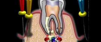
Additionally, physiotherapy is prescribed to speed up the healing and recovery process. This is usually calcium chloride electrophoresis or phototherapy. When diagnosing the malignant nature of formations in the oral cavity, surgical intervention may be prescribed. In the last few years, cryotherapy, electrocoagulation and laser procedures have been actively used to treat lichen planus in the mouth.
Electrophoresis is a traditional treatment method. In some cases, it is replaced by cryotherapy.
During remission, it is important to carry out a complete sanitation of the mouth to eliminate foci of infection, as well as get rid of all provoking factors: sharp edges of the teeth, poor-quality fillings or orthopedic structures.
Diet
Throughout the entire treatment period, it is recommended to refrain from hot, cold, spicy and salty foods. You should not drink alcoholic drinks or soft drinks. Usually a gentle diet is prescribed, which consists of soft foods to avoid damage to the mucous membranes of the oral cavity.
Normalizing the natural intestinal microflora allows you to avoid complications and subsequent relapses. To do this, probiotics and prebiotics are selected individually; it is recommended to consume fermented milk products (kefir, cottage cheese or fermented baked milk) daily.
Stress and negative emotions can worsen the course of the disease. During treatment and after full recovery, it is advisable to avoid strong psycho-emotional overloads.
Drug therapy
The main actions in the treatment of lichen planus are the elimination of the causes that caused it: allergies, systemic pathologies, replacement of low-quality dental prostheses.
LLP therapy lasts from 2 – 3 weeks to a couple of months, depending on the form of the disease and severity. The patient is prescribed:
- Hormonal drugs based on chloroquine.
- Antihistamines: Loratidine, Zyrtec, Suprastin, Cetrin.
- Sedatives to normalize the nervous system.
- Vitamins that promote epithelial healing – A and E.
- Preparations for normalizing oxygen metabolism and accelerating cell regeneration: “Etrethionate”, “Fenoro”, “Tigazon”, “Actovegin”, “Cyto-Mac”.
- External treatment products: solutions based on citric acid, menthol, Solcoseryl ointment.
- Physiotherapy: exposure of affected areas to ultraviolet irradiation.
- Cryodestruction or electrocoagulation of necrotic areas.
- Medicines to strengthen the immune system: Reaferon, Neovir, Interlock.
Treatment of lichen is always complex and includes taking several types of medications.
Possible complications
Lichen planus is chronic in nature. Therefore, after diagnosis, the patient is registered at a dermatovenerological dispensary, which makes it possible to monitor his subsequent condition. If you follow all the doctor’s prescriptions, the prognosis for recovery is positive. After treatment, it is important to follow simple rules of prevention and undergo a full medical examination annually, as well as promptly treat and prevent caries.
The ulcerative form is the most difficult to treat
Complications arise when the disease transforms into an erosive-ulcerative form. In this case, the most powerful medications are selected, as well as procedures to reduce discomfort and the risk of capillary bleeding.
If you do not contact a specialist in a timely manner, complications in the form of bacterial stomatitis may occur. Transformation into a malignant tumor occurs extremely rarely. This usually occurs against the background of provoking factors, which is why it is so important to observe prevention.
Treatment with folk remedies
To speed up recovery, you can use traditional medicine. In the treatment of lichen planus the following is used:
- Rosehip or sea buckthorn oil. A cotton swab is moistened in the product and application is made for 40 minutes. You can also drink 1 tsp. sea buckthorn oil on an empty stomach.
- Cranberry juice or apple cider vinegar. Gauze or bandage is moistened in liquid and applied to the affected area for 10 minutes. The number of procedures should not exceed 6 times a day.
- Herbal infusions. Pharmacy collections of calendula flowers (1 tbsp), burdock roots (2 tbsp) and hop cones (2 tbsp) are poured with hot water (500 ml). Boil in a steam bath, filter and add 2 tbsp. l. Vaseline. The prepared mixture is treated with the oral mucosa several times a day.
Sea buckthorn oil effectively heals affected areas of the mucous membrane.
Important! All folk remedies are used only as an additional method to drug therapy and after consultation with the attending physician.
Bottom line
Some patients turn to a specialist complaining of the appearance of nodules in the mouth and various unpleasant symptoms. Most likely it is lichen planus, which appears on the oral mucosa. The disease is quite rare and the causes of its occurrence have not been fully established, but there are many factors that provoke the appearance of papules.
As soon as you notice one of the symptoms, you should definitely consult a doctor and start treatment, also not forgetting about preventive measures. All this is important not only to get rid of discomfort as quickly as possible, but also to avoid consequences.