What is average caries
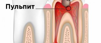
Caries manifests itself in a tooth in different ways. Here you can recall the structure of the tooth - on top there is a thin but very strong layer of enamel, under which there is dentin - its structure is softer and penetrated by dentinal tubules (through which nutrients are delivered to the enamel). Inside the tooth there is a cavity filled with nerve endings, blood vessels and connective tissue - the pulp.
When the carious process has destroyed the enamel and penetrated into the dentin, but not deeply (not coming close to the pulp) - this is medium caries. And since dentin has a large number of tubules, infection quickly occupies large volumes and gets close to the pulp.
Interesting fact! This disease is most often diagnosed in patients over 20 years of age. But it happens that the milk bite is also affected. In children, the structure of dental tissues is more fragile and porous, so the infection develops rapidly. Therefore, parents should always be vigilant and listen carefully to the child’s complaints.
Causes of the disease
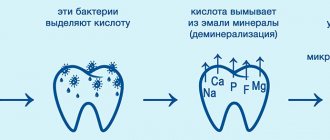
The main cause of average caries is the instability of the enamel before the attack of microbes, of which there are a lot in the oral cavity. In the normal state, bacterial acids are neutralized - washed off with saliva or removed with a toothbrush. But if the enamel is not strong enough (due to hereditary factors or lack of minerals in food), it will gradually begin to deteriorate under the influence of streptococci and staphylococci. Gradually, bacteria penetrate into the resulting cavity and infect the surrounding healthy tissue.
On a note! For such an insidious disease as caries, timely and accurate diagnosis is very important. But only high-tech equipment, for example, a dental microscope, can detect pathology in the early stages. Thanks to multiple magnification, the device not only diagnoses caries at the earliest stages, but also allows you to carefully and precisely treat it, as well as install a filling with maximum preservation of healthy tissue. After treatment of caries under a microscope, patients note an excellent aesthetic result, since artificial materials are ideally selected and fully match the color of natural enamel.
Poor nutrition also provokes caries. If a child or adult consumes a large amount of sweets, baked goods (in general, any products with sugar), then a favorable environment for the growth of microbes is created in the mouth. Accordingly, they will produce more toxins.
The reasons include malocclusion - when the teeth grow crowded, a large amount of plaque and food debris accumulates between them, where bacteria develop.
Diagnosis and Treatment of Dentin Caries
According to international statistics, one of the most common diseases among the population is caries. Caries affects the teeth of about 95% of the population. Caries, according to the international classification, depending on its location/limitation, can be within the enamel, within the dentin, or within the cement. This article will focus specifically on dentine caries, since patients often complain about existing dentine caries (the previous stages do not cause much pain, there may be discomfort and aesthetic dissatisfaction), as well as on the diagnosis and treatment of dentine caries.
First, let's get acquainted with the position of dentin caries in generally accepted classifications.
Main symptoms and complaints
With average caries, characteristic symptoms and complaints arise, knowing which you should contact your dentist as soon as possible:
- the pain is aching and does not last long,
- the patient can clearly point to the diseased tooth,
- pain occurs from chemical irritants: sour and sweet foods and drinks. Even if the toothpaste contains sweeteners, a painful reaction will appear,
- pain from mechanical irritation: pressure on the tooth from closing the jaws while eating or talking, chewing food, pressing with a brush or tongue,
Good to know! The pain goes away very quickly after removing the irritating factor. For example, if you rinse your mouth with plain water after eating a sweet meal or stop chewing on the sore side. If the pain does not go away for a long time, then there are complications - pulpitis or periodontitis.
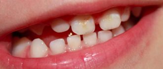
- darkening or hole in the tooth: may be clearly visible. But there are often cases when there is a small gray spot on top of the enamel (where the “entrance” to the decaying cavity is located), and under the enamel there is a large amount of darkened, softened dentin. Such a defect can only be identified at an appointment with a dentist,
- Bad breath: caused by a large accumulation of bacteria and necrotic dental tissue in the carious cavity.
Average caries can have an asymptomatic course - when nothing bothers the patient. In this case, the disease can only be detected during a routine dental examination.
Diagnosis and Treatment of Dentin Caries
According to international statistics, one of the most common diseases among the population is caries. Caries affects the teeth of about 95% of the population. Caries, according to the international classification, depending on its location/limitation, can be within the enamel, within the dentin, or within the cement. This article will focus specifically on dentine caries, since patients often complain about existing dentine caries (the previous stages do not cause much pain, there may be discomfort and aesthetic dissatisfaction), as well as on the diagnosis and treatment of dentine caries.
First, let's get acquainted with the position of dentin caries in generally accepted classifications.
Classification of the pathological process
In modern dentistry, there are about 20 forms of caries. To systematize them, several classification methods were created. Let's look further at the most common types.
By location:
- cervical: pathological foci are located directly near the gums,
- approximal (or interdental): at the points of contact of two adjacent teeth,
- fissure: in the dental grooves (fissures or depressions) on premolars and molars.
By process stage:
- superficial: white lesions are found on the enamel - destruction of hard dental tissues occurs under the influence of bacteria. The stage is asymptomatic and a person often does not attach importance to “chalky spots” on the enamel,
- initial: the enamel is destroyed almost throughout its entire depth, but has not reached the border with dentin,
- medium degree: the “hero” of today’s article,
- deep: the process has approached the border with the pulp. This form quickly develops into pulpitis.
By current speed:
- acute: the process proceeds rapidly. From the superficial stage to the deep stage it can take less than 6 months. Often several teeth are affected at once,
- chronic: delayed version. Dental tissue actively resists destruction, so the pathological process continues for years.
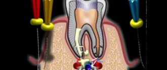
Treatment of deep caries in two visits
On the first visit, pain relief (local anesthesia) is performed. Under anesthesia, the carious cavity is opened and all affected tissue is removed until the cavity has hard, dense walls and a bottom. Then - the application of a therapeutic pad and temporary filling.
Next - observation from several days to several weeks. If there are no complaints, on the second visit the temporary filling is replaced with a permanent one. Appear 6 months after applying the filling to ensure the effectiveness of the measures: no complaints, living pulp, healthy bone near the apex of the tooth root.
Diagnosis and differentiation from similar pathologies
Diagnosing average caries is not difficult for a professional dentist. To begin with, the doctor will listen to the patient’s medical history in order to have an idea of the nature of the complaints and the duration of pain symptoms. Then an instrumental examination is carried out. Probing reveals a cavity with a hard bottom filled with softened dentin. Probing may be painful.
For differential diagnosis, X-rays are used (to exclude pulpitis or periodontitis). Electrodiagnostics EDI is also connected - average caries gives readings from 2 to 6 mA. Elevated values indicate complications.
If there is a suspicion of non-carious lesions (fluorosis, wedge-shaped defect, tetracycline teeth), then special markers based on methylene blue are used. They stain only carious defects. After a complete diagnosis, the dentist will already know which treatment method to use.
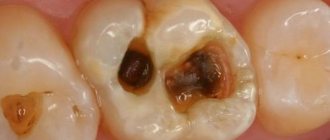
Clinical picture of moderate carisogenic lesion
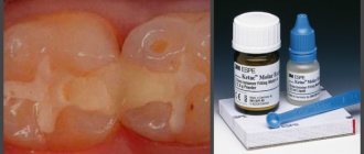
Medium caries on molars in the fissure area
This form is characterized by the formation of a cavity with the destruction of all layers of enamel and surface dentin. Its layer remains large enough, so patients show a minimum of complaints. In order for the differential diagnosis of moderate caries to be successful, it is necessary to clearly know how it manifests itself.
The main symptoms of the middle stage are:
- virtually no complaints;
- quickly passing unpleasant sensations appear rarely and only from strong food irritants;
- the carious cavity is not deep;
- probing is painful along the enamel-dentin border;
- the bottom and walls of the defect are dense, the entrance hole is usually wide;
- minimum food residues and their decay in the cavity;
- the edges of the lesion are fragile and sharp.
Dentists often use the electroodontometry method during research. With average caries, the pulp response to electric current is normal (2-6 µA). By watching the video in this article, you can get acquainted with modern methods of identifying and treating secondary caries.
Table. Differential diagnosis of diseases of hard dental tissues (moderate caries and non-carious lesions):
| Disease | When appears | Main causes | Features of the flow | Objective data |
| More often after teething. | Poor oral care, low resistance of hard dental tissues. | Constant, progressive. | Does not have a clear shape, presence of pigmentation. |
| Before teething. | Systemic diseases of the mother and child. | Stable. | Grooves, depressions, walls without color change, the bottom is hard, yellowish, shiny. |
| Before teething. | Excessive intake of fluoride compounds into the body from drinking water and food. | Stable. | The hard tissues of the tooth are chalky in color, there are extensive defects and pigmentation. |
| After teething. | Like an occupational hazard. | Developing quickly. | The exposed dentin has a dirty gray color, the enamel is almost completely absent, and the crown takes on a conical shape. |
| After teething. | Not completely clear | Progresses slowly. | A wedge-shaped defect, yellow in color. |
Differential diagnosis with fluorosis
It is usually carried out with the erosive form of fluorosis. At the same time, all the enamel has a chalky tint. In addition, individual areas of brown or dark pigment spots are observed on its surface.
The erosions themselves look like wide and deep defects resembling a carious cavity. However, this manifestation is observed against the background of greatly altered hard tissues of the teeth.
With moderate caries, the enamel loses color only in the affected area. Fluorosis is characterized by the involvement of large areas of the dental arch, with a predominant localization on the vestibular surface.
Medium caries and wedge-shaped defect
Here again the peculiarity of the localization of the pathology and its form come to the fore. The defect is most often found on the vestibular surface and in the neck of the crown. Usually canines and premolars are affected; other teeth are less often involved in the process.
The specific shape of the lesion is formed by the convergence of two planes of the cavity at an angle. The affected area has signs of sclerosis. During probing, the walls and bottom of the cavity are smooth, without signs of demineralization.
Pathological abrasion and caries
Abrasion of the frontal area of the upper jaw due to improper prosthetics
This phenomenon is usually observed due to external influence on the hard tissues of the tooth with their significant weakening.
The main signs of enamel wear are:
- unusual form of the defect (it corresponds to the structure of the traumatic factor - a pencil, a mouthpiece, an antagonist artificial tooth);
- the affected area is smooth and hard;
- pigmentation is almost always absent;
- pain rarely occurs (but increased sensitivity may be present especially when exposed to an irritant);
- usually the defect is reflected on the antagonist tooth;
- The convex parts of the crown are primarily affected.
Deep stage of cariogenic damage
Unlike average caries, at this stage there are quite serious pain sensations when food irritants enter. But after they are eliminated, the unpleasant sensations gradually subside.
The carious cavity with deep caries is usually highly pigmented, filled with necrotic decay and food debris. In permanent teeth, especially in premolars and molars on the chewing surface, the acute course of the pathology has a specific form.
The entrance to the carious cavity is narrow, and the bottom is very wide. Treatment of such a pathology should be immediate.
The price of carelessness is the development of complications leading to tooth loss. In primary occlusion, damage occurs differently. The crown is severely destroyed, but such teeth are not affected by pulpitis for a long time.
Disease prevention
Preventive measures are based on the “three pillars” - hygiene, regular dental examinations and maintaining the mineral composition of the enamel. If you want to keep your teeth strong and healthy for longer, then do not forget to brush them regularly and correctly, visit your doctor at least 2 times a year, and also watch your diet. Minimize unhealthy and sweet foods, and eat foods with calcium and phosphorus daily - cheeses, dairy products, brown bread, cereals, legumes, orange and green vegetables.