Parietal hyperplastic sinusitis is a complication of inflammation of the maxillary sinus, which is accompanied by increased growth of mucosal epithelial cells. Develops against the background of advanced sinusitis, rhinitis or improper treatment. This form of maxillitis is characterized by deterioration in breathing, constant congestion and headache. As a result, the physiology and structure of the epithelium is disrupted, a chronic form of the disease is formed, and a persistent increase in the volume of the cavity is formed. The pathology is difficult to treat in the later stages, but is diagnosed using radiography. A complex of conservative and surgical methods is used for therapy.
Features of a chronic disease, code according to ICD-10
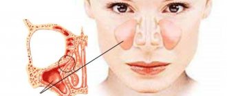
The maxillary sinus, or cavity, is the largest adnexal region, which occupies a large space of the upper jaw. Inside, it is lined with a mucous membrane of ciliated epithelium and filled with air.
Location of the maxillary sinus in the maxillary bone.
Due to the fact that their cells do not contain nerves and blood vessels, sinusitis is often asymptomatic. Against the background of prolonged inflammation and infectious processes, tissues begin to increase in volume and then grow chaotically. It is this mechanism that underlies the disease.
Parietal sinusitis is a complication of advanced or chronic sinusitis. In the ICD-10 disease classifier, it has code J32.0.
Causes of development of hyperplastic sinusitis
There are several reasons that provoke the development of parietal sinusitis with pronounced tissue proliferation.
The main ones:
- chronic sinusitis or rhinitis;
- allergic reaction to irritating substances;
- bacterial, fungal and viral infections;
- acquired and congenital pathologies of anatomical development;
- multiple polyps in the nasal mucosa;
- neurovegetative pathologies.
Abnormal growth of the epithelium sometimes occurs during the eruption of baby teeth, with long-term use of hormonal drugs and antibiotics. Strong provoking factors are carious formations in the nasal cavity and vitamin deficiency .
You can find all the information about viral sinusitis here.
What are the sinuses and the ethmoidal labyrinth?
The sinuses are fairly large cavities located in the upper jaw area that lighten the weight of the bone. The area of these sinuses is located directly under the eyes. When examining x-rays, you can see a darkening in this area, indicating the absence of any abnormalities.
Experts today have not been able to establish the significance of the paranasal sinuses in human evolution, but they suggest that the cavities are necessary to increase the volume of the maxillary bone and reduce the overall mass of the skull bones. Air enters the paranasal sinuses through the ethmoid labyrinth, which has the appearance of a bone with many small round holes.
The ethmoid labyrinth is located on the bone of the same name, which is localized in the area of the bridge of the nose and stands slightly higher than the maxillary.
It is worth noting that the bone really has the appearance of a labyrinth with several curls, and there are holes on them.
Both the maxillary sinuses and the ethmoidal labyrinth are paired bones that play a critical role in the breathing cycle, as well as in the formation of the timbre of the human voice.
What is “parietal thickening of the maxillary sinus mucosa” - symptoms
Against the background of prolonged sinusitis, swelling or thickening of the mucous membrane of the maxillary sinus appears. The epithelium layer increases, the natural relief is smoothed. The mucous membrane thickens and becomes covered with thick sputum. There is an active accumulation of serous exudate, which leads to blockage of the pores. Symptoms in this form of sinusitis are less pronounced than in the acute phase.
The following signs are noted:
- nasal congestion on one or both sides;
- constant and weak rhinorrhea with purulent contents;
- aching pain in the upper jaw;
- swelling of the nose, cheeks and eyelids;
- dry cough, worse in the morning and evening.
The pain is often transmitted to the frontal part of the head and temples. In addition, the risk of developing otitis increases, congestion in the ear canals and hearing loss appear. Conjunctivitis, keratitis and other eye diseases are rarely diagnosed.
The disease often develops in the autumn-winter period, when the body is weakened. Against the background of reduced immunity, sinusitis can be complicated by acute respiratory viral infections and acute respiratory infections. This disease needs to be treated comprehensively - antiviral, symptomatic drugs.
Symptoms of tissue thickening
Signs of changes in the condition of the maxillary sinus mucosa are not specific only to this case.
Important: the constant inflammatory process changes the mucous membrane, cracks form in it, and polyps grow.
The symptoms are similar to most symptoms of respiratory diseases.
The chronic condition is characterized by the following complaints from patients:
- Breathing is difficult both when inhaling and exhaling.
- Constant nasal discharge, which has a different appearance and consistency.
- Often the discharge smells like pus. It can be felt even when the patient breathes.
- It is difficult for the patient to blow his nose; there is a feeling of mucus, but it is difficult to remove it from the nasal cavity.
- This condition is characterized by a deterioration in the sense of smell, up to its complete loss.
- The patient's voice becomes nasal, the person speaks “into his nose.”
- There may be complaints of congestion, pain and tinnitus.
- If an increase in mucous occurs due to the development of sinusitis, then in the initial stages the mucus will be liquid. At this stage, sinusitis is catarrhal. As the disease progresses, it will become thicker and thicker. The development of a purulent stage of the disease is possible.
- Sometimes, when the mucous membrane of the maxillary sinuses thickens, a person’s eyelids swell and their eyes water. Their soreness may mistakenly suggest conjunctivitis.
- Headaches come in varying intensity, nature and location.
- Allergic reactions are accompanied by a liquid state of mucus discharged from the nose.
- In the cheek area, on the sides of the nose, pain appears, increasing with pressure.
- During an exacerbation, the body temperature rises and the condition worsens sharply.
When the acute stage passes, even if the condition has already become chronic, the patient begins to feel better. The satisfactory condition lasts until the next onset of the acute stage.
The patient gets used to his condition and the alternation of these periods; he does not consult a doctor.
Mechanisms of changes in the structure of the mucosa
Thickening of the internal tissues of the maxillary sinuses occurs according to the following principle:
- When the disease occurs, swelling and thickening occur. The ducts that remove mucus from the sinuses are located on the surface of the mucous membranes, which means that when there is swelling, they are blocked. The mucous membrane performs its function of producing mucus. In case of illness, it is produced more than in a normal situation.
- Excess mucus remains in the sinus, causing pressure. The walls of the sinus begin to stretch.
- With increased pressure, cysts grow. These formations are subject to uncontrolled growth in size.
- If pathogenic organisms penetrate the sinus cavity, the cyst tissue becomes inflamed.
- The cells begin to produce pus, which has no way out of the cavity.
Diagnostics: darkening, impaired pneumatization
Sinusitis with epithelial proliferation can only be determined through comprehensive diagnostics. It includes medical history, laboratory tests and the use of instrumental methods. What to pay attention to:
- Wall darkening in the picture . It can be seen on an x-ray. To confirm the diagnosis, photographs are taken in at least two projections. The overgrown epithelium resembles dark asymmetrical spots. In addition, CT and MRI are used.
- Headache . A subjective symptom that is difficult to evaluate during diagnosis. Its location and nature can only be determined during anamnesis collection. To study the intensity of pain, special sensory scales are used.
- Violation of pneumatization of the maxillary sinuses , i.e. natural air exchange in the nasopharynx.
An important role is given to laboratory diagnostic methods - clinical analysis of blood, urine and nasal mucus.
How is sphenoiditis treated?
There are two treatment options for sphenoiditis:
- Medication.
- Surgical.
Drug treatment
Painkillers such as paracetamol or ibuprofen are suitable for treating acute pain.
Decongestants such as xylometazoline, pseudoephedrine, or phenylephrine in the form of nasal drops or nasal sprays help drain secretions from the paranasal and sphenoid sinuses.
Corticosteroids (cortisone), such as mometasone or fluticasone, may be used as a nasal spray.
If symptoms do not go away for more than 10 days, and they intensify, or a high temperature is observed, this indicates a bacterial infection. Then the doctor prescribes antibiotic therapy.
Complications: hyperplasia, bilateral form
Parietal sinusitis is not only an advanced form of sinusitis, but can also cause serious complications. Due to the active proliferation of tissues, they lead to disruption of the physiology of breathing, problems with sleep, the appearance of a profuse runny nose, and snoring. Common problems caused by the disease:
- Deformation of the sinus mucosa (hyperplasia). Leads to a change in the volume and anatomical integrity of the maxillary sinus. The tissues gradually flatten, congestion and breathing problems develop. Dangerous consequences are chronic hypoxia and regular respiratory diseases.
- Cystic proliferation (cystic sinusitis) . Single or multiple benign neoplasms form inside the maxillary sinus. They reduce the volume of this area, leading to the accumulation of mucus and difficulty breathing. In advanced cases, surgical intervention is required.
- Transition to a bilateral form , when the swelling spreads to both sinuses. This article will tell you more about bilateral sinusitis.
Against the background of hyperplastic sinusitis, nasal polyps often appear. This is a type of tumor that tends to grow. Changes the structure and volume of the ciliated epithelium.
Due to the close proximity of the brain and ENT organs, sinusitis can cause complications in the form of otitis media, chronic rhinitis and bronchitis. When an infection occurs, there is a risk of meningitis.
Treatment
Consolidation of the sinus mucosa is, in fact, one of the morphological manifestations of the pathological process. And its cause should be treated. For sinusitis, medications come first:
- Vasoconstrictors (Otrivin, Nazivin, Noxprey).
- Antiseptics (Chlorophyllipt, Dimexide, furacillin).
- Antibiotics (Augmentin, Xefocam, Sumamed).
The drugs are prescribed in forms for local (drops and sprays, rinsing solutions) and systemic use (tablets, injections). But any medications should be taken only on the recommendation of a doctor to avoid negative consequences, including drug-induced rhinitis. For atopic rhinitis, antihistamines (Allergodil, Tavegil) and topical corticosteroids (Flixonase, Nasonex) are required. In this same situation, it is extremely important to completely eliminate contact with the causative allergens.
Inflammation in the maxillary sinuses can be treated by washing with a YAMIK catheter and physiotherapy (UHF, EF, laser therapy). If the effect of conservative measures is not enough, then a puncture is performed for sinusitis, evacuating pus from the affected sinuses and washing them. And the hypertrophic process requires surgical manipulation - endoscopic or radical sanitation (according to Caldwell-Luc).
When the results of the initial examination reveal that the mucous membrane of the sinuses is thickened, it is necessary to first understand the cause of the changes. This process can be observed in inflammatory and hypertrophic disorders, allergic and neurovegetative disorders. And only after the patient undergoes additional diagnostics will it become clear what he is dealing with and how to cure the disease.
Treatment in adults
To combat parietal sinusitis, complex therapy is used. It includes the following methods:
- drug elimination of inflammation;
- rinsing to remove exudate and clear mucus;
- treatment of sinuses with antiseptic drugs;
- radical removal of overgrown tissue.
Additional methods include physiotherapy, as well as adherence to daily routine and nutrition. In advanced cases, surgery is resorted to.
How to treat with antibacterial therapy
Prescribed when a bacterial infection is detected. The course of treatment takes from 5 to 12 days, after which it is necessary to replace the drug or discontinue therapy. Products in the form of a spray or nasal drops are especially effective - they penetrate directly into the sinuses. Antibiotics for sinusitis:
- Augmentin . It is prescribed in tablet form. The active substance is amoxicillin. Effective against most pathogenic microbes. The recommended dosage is 1 t 3 times a day. For children, Augmentin is used in the form of a suspension, taken based on body weight.
- Isofra. Antibiotic in the form of a spray, the active substance is framycetin. Effective against gram-positive and gram-negative bacteria. Used as part of complex therapy, 1 injection up to 4 times a day. The duration should not exceed 7 days.
- Polydexa . Nasal spray with an antibiotic and a vasoconstrictor. Recommended for severe swelling and inflammation accompanied by congestion. Apply one spray into each nostril 3-4 times a day. The course of treatment is 5-10 days.
Price – from 315 rub.
Find out which is better Isofra or Polydex here.
Antibiotics are prescribed to children and during pregnancy with caution. They can lead to side effects, such as allergic reactions and hives. The most effective drug can be selected only after analyzing the mucus for bacterial culture.
Washing at home
An effective procedure for cleansing the sinus mucosa, removing mucus and pathogens.
Pharmaceutical preparations are often used for rinsing:
- Furacilin;
- saline solution (sodium chloride);
- products based on sea water (Dolphin for nasal rinsing, Aqua Maris, Aqualor, Marimer).
Price – from 253 rub.
During recovery and to prevent relapse in the autumn-winter period, you can also use home methods. For rinsing, a saline or soda solution (1 tsp per 200 ml of water), diluted aloe juice, Kalanchoe or distilled water is suitable. Regularity – 3-4 times a day before meals.
What to do if your ear hurts after rinsing your nose is described in this material.
Rinsing at home can be done using a teapot with a long spout, a syringe or a large syringe. For children, the procedure is carried out only under the supervision of an adult.
Puncture and YAMIK procedure
Both methods are based on the same mechanism - cleansing the sinuses mechanically. For this purpose, use a syringe with a long needle, a sinus catheter . The procedures are performed under local anesthesia to relieve the patient of pain. Puncture allows you to avoid surgical intervention in advanced cases. It is prescribed in the presence of a large volume of pus, but in the absence of neoplasms in the maxillary sinus.
Surgery
There are a number of surgical methods to get rid of advanced parietal sinusitis. They are used if conservative therapy does not bring the desired result, and relapses of the disease are observed. The following methods are used:
- Endoscopic micromaxillary sinusotomy . Access to the sinus is through a small puncture under the upper lip. General anesthesia and a 24-hour hospital stay are required.
- Endoscopic endonasal microsinusrotomy. No puncture is required; access to the sinus is possible through natural openings in the nasal cavity.
- Classic maxillary sinusotomy . A radical procedure in which an incision is made and a hole is made in the bone tissue. After excision, a small suture is applied, there is a risk of relapse.
The main goal of all methods is to deeply cleanse the sinuses of mucus, cleanse its walls and remove tumors, if any. Depending on the method, the operation lasts from 10 to 60 minutes. The choice of a specific method is determined by the qualifications of the doctor and the equipment of the clinic.
Read about the possible occurrence of allergic sinusitis at this link.
Types of thickening of the mucous membrane of the maxillary sinuses, cells of the ethmoidal labyrinth of the nose
Thickening of the mucous membrane of the maxillary sinuses and cells of the ethmoidal labyrinth is divided depending on the changes in the tissues, as well as taking into account the course of the pathological process.
Depending on the course, acute, subacute and chronic thickening is distinguished. In the first case, the symptoms are pronounced and appear immediately after exposure to the predisposing factor.
In addition, no more than 3 weeks pass from the onset of the disease to its transition to an advanced stage. In the subacute course, the signs are less pronounced, and the symptoms increase gradually. After approximately 4 weeks, progression of the pathology is observed.
The chronic course of the disease is accompanied by mild symptoms that can persist for 6 weeks or more. In this case, the patient may not suspect the presence of the disease and considers the manifestations to be a signal of a developing cold.
Taking into account changes in tissues, several types of disease can be distinguished:
| Type of pathology | Peculiarities |
| Catarrhal type | This type of hypertrophy is considered the most common and manifests itself in the form of severe tissue swelling and filling with serous exudate. In this case, nasal congestion and other symptoms are observed, which patients confuse with simple rhinitis of colds and allergic origin. |
| Purulent | With purulent inflammation, swelling is less pronounced, but a large amount of purulent masses filling the nasal passages and sinuses leads to nasal congestion. In addition, microorganisms multiplying in purulent masses lead to damage to the mucous membranes. |
| Polyposis | With polypous hypertrophy, compactions form on the mucous membranes, which after some time turn into polyps. These formations are quite dense and disrupt the passage of air masses through the lattice labyrinth. A feature of this type of pathology is the impossibility of treatment without surgery. |
| Purulent-polyposis | The most severe form of the disease, which is accompanied by the appearance of polyps and purulent masses on the mucous membrane of the nasal sinuses and ethmoid labyrinth. In this case, the patient’s condition worsens, since the polyps prevent the flow of air, and the purulent masses provoke a deterioration in the condition of the mucous membrane. |
Additionally, experts distinguish between closed and open forms of pathology. In the first case, hypertrophy is accompanied by inflammation, but the microorganisms do not spread beyond the sinuses and ethmoid labyrinth. In the second case, inflammation and hypertrophy are the result of microorganisms entering from another cavity, for example, the oral cavity.
Conclusion
- Parietal sinusitis is a complicated form of sinusitis, which is characterized by the proliferation of the mucous membrane in the sinus.
- It develops against the background of infection, advanced maxillitis, anatomical defects and allergies.
- The main symptoms are nasal congestion, excessive secretion of thick mucus, swelling of the cheek and headache.
- Radiography is an informative diagnostic method. Additionally, tests, medical history, and pain assessment are used.
- Treatment is a complex of conservative methods (taking medications, possibly rinsing the nose with Miramistin and the YAMIK procedure). In difficult cases, surgery is prescribed.
The mechanism of changes in the structure of the mucosa
Thickening of the mucous membrane develops gradually, especially in patients whose immunity is not weakened. When the maxillary sinuses are affected, the damage begins with the development of swelling of the mucous membrane, as well as blocking of the ducts through which mucus is excreted.
Thickening of the mucous membrane of the maxillary sinuses
These secretions cannot be removed through the ducts, so the swelling intensifies, which worsens the patient’s condition. Pressure increases in the sinuses, which leads to the formation of cysts and polyps. If, before the development of edema, pathogenic microorganisms enter the sinus cavity, pus is formed when the ducts are blocked and the mucous membranes swell.
This development mechanism is observed in most cases. When the pathological process is mild, there are no microorganisms in the cavity, which reduces the likelihood of complications.