Description
This is a form of acute inflammation of the lymph nodes of nonspecific or specific etiology, accompanied by the formation of purulent exudate. It is characterized by enlargement, thickening, soreness of the affected area, redness of the skin, the appearance of symptoms of fluctuations, fever and other signs of intoxication of the body. Diagnosis is carried out using a clinical examination, laboratory and instrumental techniques (ultrasound, CT of lymph nodes, puncture). Complex treatment - surgical opening and debridement of the lesion, drug therapy, physiotherapy.
Forecast
The likelihood of complications increases significantly in the absence of timely assistance.
If you seek medical help in a timely manner, the prognosis is favorable.
The pathology can be successfully treated surgically, and with the help of auxiliary drug therapy, the symptoms are eliminated and the general condition of the patient is normalized.
The likelihood of complications increases significantly in the absence of timely assistance.
In such situations, phlegmon can cause conditions that threaten the patient's life.
Possible complications
Possible complications of phlegmon in the maxillofacial area include:
- Blood poisoning and septic shock;
- Impaired brain activity due to intoxication;
- Asphyxia;
- Impaired cerebral circulation due to compression of blood vessels;
- Thrombosis of the neck veins;
- Development of a brain abscess.
Important to remember! In the absence of timely treatment, phlegmon can cause a skin defect that will persist even if treatment is successful.
Additional facts
Among soft tissue pathologies, acute lymphadenitis occupies one of the first places. The appearance of purulent forms accounts for 20–35% of the total number of inflammatory diseases in the maxillofacial area. 46.5% of children undergo hospital treatment with a complicated course of lymphadenitis, which is associated with the structural and functional immaturity of the lymphatic system and diagnostic errors. The nonspecific process is characterized by autumn-winter and spring seasonality. The distribution of specific lymphadenitis (late rot, tularemia, tick-borne encephalitis) has a clear geography (Far East, Central Asia, China, Africa).
Purulent lymphadenitis
Types and classification of inflammation
An abscess occurs due to decay in the lower teeth
Inflammatory processes are divided into maxillary and mandibular (including submandibular), since their sources of origin are located in the same area.
- Maxillary abscess mainly develops due to the spread of infections from wisdom teeth. Characterized by difficulty swallowing and opening the mouth.
- An abscess of the lower jaw originates from the molars and premolars - the lower molars, causing pain when chewing and swallowing food.
- A submandibular abscess is characterized by severe swelling in the submandibular triangle, which leads to a noticeable distortion of the oval of the face.
Based on education, dental surgeons classify abscesses as follows:
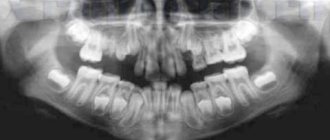
- odontogenic, occurring as a result of the spread of pathogenic flora from the root of a diseased tooth (wisdom, molars, premolars);
- intraosseous are a consequence of ENT diseases (otitis, sinusitis), suppuration of cystic formations, osteomyelitis, inflammation of the periosteum;
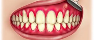
- gingival, originating in the inflamed tissues of the gums and periodontal structures;
- salivatory, characterized by a purulent-inflammatory process in the salivary glands.
Severity:
- mild – the abscess is located in one zone;
- medium - the abscess affects several parts of the face;
- severe – the soft tissues of the neck and face, and the floor of the mouth are affected.
Most purulent inflammations affecting the maxillofacial area are odontogenic in nature, forming due to the spread of infection from diseased teeth.
Causes
The most significant pathogen (in 94% of cases) is staphylococcus aureus, epidermal - in the form of a monoculture or in combination with streptococci, Escherichia coli, and anaerobes. The etiological characteristics of the disease are constantly changing, which is associated with the emergence of new strains and increasing resistance to antibiotics. A more detailed study of the contents of the lymph nodes allows us to identify viruses (Epstein-Barr, cytomegalovirus), chlamydia. Specific variants are found with the participation of mycobacteria, Treponema pallidum, Toxoplasma, and fungal flora. Purulent lymphadenitis is almost always secondary in nature, caused by the spread of infectious agents from the main focus through the lymphatic or blood vessels. The source of the inflammatory response is pathological conditions from several places: • Dental infections.
Odontogenic disorders, as the most common etiological factor, account for 47% of nonspecific forms of pathology.
In the dental office, damage to the submandibular region and chin is the result of alveolitis, periodontitis, periostitis and osteomyelitis of the jaw. • Diseases of the ENT organs.
They are the second most common cause: tonsillogenic and otogenic processes account for a quarter of cases.
Cervical and submandibular lymph nodes become inflamed in patients with angina (lacunar, complicated by paratonsillitis, periglottal abscess), pharyngitis, adenoiditis. Abscesses are also accompanied by otitis media, mastoiditis, and acute sinusitis. • Pathology of the skin and soft tissues.
In dermatology, lymphadenitis occurs as a result of microbial eczema, pyoderma (boiling, carbuncle, ecthyma), trichophytosis of infiltrative suppuration, erysipelas.
Surgeons are faced with purulent lesions of the lymph nodes in infected wounds, abscesses and phlegmon, felon. This reaction is typical for thrombophlebitis, osteomyelitis (if the pus ruptures with the formation of fistulas). • Diseases of the genitourinary system.
Purulent inguinal lymphadenitis most often indicates sexually transmitted diseases. It is part of the symptoms of inflammatory pathology of the pelvic organs (chlamydia, ureaplasmosis, gonorrhea), genital organs and perineal skin (vulvitis, balanitis, herpes). In children, there is a connection between purulent inflammation and acute respiratory viral infections, scarlet fever and mononucleosis. In a certain number of patients, the cause is a specific infection: tuberculosis, syphilis, toxoplasmosis. Lymph nodes are affected by tularemia, plague, actinomycosis and a number of other diseases. Compared to infectious factors, the role of traumatic factors is insignificant - if microbes enter directly into the node through an open wound, then primary lymphadenitis is checked. The risk of abscess formation increases in individuals with weakened general reactivity of the body (hypothermia, frequent and prolonged stress, immunodeficiency, chronic diseases).
Description and code according to ICD 10
The boil begins to abscess (when a secondary infection joins the inflammatory process) if there is no bubble on its surface necessary for the evacuation of necrotic elements. The pus begins to soften the tissue structures, melting them and provoking extensive inflammation of the deep layers of the skin. An abscessed boil can lead to infection with purulent masses of muscles, fatty structures and blood vessels.
Most often, such ulcers form on skin areas that are subject to increased contamination and friction: thighs, nose, armpits, forearms, chest, right or left lower leg, neck, lower back. The most unpleasant place for an abscess to be localized is the perianal area (the area of the anus). Such formations are accompanied by particularly unpleasant symptoms.
Have you ever had an abscess boil:
Yes
71.43%
No
28.57%
Voted: 7
In the international classification of diseases, the boil is assigned the code L02. But since ulcers can appear on various parts of the body, ICD-10 codes are determined based on the location of the formation:
- on the face – L02.0;
- on legs and arms – L02.4;
- on the neck – L02.1;
- on the buttocks – L02.3;
- on the body – L02.2;
- on other parts of the body – L02.8;
- with unspecified location – L02.9.
It should be understood that a boil and an abscess are different phenomena. The difference between a boil and an abscess is that the latter is an accumulation of pus caused by a focal infection that destroys the affected tissue.
Abscesses form not only in the subcutaneous tissue, but also in bone and muscle structures, and internal organs. A boil appears on a hair follicle, and there is a high probability that it will “ripen” and go away on its own (which cannot be said about an abscess).
Pathogenesis
Inflammation is evidence of the barrier (protective) function of the nodes. At first, the process is reactive or serous in nature, accompanied by edema, vascular congestion and lymph retention. Further development of the infection leads to penetration of the pathogen into the lymphoid structures. Proliferation of cellular elements is observed: the number of lymphocytes (mainly immature), neutrophils and macrophages increases. Microbes and exotoxins stimulate the chemotaxis of leukocytes and their death (including during phagocytosis), which is accompanied by the formation of pus. As a rule, morphological changes are limited to the capsule, but there is a risk of destructive forms involving surrounding areas under the influence of proteolytic enzymes.
Classification
The classification of acute purulent lymphadenitis used in practical surgery should reflect the nature of the pathological condition in clinical diagnosis. To obtain comprehensive information about the changes taking place, the following criteria are taken into account: Due to their origin, a secondary (infectious) and primary (traumatic) process is distinguished. In turn, microbiological forms are nonspecific and specific. The latter are represented by tuberculous, syphilitic, and fungal varieties. • Path of entry.
Depending on the location of the source of the infectious disease, lymphadenitis is divided into odontogenic (as a result of damage to the teeth) and non-odontogenic.
The latter include stomatogenic, otogenic, tonsils, rhinoceroses, dermatogenic (respectively affecting the mucous membrane of the mouth, ears, tonsils, nose, skin). • Prevalence.
Since the spread of purulent lymphadenitis is isolated (local), regional (includes several nodes in one or neighboring areas) and generalized (affects three or more groups).
The pathological condition can cover several areas: cervical, submandibular, axillary, inguinal, etc. • The degree of expansion of the lymph nodes.
When assessing the inflammatory reaction of lymphoid formations, it is customary to distinguish between several degrees of its increase: from 0.5 to 1.5 cm in diameter (at first); 1.5-2.5 cm (second); up to 3.5 cm or more (third). The disease goes through several clinical and morphological stages of development. First, simple (serous) lymphadenitis occurs, and then the inflammation becomes purulent (abscess). Without proper treatment, adenophlegmon develops.
Causes of phlegmon of the maxillofacial area
The causative agent of phlegmon is bacterial microorganisms: streptococci, pneumococci, staphylococci,
E. coli. Cellulitis of the maxillofacial area is an infectious disease.
The causative agent is predominantly bacterial microorganisms: streptococci, pneumococci, staphylococci, E. coli.
Pathogenic microflora penetrates the subcutaneous fatty tissue through small skin lesions.
Most often the cause is odontogenic, but infection through the lymphatic or circulatory system is possible.
Anaerobic bacteria (clostridia) and non-spore-forming microorganisms (peptococci, poststreptococci) also act as pathogens.
The presented microorganisms are able to reproduce in the absence of oxygen. They cause the rapid development of necrotic processes in tissues.
Factors contributing to the development of the disease:
- Reduced immunity;
- The presence of allergies with severe skin manifestations;
- Acute or chronic tonsillitis;
- Carious lesions of teeth;
- Contact with aggressive substances under the skin;
- Furunculosis;
- Use of low-quality cosmetics;
- Failure to comply with hygiene standards.
Symptoms
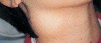
The purulent process is a continuation of the serous process, which is observed with a decrease in the body's resistance, early treatment, delayed diagnosis or incorrectly chosen therapy. The disease manifests itself in a disturbance in general health with fever up to febrile (39 ° C), chills, malaise, body pain and loss of appetite. Poisoning in a child is also manifested by pallor, dry skin and mucous membranes, lethargy, asthenia and sleep disturbances. When examining the affected area, swelling is detected without clear boundaries, which leads to visible asymmetry. The skin over the site of inflammation is hyperemic, tense and will not be wrinkled. Palpation of the nodules is painful, acquires a dense-elastic consistency and has limited mobility due to periadenitis. Melting of tissues (formation of an abscess) is determined by the phenomenon of fluctuations in the center of the swelling - fluctuations in the exudate during sudden movements. Lymph nodes do not usually blend with the skin or surrounding tissue. In some cases, among local changes, signs of inflammatory lesions of the lymphatic vessels (lymphangitis) can be identified. You can then see that a tight, painful cord runs toward the widened site of the infection's entry door, which is identified externally by a linear redness (a narrow stripe on the skin). An active inflammatory reaction in the area of primary disorders complements the clinical picture with a number of accompanying symptoms (from the mouth, throat, genitourinary tract, etc.). Lethargy. Leukocytosis. Body aches. Malaise. Neutrophilia. Chills.
Abscess of the maxilloglossal groove
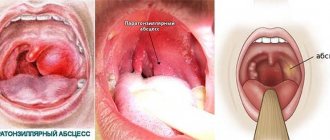
Abscess of the maxillo-lingual groove (abseessus sulci mandibulo-lingualis). The maxillo-lingual groove is the posterior part of the sublingual region, located behind the sublingual salivary gland, between the lateral surface of the tongue and the body of the lower jaw. To examine the maxillo-lingual groove, it is necessary to retract the tongue in the opposite direction using a spatula or dental mirror.
The inflammatory process spreads to this area of the sublingual region with acute purulent apical pericementitis of the lower large molars, as well as with difficult eruption of the lower wisdom tooth (pericoronaritis).
As a rule, inflammatory phenomena with an abscess of the maxillo-lingual groove develop acutely. On the 2-3rd day from the onset of the disease, mouth opening is already significantly limited, severe pain is noted when moving the tongue and swallowing. The temperature often rises to 37.5-38.5°.
During an external examination, only some patients can notice a slight swelling in the area of the submandibular triangle; the skin in this area is not changed. When palpating the tissues in this area, pain and enlargement of the group of submandibular lymph nodes are detected. Due to the spread of the inflammatory process to the lower pole of the internal pterygoid muscle, as a rule, trismus of the second or even third degree is observed.
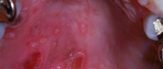
When examining the vestibule of the mouth, no changes are detected. After slowly and carefully spreading the jaws, which is convenient to do with small turns of a metal spatula, it is possible to examine the sublingual area, and by moving the tongue with an instrument in the opposite direction, also the maxillo-lingual groove. The mucous membrane in the area of this groove and in the corresponding area of the inner side of the alveolar process turns out to be sharply hyperemic. Maxillo-lingually, a fluctuating bulge is found.
Spontaneous opening of the abscess as a result of a breakthrough of the mucous membrane covering it; the groove is smoothed, the tissues in this area are infiltrated; often draining it after the incision usually leads to the rapid elimination of all painful phenomena.
As the process progresses, purulent exudate from the tissues forming the maxillo-lingual groove spreads to the sublingual region along the adjacent tissue or to the submandibular triangle along the tissue surrounding the duct of the submandibular salivary gland. Often, a purulent process from the tissues forming the maxillo-lingual groove passes into the pterygo-maxillary and even peripharyngeal space.
Treatment of abscess of the maxillo-lingual groove is surgical. In the area of greatest tissue bulging, an incision about 1.5-2 ohms long is made. In this case, the tip of the scalpel is directed towards the alveolar process to avoid injury to the lingual nerve, as well as the lingual vein and artery located near it. The operation, due to the significantly limited opening of the mouth, is more convenient to perform with a small narrow scalpel on a narrow handle.
If, after dissecting the mucous membrane, no pus is released, a slightly bent grooved probe is inserted into the incision, the deeper tissues are pushed apart and thus the abscess is opened. This surgical intervention, due to the difficulty of manipulating the syringe, is usually performed without anesthesia. Reducing pain during dissection of the mucous membrane can be achieved by first applying a small tampon moistened with a 3-5% dicaine solution to the area of the intended incision.
If the amount of purulent discharge is small, it is advisable to insert a small strip of iodoform gauze or thin rubber into the surgical wound; this prevents the edges of the cut from sticking together.
After the elimination of acute inflammatory phenomena, the tooth that served as the entrance gate for the infection is removed.
Possible complications
If acute inflammation does not stop in time, the capsule melts with pus breaking into the surrounding fibers. In this case, a reverse process is observed, called adenophlegmon. Localization in the cervical region is accompanied by a rapid course with rapid spread of pus through the interphase spaces. Breakthrough of exudate into other anatomical areas (organs, cavities) leads to the formation of fistulas, abscesses and mediastinitis. The infection can reach the venous vessels (thrombophlebitis) or enter the bloodstream with the development of septicopyemia.
Diagnostics
A preliminary diagnosis is established on the basis of clinical data - examination (complaints, medical history), examination and palpation of lymphoid formations. The area where the primary infection is likely to be located is also subject to physical examination. To determine the cause and nature of the disorders, a set of laboratory and instrumental methods is required: • General blood test.
Common signs indicating inflammatory changes in the body are leukocytosis and increased ESR.
By their level and other indicators (shift of leukocytes to the left, granularity of toxic granulocytes) one can judge the severity of infectious disorders. Bacterial pathology, according to the results of a clinical blood test, is manifested by neutrophilia, and viral pathology - by lymphomonocytosis. Ultrasound of the lymph nodes allows you to determine the size, shape, structure, contours, depth, relationship to nearby tissues and the presence of complications. According to Doppler ultrasound, purulent lymphadenitis is accompanied by an increase in size, thickening and thickening of the nodule capsule, heterogeneity of the structure with anechoic areas and the presence of zones without blood flow. • CT scan of affected areas.
This is the most accurate imaging method in clinical practice.
A CT scan can help you determine the size, location of inflamed structures, the presence of an abscess, and the spread of pus. Determines primary changes in the lungs and other organs. • Puncture of lymph nodes.
Identifying signs of abscess formation requires diagnostic puncture of the lymph nodes. The resulting exudate is subjected to microscopic and bacteriological examination to determine sensitivity to antimicrobial drugs. To rule out certain abnormalities, a piece of tissue removed by puncture or fine needle biopsy is sent for histological analysis. For a more detailed study, ultrasound of the lymphatic vessels, lymphography, and lymphoscintigraphy are prescribed. The diagnosis of purulent lymphadenitis caused by specific flora requires the use of additional techniques. Tuberculosis infection is confirmed by tuberculin tests (Mantoux, Koch, Pirquet) and syphilis by serological tests (RW, RMP, ELISA, RPGA, RIBT). The diagnosis is made by a purulent surgeon with the involvement of doctors from related fields. Given the location of the primary process, consultation with a dentist, otolaryngologist, dermatologist, etc. may be required. If a specific etiology is suspected, the patient should consult an infectious disease specialist, tuberculosis specialist, or venereologist. Differential diagnosis is carried out for chronic forms, lymphadenopathy with lymphoblastic leukemia and lymphogranulomatosis, as well as for metastases of malignant tumors. Purulent atheroma, abscesses and phlegmon should be excluded.
Types of phlegmon
Phlegmons of the maxillofacial area in dentistry are classified depending on the location.
The main types of pathology are presented in the table below:
| Localization | Characteristic |
| Phlegmon of the temporal region | It is an inflammatory formation in the subcutaneous layer in the temple area. Accompanied by throbbing pain, the intensity of which depends on the depth of the lesion. With superficial phlegmon, severe swelling is noted. In some cases, due to temporal phlegmon, the patient cannot open his mouth normally. |
| Orbital phlegmon | The purulent-inflammatory process occurs in the fatty tissue of the orbit. In most cases, the pathology is one-sided. Accompanied by intense headaches, severe swelling of the eyelids, conjunctiva, and protrusion of the eyeball. Eye movement is limited. A significant decrease in visual acuity or its complete absence is possible. |
| Phlegmon of the subtemporal space | A purulent-necrotic process occurring in the infratemporal fossa. Occurs against the background of caries of the upper teeth. It is also possible for phlegmon to spread from the area of the upper jaw and temples. Patients experience pain above the upper jaw, which radiates to the ear, temple or teeth. |
| Phlegmon of the peripharyngeal space | Phlegmon in this area occurs against the background of carious lesions of the lower teeth and infectious diseases. Accompanied by moderate pain that is permanent. There is an increase in local lymph nodes, difficulty swallowing and opening the mouth. |
| Phlegmon of the pterygomaxillary space | The pathology is localized in the area of the pterygomaxillary fold. Phlegmon occurs mainly against the background of carious lesions and other dental diseases. Infection is also possible if antiseptic standards are not observed during torusal anesthesia. There is pronounced facial asymmetry. The patient is unable to open his mouth and swallow food normally. There is hyperemia of the mucous membrane. |
| Phlegmon of the parotid region | It occurs against the background of a purulent form of lymphadenitis, the presence of carious lesions in the upper molars. Accompanied by swelling of the tissues of the parotid region. In this case, the skin color, as a rule, does not change. There is pain when moving the jaw. |
| Phlegmon of the chewing area | Localized in the area of the masticatory muscles (cheeks). Accompanied by severe swelling, facial asymmetry, and pain. Mouth movements when chewing are limited. |
| Cellulitis of the floor of the mouth | Located in the sublingual or submandibular region. Accompanied by swelling under the tongue and pain. The patient has difficulty breathing and increased salivation. The mobility of the tongue decreases due to which speech defects occur. The tissues under the tongue acquire an unhealthy shine and turn red. |
Cellulitis of the jaw space occurs against the background of carious lesions and other dental diseases
Treatment
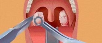
Effective treatment is achieved only with a complex effect on the pathology using surgical and conservative methods. An abscess or the presence of adenophlegmon is an indicator for opening the lesion, evacuation of pus, washing and draining the wound. Necrotic tissue and destroyed nodes are removed. The operation is performed by a surgeon in a hospital under local anesthesia. In the postoperative period, the patient needs bed rest with limited movement in the affected area and an easily digestible diet. Antibacterial and detoxifying drugs, nonsteroidal anti-inflammatory drugs, and desensitizing drugs are prescribed. Dressings are carried out with hyperosmolar and antimicrobial ointments, the skin is treated with antiseptics. The recovery period involves the use of certain physiotherapeutic procedures (UHF, electrophoresis, galvanic and magnetic therapy).
Features of treatment of cheek abscess
The doctor will help you decide on therapeutic methods, taking into account all the nuances of the development of the abscess, localization, presence of complications, assessing the risk of spreading inflammation, the patient’s immune system, age, and concomitant diseases.
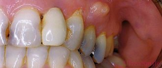
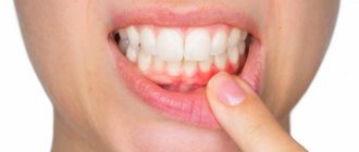
Often they resort to opening the attribute located on the cheek; if the abscess is shallow, the doctor will limit himself to conservative treatment with physiotherapy.
Surgical
Surgical treatment of an abscess on the cheek is used for deep, extensive lesions, the presence of complications, extensive symptoms, and the absence of positive dynamics with antibiotic therapy.
Need advice from an experienced doctor?
Get a doctor's consultation online. Ask your question right now.
Ask a free question
Variations of manipulations to eliminate an abscess:
- removal of a tooth affected by caries with sanitation of the oral cavity with an antiseptic solution - often used in pediatric dentistry in the absence of positive changes after treating a formation on the cheek with medications, during extirpation of incisors;
- opening the periosteum in order to evacuate purulent fluid from the abscess cavity, drain the affected area, eliminate inflammation with medications;
- a combination of two methods - tooth extraction and periostomy.
Stages of cheek surgery:
- administration of anesthetic;
- incision of the external capsule of the abscess;
- evacuation of the contents of the cavity with excision of the walls in order to prevent recurrence of pus accumulation;
- antiseptic treatment;
- drainage for large abscesses or suturing for minor abscesses of the cheeks;
- applying a cotton-gauze bandage with a hypertonic solution to provoke rejection of residual masses.
The course of antibiotic therapy after surgery is from 2 weeks, agreed with the doctor individually.
Conservative
The traditional treatment regimen for an abscess on the cheek involves a complex effect:
- Antibiotic therapy using broad-spectrum drugs or a combination of several units to suppress the activity of the pathogen, eliminate toxic waste products of bacteria, and effectively eliminate the purulent contents of the abscess. The group of penicillins with clavulanic acid and cephalosporin drugs are often used. Mostly they resort to the intravenous or intramuscular route of administration due to the severity of the chewing process.
- Local drugs with antiseptic, anti-inflammatory, and sedative effects are prescribed as an element of complex therapy for the elimination of an abscess on the cheek. Treatment with individual methods does not bring the desired results, but helps relieve acute manifestations of the disease. Levomekol, Vishnevsky ointment, Kamistad, Ichthyol liniment, Methogil denta-gel for mucous membranes are used.
- You can rinse your mouth with antiseptic solutions: Miramistin, Chlorhexidine, Chlorphyllipt alcohol.
- For the treatment of abscess, anti-inflammatory and analgesic substances are used - Ibuprofen, Paracetamol, Nimesulide. Long-term use may cause bleeding after tooth extraction.
If there is no positive dynamics in the treatment of the attribute located on the cheek, consult a doctor and adjust the therapy.
Physiotherapy
After opening the abscess in the cheek area, physiotherapeutic procedures are performed to stimulate regeneration and prevent scarring of the facial area:
- exposure to microelectric current pulses to activate cell growth in the lesion;
- UV therapy;
- electrophoresis with medications with a targeted effect on the buccal area;
- Ultrasound pulses;
- laser therapy after an abscess.
Physiotherapeutic influence to obtain a positive effect requires a course of 21-30 days, depending on the severity and extent of damage to the cheeks.
Bibliography
1. General surgery: textbook / Petrov S. V. - 2010. 2. Lymphadenitis, lymphangitis, lymphadenopathy of the maxillofacial region: textbook. -method. Manual / N. N. Cherchenko, etc.; – 2007. 3. Lymphadenopathy and lymphadenitis in children: diagnosis and treatment / M. S. Savenkova, A. A. Afanasyeva, A. K. Abdullaev, L. Yu. Neizhko // Difficult patient. - 2008 - No. 12. 4. Diagnosis and treatment tactics of cervical lymphadenitis / Skorlyakov V.V., Babiev V.F., Keshchyan S.S., Stagnieva I.V., Boyko N.V. // Young scientist. - 2020. - No. 16.
Source
Description of the disease
Phlegmon of the maxillofacial region
Phlegmon is a purulent inflammation that occurs in the soft tissues.
The pathological process does not have clear boundaries, which is why it quickly spreads to blood vessels, nerve endings and organs.
Phlegmon of the maxillofacial area mainly affects bone tissue and tendons, salivary glands, and muscle tissue.
Cellulitis is a dangerous pathological condition. Due to the purulent-inflammatory process, a large amount of toxic substances enter the bloodstream, which causes general intoxication of the body.
The disease is acute, characterized by the rapid development of symptoms, against the background of which the functions of the masticatory apparatus, swallowing, and breathing are impaired in patients.
Phlegmon in ICD 10
In the international classification of diseases, phlegmon of the maxillofacial area is included in the group of diseases of the skin and skin tissue (L00 - L99). The pathology is included in the block of infectious skin diseases and is designated in the ICD by code value L 03.2.
