Main complaints
When it comes to chewing teeth, people often go to the clinic when they experience pain. But if the front teeth, which are visible when smiling, somehow change, the person makes an appointment, even if nothing hurts. The main complaints with which people come to the doctor are:
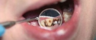
- change in tooth color or detection of stains;
- change in the shape and integrity of the tooth, even without the presence of pain;
- shortening of teeth, which makes nasolabial folds more noticeable.
Some complaints appear over time, but some problems arise even at the teething stage. Therefore, it is important to understand the reasons for the changes and find out how the problem can be eliminated.
Tooth discoloration, spots and specks: why they occur and how to fix them
The appearance of any pigmentation on teeth can be a consequence of diseases:
- appeared after tooth eruption;
- transferred before eruption.
It is impossible to determine the cause and get rid of stains at home, and trying to whiten the enamel yourself can lead to unpleasant consequences: demineralization and hyperesthesia. Therefore, if you find that the color of a tooth has changed, make an appointment with your dentist.
The specialist will find out what contributed to the staining and choose treatment tactics depending on the origin of the spots.
Teeth staining due to increased enamel permeability
Increased enamel permeability (demineralization) is a condition of hard tooth tissues with a lack of essential microelements. Typically calcium and fluoride. Demineralization is a consequence of the influence of an acidic environment on the tooth surface or the result of exposure to bacteria, which subsequently leads to carious lesions.
With increased enamel permeability, people come to the clinic with complaints:
- the appearance of white, non-shiny and rough spots on the surface of the tooth (usually in the cervical area);
- for the appearance of hypersensitivity - a painful reaction to chemical, thermal or physical irritants.
In addition to unpleasant sensations, the surface of the stains may also be stained with food coloring (when drinking tea or coffee). Since the pigment penetrates the surface rough layer of enamel, the tooth is polished to remove the color stain.
Next, to eliminate the problem and restore the enamel structure, the doctor will conduct remineralization therapy, cover the tooth with fluoride-containing varnish and advise you to change your toothbrush and purchase toothpaste with low abrasiveness and therapeutic fluoride content. Your doctor may also prescribe you to take calcium tablets.
Spots on a tooth due to fluorosis
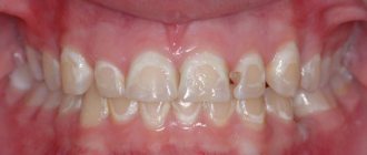
Fluorosis is a non-carious disease caused by excess fluoride. Fluoride is a substance that is part of enamel and helps strengthen teeth. However, if there is too much of this mineral, brown spots appear on the surface of the tooth, and the enamel itself becomes more fragile.
Mild fluorosis causes dull white and tan spots to appear on the teeth, causing the tooth to appear mottled or pockmarked. In some cases, the disease manifests itself only on the central incisors, but more often it affects all teeth. This is because fluoride enters the entire body in excess and is deposited in all teeth, and sometimes in human skeletal bones.
If there is too much fluoride absorbed by a person (the microelement comes from water, from food, with toothpaste when brushing teeth), in addition to stains on the teeth, erosions appear - uneven enamel defects with uneven edges, cavities that become pigmented (colored) over time.
Treatment options include:
- bleaching followed by mineralizing therapy;
- direct photopolymer restorations;
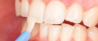
- production of permanent orthopedic structures – veneers and crowns.
The choice of treatment depends on the form of fluorosis, which only your doctor can determine.
In addition, your dentist will advise you to reduce the amount of fluoride you consume. For example, refusing to drink tap water or artesian water with a high fluoride content.
Spotted form of enamel hypoplasia
Hypoplasia is a disease that occurs during tooth formation and manifests itself in the form of a violation of the formation of dental tissues (amelogenesis imperfecta). In this case, the tooth defect is visible immediately after eruption: it grows unlike the others.
Hypoplasia develops during tooth development under the influence of:
- local factors (for example, if caries of a baby tooth was complicated by periodontitis, or as a result of an impacted dislocation of a baby tooth);
- systemic factors (previous disease or nutritional deficiency of microelements necessary for the proper formation of dental tissues).
Depending on what caused it, local and systemic hypoplasia are divided. Local appears only on one tooth, and systemic - on all teeth that were formed and erupted simultaneously, for example, on the central incisors.
The spotted form of hypoplasia looks like white, less often yellowish spots on the surface of the tooth. In this case, the enamel is not damaged, its thickness in the area of the stains is the same as in unaffected areas. The tooth remains shiny and smooth, stains are not stained with dyes, which means the enamel structure is not damaged, demineralization is not observed. The spots usually do not increase in size over time.
Since the stain is located throughout the entire thickness of the enamel, it cannot be sanded off. Treatment options include covering the spot with filling material, or orthopedic treatment - making veneers or crowns.
Change in filling color after restoration
Over time, a filling that was previously invisible may change color, darken, and begin to differ from the tooth’s own tissue. Such complaints most often appear in people who regularly drink tea, coffee, or smokers. The thing is that the filling material differs in structure from enamel and can be stained.
This is especially noticeable on fillings that have served for quite a long time. Under the influence of various factors - the action of an acidic environment in the oral cavity, stress, the influence of saliva - the structure of the filling may be damaged.
To avoid discoloration of the filling, it is important to care for it. This is extremely difficult to do at home, so it is worth visiting the dentist at least twice a year for:
- therapeutic examination;
- professional hygiene;
- polishing the surface of the filling.
It is also important to determine whether there is a gap between the filling and the tooth tissue. Violation of the fit with a subsequent change in color may indicate the development of a secondary carious process under the filling. Since the photopolymer material has a certain degree of transparency, caries can “shine through” its surface.
In this case, the filling will have to be sanded down, the infected tooth tissue removed and reinstalled. After the new filling has hardened and polished, it is recommended to abstain from food and drinks containing coloring substances for 24 hours so that the pigment does not penetrate into the filling material during the final polymerization period.
Change in tooth color after root canal treatment
Some patients complain of tooth discoloration after root canal treatment:
- loss of shine;
- gray enamel;
- the appearance of an unusual shade - pink or gray;
- to “shine through” a new, unnatural color from inside the tooth.
It is worth noting that after pulp extirpation treatment, the tooth is no longer “alive.” There are no nerves or vessels inside, which means there is no tissue trophism. Due to the fact that the nutrition of hard tissues has been stopped, the color may change somewhat, the shine may disappear, and a grayish tint may appear.
Such changes appear over time. However, complaints can arise almost immediately after root canal treatment, when the cavity in the tooth is closed with a temporary filling. In this case, there is no need to worry. A temporary change in color may just be due to the temporary filling material.
Tooth tissues have certain characteristics of transparency and light refraction. When a cavity appears in a tooth (and it inevitably appears during treatment, since the dentist needs to gain access to the pulp), and is then filled with a material with an uncharacteristic color, it can show through. Therefore, it seems that the tooth has become whiter/grayer/less shiny.
A similar effect is observed in the case of installing a metal pin for further tooth restoration. When choosing a material with a high degree of transparency and light transmittance, the metal can “shine through”.
The tooth may change color as a result of root canal treatment with resorcinol-formalin. In this case, the enamel sometimes takes on a pinkish tint. If the channels are “silvered”, the tint may be gray. In this case, you can re-fill the canals, carry out endodontic whitening, but with a high probability the tooth will require restoration or crowning to restore the natural color and cover the unnatural color.
Remineralization of tooth enamel. Clinician's perspective
July 23, 2018
| ANDREY AKULOVICH professor, clinical director of the ROCS brand |
| TATYANA KUPETS Head of the Department of Science and Medical Programs of the Group (ROCS brand) |
Toothpaste with a remineralizing effect can compensate for the lack of minerals and become a successful additional remedy in the treatment of demineralization. Introducing the new ROCS biocomplex toothpaste.
The enamel of our teeth is regularly exposed to aggressive attack, from the simplest food acids from fruits and juices to the acids used during dental procedures. In addition, there are factors that cause or simply enhance the loss of the mineral component of enamel endogenously: these are diseases associated with impaired mineral metabolism, and a number of hormonal disorders or conditions.
The disappearance of minerals from tooth enamel is called “demineralization” (“decalcification”). At the same time, in our body there is a constant process of compensation for the demineralization process - this is physiological remineralization, which is provided by the protein fractions of saliva, as well as a number of its other properties. But very often locally (in the area of several or a whole group of teeth), as a result of excessive demineralizing load, processes arise that become decompensated, and then chalky-colored spots or stripes appear on the teeth. A symptom that often accompanies demineralization is enamel sensitivity. In this situation, the teeth already need specialized help - remineralizing therapy (abbreviated as remotherapy).
Various manufacturers have already proposed many products and preparations for remotherapy, in particular those based on casein/amorphous calcium phosphate, silicon compounds, tricalcium phosphate and other components. All of them either have restrictions for use or insufficient clinical effectiveness, or are difficult to access for mass consumption. Balanced mineral complexes for self-use by patients are rightly considered today to be the most logical, very accessible, without contraindications and time restrictions for use, and at the same time effective for remotherapy.
ROCS Medical Minerals is , without exaggeration, a unique and most sought after mineral complex by specialists, created taking into account the clinical logic of enamel remineralization. The main active component of the complex is calcium glycerophosphate - a compound based on two components that are most important for saturating the enamel with minerals - calcium and phosphorus.
Calcium glycerophosphate (GP) is a compound with a high level of bioavailability, since it is a direct substrate for salivary enzymes. It is the ability of GF to integrate into the physiological process of mineralization that allows for the rapid restoration of demineralized tooth enamel. The remineralization process is accelerated by the inclusion of magnesium salt, an activator of salivary enzymes, in the gel. Another basic component - xylitol - performs an anti-carious function. It is included in the ROCS Medical Minerals gel.
|
|
The ROCS Medical gel line is represented by the following options
| A very important feature of the ROCS Medical gel is the absence of fluorine compounds in its composition. When identifying foci of enamel demineralization, dentists very often prescribe fluoride-containing preparations, forgetting that the film formed by fluoride on the surface of the tooth prevents not only the release of calcium from the tooth enamel, but also the penetration of ions from saliva into the demineralization focus. That is why fluoride is needed not at the stages of the course of remotherapy, but after its completion. In this case, fluoride preparations are prescribed to stabilize the results of remineralization with calcium and phosphate. The presence of fluoride in remotherapy formulations most often reduces their effectiveness and limits their use. Fluoride-containing preparations cannot be recommended in areas of endemic fluorosis and for the prevention of early childhood caries. |
Classic ROCS Medical Minerals , which is a gel for remotherapy and has a wide range of indications for use. The main situations for its use: caries in the white spot stage, hypoplastic processes in tooth enamel, demineralization of enamel after teeth whitening, in a complex scheme with methods of microabrasion and composite infiltration of enamel, complex treatment of dental fluorosis, focal demineralization after removal of braces, stabilizing and therapy that improves the results of clinical teeth whitening, as an alternative to teeth whitening in the case of a number of contraindications to it, and others.
ROCS Medical Sensitive , unlike the classic gel, contains a special component - potassium nitrate, which is a traditional substance for drugs that relieve the sensitivity of hard dental tissues due to impaired conduction of pain impulses. Due to its sensitivity-compensating properties, ROCS Medical Sensitive is recommended as a gel with the same remineralizing properties, but for those clinical situations where there is also an additional factor of tooth sensitivity, which quite often accompanies local loss of minerals in the teeth. In addition, the gel is also recommended as a means to relieve normal tooth sensitivity.
Since remotherapy can and should be done at any age, starting from the moment of teething, a special ROCS Medical minerals with strawberry flavor . Due to the flavoring additive, it is more pleasant for children and adolescents.
For difficult situations, when there is a need for a particularly active remineralizing effect, there are a number of products that can enhance the effect: special mineral rinses, mineral tablets (such as ROCS Medical chewable tablets ).
And, of course, during the days and subsequently after remotherapy, it is extremely important for the patient to use a special toothpaste with special remineralizing activity. ROCS Biocomplex Active Protection toothpaste is ideal for this purpose and contains the proteolytic enzyme bromelain and its peptide components, calcium glycerophosphate, magnesium chloride and xylitol (6%). Each component of the complex, performing its task, increases the efficiency of others. The enzyme, by breaking down plaque proteins, promotes its high-quality removal from the surface of the teeth. Bromelain and peptides are essential factors in plaque control. They provide the prolonged action of toothpaste against plaque. According to the subjective assessment of consumers, teeth remain smooth and clean throughout the day.
| BASIC RULES REMINERALIZING THERAPIES It is important to remember some basic rules for remineralizing therapy (regardless of the nosological form): • remotherapy is carried out only by applications in an individual or disposable mouth guard • optimal application time is about 30 minutes • one application per day after brushing your teeth is enough • the optimal course of remotherapy is 30 days, the minimum is 14 days |
Slowing down the formation of plaque provides teeth with almost constant availability of the mineral components contained in toothpaste and saliva. As a result, mineralization occurs of both healthy enamel and the initial foci of carious lesions.
For successful mineralization of teeth, it is desirable to have factors that modulate the permeability of tooth enamel. For this purpose, the active complex of the paste - MINERALIN® - uses xylitol in a concentration of 6%. Research by American and Finnish specialists has found that in this concentration xylitol significantly enhances the mineralization process, which is associated with its ability to form complex compounds with calcium. In the composition of toothpaste, this substance performs several functions: being a sweetener, xylitol improves its taste characteristics; Being a polyhydric alcohol, it functions as a moisture-retaining component. The mechanism of involvement of this substance in the biochemical metabolism of streptococci is characterized as lethal synthesis, and therefore the activity of cariogenic bacteria is reduced. Experiments have established that the effect of xylitol against cariogenic streptococci appears at a concentration of 2–2.5%.
ROCS Biocomplex Active Protection toothpaste with ROCS Medical Minerals , you can achieve the maximum rate of remineralization of tooth enamel.
Tags: ROCS
More on the topic
ROCS® Innovation Collection
There are practically no effective hygiene products in the world that would solve the problem of dry oral mucosa. The group, one of the world leaders in the production of quality oral care products, introduced to the market a unique ROCS® PRO Moisturizing toothpaste (moisturizing). We present this and other interesting global innovations of the company in our review.
New remineralizing gel ROCS Minerals BIO
ROCS presents a new product - a remineralizing gel for strengthening teeth of a new generation. ROCS® Minerals BIO combines one of the most effective remineralizing systems and advanced biotechnology. The product does not contain fluoride and is safe to swallow; Suitable for use by both adults and children from infancy.
Innovative product from ROCS: Active Magnesium toothpaste
The formula includes high concentrations of magnesium, a vital chemical element necessary for the functioning of more than 300 enzymes in the human body, including the mineralization of teeth and bones.
ROCS® PRO MOISTURIZING – global innovation in oral hygiene products
There are very few effective hygiene products in the world that would effectively solve the problem of dry oral mucosa. The ROCS team of experts and developers presents a unique ROCS PRO Moisturizing toothpaste. The patented formula of the product has been tested many times in clinical practice and has shown high effectiveness.
ROCS presents a bright summer novelty for children
ROCS specialists have developed a beautiful and comfortable ROCS Junior toothbrush especially for children aged 6 to 12 years. The original design and rich color combinations of the handle and bristles will delight the child and allow him to choose a unique color to suit his mood.
The Russian brand ROCS is available for order on the global iHerb platform
ROCS became the first Russian brand represented by iHerb internationally. The global online store announced the launch of sales of more than 20 ROCS products in all regions of its trading platform presence.
Beauty vibes from Femegyl
Nature gives beauty. Women knew about this many hundreds of years ago. Back then, they collected plants to make extracts and oils for skin care. Now professionals do this for women: cosmetologists, chemical technologists and doctors. Using natural ingredients, they create such effective skin care products as Femegyl Velvety Skin Fluid Serum.
Act outside of the mold
If marketers had been asked in 2005 whether it was worth launching a brand with a concept like ROCS, they would have heard the answer: it’s a crazy idea, people won’t understand. High-tech formulas, expensive production - why all this when there are proven business schemes? Svetlana Matelo, Ph.D., talks about how to achieve success by acting contrary to patterns. Team Leader (ROCS).
FEMEGYL presents a new product
For those who, when choosing skin care products, doubt whether gel or foam is better, the universal product FEMEGYL Gel Foam Balance has been invented. Suitable for all skin types, it combines the best properties of both gel and foam.
The ROCS brand was recognized as the leader of the pharmaceutical market in 2020.
At the end of December, the results of the “National Pharmaceutical Rating 2019” were announced in Moscow. Its goal is to recognize the best in the industry and tell the professional pharmaceutical and medical community, as well as a wider audience, about those who achieve outstanding results in their work to provide the country with quality medicines and more. ROCS, following a good tradition, became the winner in the Toothpaste Brand category. Return to section
Change in tooth shape: what could be the cause and how to eliminate the defect
A change in shape is the appearance of chips, cracks, irregularities, and chips in visible areas of the tooth. At the same time, a violation of integrity is not always accompanied by pain. Often the tooth is destroyed gradually, and in the process the tissue is rebuilt so that irritants do not affect the pulp.
The absence of pain is not a reason to hesitate. The sooner you see a dentist, the less tissue the tooth will have to lose. Accordingly, the simpler and cheaper the treatment will be.
Let's look at the main causes of defects in enamel and dentin. Let's look at how they can be eliminated.
Wedge-shaped defect
A wedge-shaped defect is a violation of the integrity of the enamel, which is characterized by a specific shape and location: it occurs in the cervical area, usually on symmetrical teeth (for example, canines or premolars). The main reason for the appearance of a wedge-shaped defect today is considered to be mechanical damage, which can be aggravated in an acidic environment.
The appearance of a wedge-shaped defect is a process extended over time. Therefore, the patient, as a rule, does not experience pain. But sometimes there may be complaints of a rapid pain reaction to cold, hot or sour.
Depending on the depth of the defect, several treatment options will be offered:
- remineralizing therapy;
- restoration of tooth shape using composite filling material;
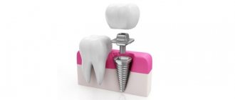
- making a crown.
Please note that the wedge-shaped defect may deepen over time. If you do not consult a dentist in a timely manner, serious aesthetic and functional complications are possible.
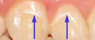
Enamel erosion
Erosion is a disease caused by a combination of physical and chemical effects on the tooth surface in conditions of insufficient mineralization. Erosion most often appears on the anterior group of teeth in the upper jaw and does not occur on the lower teeth.
The defect appears on the labial surface of the tooth and looks like an oval depression that can increase over time. The bottom of the recess is smooth, and the enamel does not lose its shine. But the deeper the depression becomes, the more colored its bottom becomes. The reason for this is the more “loose” structure of the tissues underlying the enamel.
In addition to the type of tooth, a patient with erosion often complains of a painful reaction to thermal and chemical influences.
Having established a diagnosis of “tooth erosion”, the dentist will offer several types of treatment, which depend on the depth of the lesion:
- Polishing the pigmented surface of the tooth and further remineralization using applications, electrophoresis and the administration of calcium and fluoride preparations for oral administration.
- Filling with composite material;
- Making an artificial crown.
In addition, the dentist may advise you to contact an endocrinologist and determine the condition of the thyroid gland. In patients with endocrine diseases, erosion occurs twice as often.
Chips, chips and cracks due to tooth injuries
Tooth trauma is damage caused by applied force. If the force is of high intensity and is applied simultaneously, we are talking about an acute injury. If we are talking about a low-intensity force that acts on a tooth over time, the injury will be chronic. You can read more about what acute and chronic dental injuries are in the corresponding article on the Ukrdental website.
If the tooth was not lost as a result of the impact, a number of complications may still arise:
- Pink staining of the tooth due to hemorrhage into the pulp;
- Graying and loss of luster as a result of pulp necrosis;
- Fracture of part of the crown or the entire supragingival part of the tooth.
Depending on the type and cause of the changes, the dentist selects the appropriate treatment:
- when staining a crown after an injury, intracanal bleaching can be performed;
- color and shape can be “corrected” using direct restoration with composite material;
- You can restore the integrity of the tooth and give it a natural color by making an artificial crown.
If after an impact the tooth remains intact but has changed color, you should not try to whiten it yourself or use highly abrasive pastes. It is necessary to contact a dentist, who, using various diagnostic methods, will determine the cause of the color change and eliminate it.
Hypoplasia and fluorosis
Hypoplasia is a disease that occurs when tooth tissue is exposed during its development. Fluorosis is also a problem that appears even at the stage of formation of hard tissues. In addition to the “harmless” spotted forms, these non-carious diseases also have more pronounced varieties.
Thus, the extreme form of hypoplasia is aplasia. In this case, the tooth is simply not covered with enamel. In less radical forms, enamel may cover only part of the tooth. Fluorosis can also manifest itself in more than just spots. Pits, grooves, and pockets of erosion may appear on the tooth.
In this case, remineralizing therapy will not be enough. To preserve the aesthetic appearance and functionality of the tooth, you will be offered a direct restoration or covering the tooth with a crown.
The role of enamel light transmission in aesthetic dental restoration
Dental restoration with modern ceramic and composite materials is closely related to understanding the aesthetic function of the tooth, which is characterized by optimal size, shape, relief, as well as the optical properties of enamel and dentin. Hard tissues reflect, transmit, and scatter light rays, forming color shades, opalescence, and fluorescence of the tooth. A pronounced feature of enamel is the ability to combine these optical effects [1, 2].
One of the most important optical characteristics of a material is the ability to transmit light, allowing you to see objects behind it - transparency: light in such cases is practically not reflected from the surface. The ability of an object to partially transmit and partially reflect light is called light transmittance. Transparent materials create their own effects: the perception of color is mixed with the appearance of volume (it seems to glow from within). If a colored area is given blurred boundaries, it will appear translucent [2, 4, 6]. The ability of tooth enamel to partially transmit and partially scatter light rays characterizes its light conductivity. The latter depends on the composition and structure of the fabric. Thus, in contrast to dentin and pulp, there are practically no pigments in intact enamel, but the inherent property of light transmission of enamel allows rays selectively reflected from dentin pigments, the enamel-dentin junction and pulp to pass through the enamel and be perceived by the eye as the color of the tooth. The thinning of the enamel layer contributes to more intense translucency of dentin. In certain areas of the tooth, the enamel does not have underlying dentin and is perceived as “transparent”. We are talking about the cutting edge and proximal surfaces.
In the clinic, there are four main types of enamel transparency on the vestibular surface of the tooth (Fig. 1). The first option is a uniform distribution of the transparent layer over the entire surface of the crown, the second is a predominantly pronounced transparency of the cutting edge, the third type is a transparent cutting edge and proximal surfaces, the fourth is characterized by transparency of only the lateral surfaces.
Rice. 1. Types of enamel transparency: 1 - teeth with a transparent layer over the entire surface, 2 - with a transparent layer in the area of the cutting edge, 3 - with a transparent layer in the area of the cutting edge and proximal surfaces, 4 - with a transparent layer only in the area of the proximal surfaces.
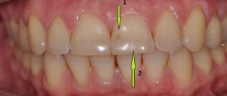
In cases where it is necessary to simulate a structure that includes the restoration of initially missing parts of the dentition (a tooth or part of it), the choice of color remains with the dentist [3, 5]. Indications for the use of color-correcting techniques are the presence of a wide gap between the teeth (diastema, trema); tooth reduction; low height, abrasion, chipped crown (Fig. 2).
Rice. 2. Wide gap between the central and lateral incisors, wear of the cutting edge of the 11th and 21st teeth.
A prerequisite for high-quality execution of aesthetic restorations is planning their size and shape (Fig. 3).
Rice. 3. Planning the size, shape and type of transparency of the teeth.
For these purposes, the height, thickness and mesiodistal dimensions of the teeth are measured (Fig. 4).
Rice. 4. Odontometry of teeth.
A diagram is created in the medical record (or on a computer monitor) that reflects the existing shape, size of the tooth and parameters of the planned restoration. Before starting work, you can model the future structure directly on your teeth. Before removing plaque and preparing teeth, filling material of non-traditional shades is applied to the contact surfaces (Fig. 5).
Rice. 5. Modeling a restoration made of a “non-usable” composite on the teeth.
The composite is quickly modeled, simulating the future restoration, photo-cured without additional processing, and assessed visually. This stage helps the doctor imagine the expected shape of the teeth, clarify the preparation areas, and also demonstrate to the patient the possible result of treatment. After discussing the action plan, the filling material is easily chipped with a trowel. Mechanical cleaning of all surfaces of the central incisors from plaque is carried out, since the restoration can extend to the proximal and palatal surfaces. The choice of material shades plays an important role in the predicted quality of the work performed, since the restoration will look natural only if the color scheme is optimally formed. The opaque composite is selected to fill the main volume of the restoration. The selected enamel composite will cover the dentin layer. A zone of at least 1.0 mm will be formed on the mesial surface using a transparent shade. A wide “transparent” layer will visually reduce the transverse dimensions of the restored tooth.
The medical record indicates the selected shades and the expected changes in volumetric parameters: an increase in mesiodistal dimensions, a transfer of the geometric shape of the crown from rectangular to square, a shift of the dentogingival dome and the transparency zone in the mesial areas to the midline of the jaws.
Tooth preparation is not required in the following cases: with palatal position of the teeth; with tenon-shaped lateral incisors; when the vestibular enamel is thinned due to repeated bleaching or abrasion; if the veneer is a temporary construction. It is mandatory to grind off the surface non-prismatic layer of enamel. The veneer border is lowered 1 mm below the gingival margin if the tooth is slightly discolored or the enamel defect does not extend under the gum. The border is drawn at the gingival margin or advanced 0.5-1.0 mm under the gum in case of significant pigmentation or destruction of tooth tissue. The shape of the cutting edge after preparation depends on the type of bite, the size of the defect, and the degree of thinning of the enamel. A beveled incisal edge is usually created when the enamel in that area becomes thinner. Finished preparation with the creation of a semicircular ledge on the vestibular slope of the cutting edge of the tooth is indicated for chipping. The formation of a semicircular ledge extending onto the cutting edge of the tooth is used for abrasion. Thinning of the cutting edge of the tooth is used for low crown heights. Preparation with overlapping of the cutting edge of the tooth and grinding off about 0.5 mm of the cutting edge to the palatal surface is indicated for high chewing loads.
In the presence of a diastema, the preparation is minimal: on the vestibular surface from the midline to the mesial edge, an enamel bevel is made, which will mask the tooth-filling transition; the mesial areas of the incisors are also prepared. The entire “interested” surface is treated with a fine-grained diamond bur, washed with a stream of water, and dried. The adhesive system is used in accordance with the instructions. Immediately after photopolymerization of the adhesive, the modeling of the restoration begins. The first portion of opaque material is applied with a medium-sized trowel to the medial surface of the tooth in the equator area (Fig. 6).
Rice. 6. The opaque base of the restoration is modeled.
Pressing the composite against the tooth, smooth it out towards the prepared surface. If necessary, the opaque composite is used to model the signs of the side: mesial convexity, sign of the crown angle. On the vestibular areas of the tooth, opaque is used to cover transparent areas of enamel. The transparent enamel imitated by the photopolymer subsequently moves mesially (Fig. 7). As a result, the opaque composite occupies such a volume that the enamel layer is 0.5–0.7 mm on the vestibular surface and 1.0–1.5 mm on the mesial surface. The opaque base is then formed in a similar manner on a symmetrical tooth.
Rice. 7. Opaque is coated with an enamel layer.
Enamel shades model individual macro- and microrelief: for example, the shape of the cutting edge approaches a straight line, the gingival contour deviates distally, the length of contact of the mesial surfaces is significant (Fig. 8). The transparent layer (I) covers the opaque and enamel tint layers to a thickness of 0.5 mm. The width of the transparent layer on the mesial surface is 1.0 mm. Processing of the restoration (removal of the hybrid layer, enhancement of contours, polishing) and coating of teeth with fluoride varnish is carried out in the usual way.
Rice. 8. Finished work: the shape of the teeth has been changed, diastema and trema have been eliminated, the cutting edge has been restored, and the type of transparency of the teeth has been recreated.
We present a clinical case of a combination of a wide gap between the central incisors with a change in their color and uneven wear of the cutting edge. The presence of a diastema and worn cutting edge of the central incisors (Fig. 9) gives the dentist the right to choose shades of color, type of transparency, optimal sizes, shapes and relief of the restoration.
Rice. 9. A combination of a wide gap between the central incisors with a change in their color and uneven wear of the cutting edge.
Changing the shade of incisor enamel requires the use of color neutralizing techniques. In order to plan the size of restorations, the height, vestibulo-oral and mesiodistal dimensions of the teeth are measured and entered into the medical record. Odontometry shows that the width of the tooth in the equator region approaches the height of the tooth, which is 9.0 mm in 1.1 teeth, 8.8 mm in 2.1 teeth (Fig. 10).
Rice. 10. The vertical dimensions of the teeth were measured with a micrometer.
The planned vertical dimensions of the incisors are 9.3 mm for the right incisor and 9.4 mm for the left (the height of the teeth varies due to different positions in the jaw). A diagram of the planned veneers is drawn on the computer monitor. The expected shape of the vestibular surface is rectangular (approaching square). Signs of belonging to the side are weakly expressed: the mesial angles in relation to the distal ones are somewhat smaller in size and the mesial region of the crown is slightly convex. It is planned to recreate the contact surfaces from the tip of the interdental papilla to the incisal edge. The cutting edge is smooth. The gingival dome deviates distally. The relief of the vestibular surface is smoothed. Mechanical cleaning of all tooth surfaces from plaque is carried out using Klint paste, which does not contain fluoride and a fatty base.
The next stage - assessment of the color characteristics of the tooth - is carried out under optimal light conditions. The patient is in a sitting position, facing the window - a source of natural light. To select shades of the composite, it is necessary to compare the cutting edge of the tooth with a standard close up; in the same way, the cervical area and the equator area, the lateral surfaces of the teeth are assessed (Fig. 11).
Rice. 11. The color of the teeth is determined.
For color neutralization, the hybrid composite material Amaris (VOCO) was chosen, the set of which contains tint syringes (Opaque O1 and O2) that allow this technique to be carried out. The opaque (O2) composite is selected to fill the main volume of the restorations, the O1 shade is selected in the incisal area. The dentin layer will be covered with a matching enamel color (TN - neutral). A wide zone (up to 1.0 mm) will be formed on the mesial surface with a transparent shade of the composite: enamel transparency type 3. Modeling of a transparent cutting edge is also expected.
Veneer production involves preparation of the vestibular and mesial surfaces with NTI diamond burs. The prepared surfaces are treated with a fine-grained NTI diamond bur (No. 850 L), washed with a stream of water, and dried (Fig. 12).
Rice. 12. Central incisors after preparation.
Preparation is made to the thickness of the veneer, which provides space for the application of the composite, enhances the strength of its adhesion to the teeth, eliminates pigmented spots, and allows rational distribution of stress in hard tissues. The work begins with marking the boundaries of the future restoration. To do this, a small spherical diamond bur is used to create a groove up to 0.5 mm deep, the boundaries are extended to the lateral surfaces. The prepared lingual surface is smoothed with a pear-shaped, proximal - with a thin bur (“mosquito sting”). Fine-grained diamond tools are used.
Next, acid etching of the enamel is carried out, for which Vococid gel is applied to the entire prepared surface and washed off after 30 seconds. The adhesive system is applied to the pre-dried enamel surface in accordance with the instructions. After photopolymerization of the adhesive for 20 seconds, modeling of the restoration begins immediately. A portion of the material with increased opacity (O2) is applied to the medial surface of the tooth in the equator area. Pressing the composite against the tooth, smooth it from the center to the periphery. The second portion of the composite is applied in the same way. An opaque composite is used to model the sign of the crown angle: the mesial angle is slightly smaller in size than the distal one. The sign of crown curvature is formed as follows: a small portion of the opaque composite is applied to the vestibular surface in the form of a roller and is shifted closer to the mesial edge (Fig. 13).
Rice. 13. An opaque base is formed and color neutralization is carried out.
The opaque composite ultimately occupies such a volume that the enamel layer is 1.0 mm on the vestibular surface, and 1.5 mm on the mesial wall and in the area of the incisal edge. Then the opaque base is formed in a similar way on a symmetrical cutter. Portions of the high-opacity composite O2 and O1 are distributed over the vestibular surface, providing “neutralization” of the color of pigmented teeth. An enamel layer is applied to the opaque base (Fig. 14).
Rice. 14. An enamel layer of TN (neutral) shade has been applied.
Shades (TN and TL) model the individual macro- and microrelief: the shape of the cutting edge is slightly arched, the gingival contour deviates distally. Then the mesial convexity (a sign of crown curvature) and the angles at the cutting edge of the crown are modeled: the mesial one is slightly smaller in size than the distal one (Fig. 15).
Rice. 15. Enamel layers are evenly distributed along the vestibular surface of the central incisors: in the cervical and equatorial regions TN (neutral), in the area of the cutting edge and proximal surfaces TL (light).
The length of contact of the mesial surfaces is the distance from the interdental papilla to the cutting edge. The transparent layer covers the opaque and enamel tint layers to a thickness of 0.5 mm (Fig. 16).
Rice. 16. Contact points between teeth have been restored.
The width of the transparent layer on the mesial surface and at the incisal edge is 1.0 mm. At the final stage, odontometry is carried out: the vertical dimensions of the teeth after restoration exceed the horizontal ones and amount to 9.3 mm and 9.4 mm for the right and left incisors, respectively (Fig. 17).
Rice. 17. The vertical dimensions of the teeth were measured.
The veneer is processed immediately after production: the surface hybrid layer is removed, the relief is contoured, and occlusal contacts with antagonist teeth are verified. Diamond (with a red ring) or carbide burs of cylindrical, conical shape emphasize anatomical formations, including the classic signs of angle, crown curvature, as well as the individual characteristics of the patient’s teeth. Yellow ring diamond burs (15 µm grit) are then used. After this, polishing is carried out with finishing ultra-fine diamond burs (with a white ring) with a grain size of 8 microns, carbide burs (30 cuts). The antagonizing areas of the palatal surface are carefully processed so that the veneer bears minimal chewing load. The proximal-gingival area is smoothed with a thin bur (in the form of a “mosquito sting”). Strips are used to finish the proximal surfaces. Polishing the surface of the veneer is also done with discs without significant pressure on the surface of the restoration. Polishing heads containing fine aluminum oxide powder as an abrasive are used, sponges and polishing pastes are used. When illuminated with short-wavelength light, the restorations have the same spectrum and intensity of fluorescence as the hard tissues of the tooth (Fig. 18). The finished work is shown in Figure 23. Treating the tooth with fluoride-containing preparations reduces the risk of carious tissue damage.
Rice. 18. When illuminated with short-wave light, the restorations fluoresce in the same way as the hard tissues of the tooth.
Conclusion. Aesthetic restoration of teeth is largely associated with the characteristics of the optical parameters of enamel, including the property of light transmission. In particular, when making veneers, it is necessary to take into account the type of transparency of the enamel, as well as the characteristics of the dental materials used to recreate the natural appearance of the tooth. The use of hybrid photocurable composites and nanocomposites, provided that adequate shades are selected, ensures the achievement of the set goal - improving the quality of aesthetic restorations.
Rice. 19. Completed incisor restorations.