Causes of hypertrophic gingivitis. Etiology and pathogenesis
In dental practice, hypertrophic growth of gums has a second name - hyperplastic gingivitis . The disease occurs in both adults and children.
Hyperplastic gingivitis is accompanied by a rapid increase in the basal cells of the gingival epithelium. The following factors can provoke the development of pathology:
- active activity of pathogenic and conditionally pathogenic microflora (with very poor oral hygiene);
- multiple dental deposits (soft pigmented plaque, mineralized stones);
- hormonal disorders;
- luring out the fetus;
- menopause;
- long-term use of oral contraceptives;
- long-term use of drugs containing sodium diphenylhydantoin;
- hypovitaminosis;
- diseases of the cardiovascular system;
- pathological bite (deep, prognathia, progenia);
- improper filling of a carious cavity, leaving a sharp edge of the filling;
- errors in prosthetics;
- thermal, mechanical and chemical damage.
Since the disease occurs during pregnancy in 49% of cases, experts call hypertrophic growth of gums pregnancy gingivitis. The occurrence of hypertrophic gingivitis in children occurs most often at the age of 12-16 years due to hormonal changes in the body (juvenile gingivitis).
Forms of hypertrophic gingivitis and clinical signs
In dentistry, there are 2 forms of hypertrophic gingivitis: edematous and fibrous.
| Edema form | Fibrous form |
| Bleeding gums while eating and brushing teeth. The gums increase in size, thereby causing aesthetic discomfort. The crown part of the tooth is covered with inflammatory gum at 1/3 of its height. | At the early stage of the disease there are no significant signs. In the moderate and severe stages of the disease, cosmetic defects in the gums are a concern. The crown of the tooth is covered with gum by 1/2 - 2/3. |
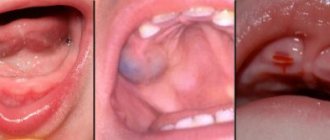
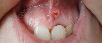
From the photo of hypertrophic gingivitis, you can see the external differences in the forms of the inflammatory process.
Fibrous form
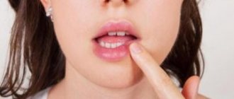
The fibrous form of hypertrophic gingivitis has a second name – “ fibrous gingival hyperplasia ”. The disease is manifested by periodontal growth, while the papillary-marginal zone of the gums becomes dense upon palpation.
There is no bleeding in this form of gingivitis, but the course of the disease and its further treatment are complicated by false periodontal pockets. When food debris gets stuck in hard-to-reach places, uncleanable plaque and mineralized deposits appear, aggravating the course of the disease.
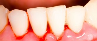
Edema form of hypertrophic gingivitis
The edematous form is characterized by swelling of the papillary-marginal-alveolar gum. The gums become hyperemic and cyanotic in the area of the gingival papillae.
When examining the oral cavity, the following are observed:
- involuntary bleeding;
- enlargement of periodontal pockets;
- multiple dental deposits.
As a rule, the hygiene index for the edematous form of hypertrophic gingivitis is 3.4–5 points. The disease can occur in a generalized (widespread) or localized (about 2-3 teeth) form.
Hypertrophic localized gingivitis, edematous form.
Ticket number 1.
Girl 13 years old. She complained of an increase in gum volume, which was accompanied by profuse bleeding. Such symptoms recur periodically. Brushes teeth irregularly. He explains this by saying that it spares the gums. Upon examination, bleeding gums were revealed, the dentogingival papillae were increased in volume and deformed. Teeth crowding in the anterior region is determined.
Determine the diagnosis.
Make a treatment plan.
Hypertrophic localized gingivitis, edematous form.
To carry out a differential diagnosis, it is necessary to conduct an examination: a more accurate history, probing with a graduated probe, identifying and determining the depth of periodontal pockets, assessing the condition of the oral mucosa (Pisarev's Shiler test, RMA Index), assessing the condition of the teeth.
Differential diagnosis is carried out with: localized catarrhal gingivitis, fibromatosis of the gums, with periodontitis (the presence of periodontal pockets, tooth mobility, changes in the interradicular septum on an x-ray), aggressive rapidly progressive periodontitis (tooth mobility, generalization of the process, etiological factor, uneven destruction of bone tissue, periodontal pockets) ,ulcerative-necrotizing gingivitis (necrotization of the apexes of the papillae, general condition of the child, cause of occurrence)
Complex treatment.
Elimination of local predisposing factors
2. Professional oral hygiene, training in methods of individual oral care, hygienic control over their implementation and recommendations on the choice of hygiene products.
3. Antiseptic treatment (0.02-0.05% Chlorhexidine solution, Stomatidine, Givalex, Rotokan, Furagin)
Anti-inflammatory therapy (“Romazulan”, “Metrogil-denta” Infusion of calendula, chamomile, eucalyptus, non-steroidal anti-inflammatory drugs: “Diclofenac”, “Lizbakt”)
Preparations of the nitrofuran series, decoctions of medicinal herbs, hypertonic solutions
Therapy aimed at tissue regeneration (seed buckthorn, peach, kyzyl-may oil, heparin ointment “Salvin”, “Maraslavin”)
General restorative therapy (Aevit, Ascorutin, Revit, Multivit)
Ticket number 2.
A 10-year-old girl complains of bleeding gums when brushing her teeth and a feeling of itching in the gums. Upon examination, the gingival papillae of the upper and lower incisors are swollen, bleed upon probing, and in the anterior region there is crowding of teeth. A year ago, treatment was carried out, after which there was an improvement in the condition of the gums.
Determine the diagnosis.
Make a treatment plan.
Chronic catarrhal gingivitis
Treatment of catarrhal gingivitis in children, both acute and chronic, primarily consists of eliminating local irritants (removing tartar, sharp edges of carious teeth, tooth roots, unsatisfactory fillings, etc.). Particular attention must be paid to the removal of tartar, supragingival and subgingival. In some cases of chronic gingivitis, careful removal of local irritants leads to the elimination of inflammatory phenomena. However, the elimination of local irritants does not always ensure complete recovery of the gums. To treat catarrhal gingivitis, local and general medications are used. The use of cauterizing agents as a therapeutic agent is contraindicated, as they cause the death of tissue cells. Therapeutic measures should be aimed at increasing local immunity of the mucous membrane, primarily at improving blood circulation in the gums. For these purposes, we use a 2% solution of sodium bicarbonate and a 1% solution of sodium chloride, etc.
Ticket number 3.
An 11-year-old girl went to the dentist with complaints of decayed front teeth, painful and bleeding gums when brushing her teeth, and pain when eating. The examination revealed multiple carious cavities, abundant deposits of soft plaque, swelling, hyperemia and bleeding of the gums.
Determine the diagnosis.
Make a treatment plan.
Exacerbation of chronic catarrhal gingivitis.
Basic methods. A detailed survey, medical history, assessment of the child’s appearance. Oral cavity. Assessing the condition of hard dental tissues by probing, percussion, palpation. Assessment of the condition of the oral mucosa, color, determination of bleeding, identification of periodontal pockets and determination from the depths, the presence and nature of exudate.
Treatment plan.
Terms of medical examination
The following terms of repeated observations of children with periodontal diseases are recommended:
• examination of children who do not have pronounced symptoms of pathology, but with an increased risk of disease, after eliminating the cause, is carried out once a year;
• examination of children with gingivitis of all forms and stages, as well as those operated on for various types of periodontal disease, is carried out 2 times a year;
• children with generalized and localized periodontitis are examined 3 times a year and treated if necessary;
• children with idiopathic periodontal diseases and severe forms of periodontitis and periodontal disease against the background of general somatic diseases (diabetes, blood diseases, etc.) are examined 3-4 times a year. Together with the pediatrician, they are given intensive general and local treatment, including pathogenetic therapy agents in the treatment complex.
The main criterion for removing children with periodontal diseases from dispensary registration is the child’s complete recovery as a result of the elimination of causative and predisposing factors or a stable remission lasting many years.
Ticket number 4.
The parents of a 4-year-old child complained of pain in the child in the area of the mucous membrane of the cheek on the right, which intensified when eating and talking. From the anamnesis it was found that when the child fell, he bit his cheek. They did not go to the clinic; they applied tinctures of medicinal herbs. There was no improvement. Upon examination, erosion was revealed, covered with a dense coating of fibrin, painful on palpation.
Determine the diagnosis.
Make a treatment plan.
Traumatic erosion
2) long-term non-healing ulcers of a specific nature (tuberculosis, syphilis, AIDS), trophic disorders with an uncompensated condition of internal organs. To clarify the diagnosis, special techniques are used, including cytological studies of the contents of ulcers and bacteriological study of secretions.
Ticket number 5.
A 14-year-old teenager complained of pain in his upper lip while eating. Upon examination, erosion was revealed along the transitional fold of the upper lip on the right, sharply painful on palpation. From the anamnesis it was found that 2-3 times a year the same painful ulcers appear on the oral mucosa, which heal within a week. Since the age of 8 he has suffered from gastroduodenitis.
Determine the diagnosis.
Make a treatment plan
Following a strict diet.
Ticket number 6.
A 15-year-old child went to the dentist with complaints of burning and pain in the mouth that appeared while eating. From the anamnesis it was revealed that the child suffers from colitis. According to the mother, the child noted similar complaints of pain in the mouth a year ago, but no treatment was carried out. Currently, the disease has arisen against the background of exacerbation of chronic colitis. There are carious teeth in the oral cavity. Upon examination, 2 oval-shaped erosions were revealed on the hyperemic mucous membrane of the palate.
Determine the diagnosis.
Make a treatment plan
Ticket number 7.
A 7-year-old boy came to the clinic with complaints of pain in the oral cavity that appeared while eating, the appearance of erosion on the oral mucosa, an increase in body temperature to 38.5 C. Upon examination, large blood crusts were revealed on the red border of the lips, hyperemia and swelling of the mucous membrane membranes of the oral cavity, erosion on the palate. There are cockades on the skin of the back of the hands.
Determine the diagnosis.
Make a treatment plan
Specify the cause of the disease - decreased reactivity of the body, toxic-allergic disease, is a hyperergic reaction of the body to drugs (antibiotics, salicylates, amidopyrine, etc.).
Differential diagnosis: OGS, pemphigus. For pemphigus - Nikolsky's symptom is always positive (with MEE - negative). Most often, erosions are localized on the mucous membrane of the cheeks (especially in the retromolar region), the lower surface of the tongue, palate and the floor of the mouth. (with MEE - more often in the anterior sections - lips, cheeks, tongue, palate.), On the skin, blisters with pemphigus appear in places of friction with clothing, pressure, maceration - the abdomen, back, inguinal folds (With MEE - the dorsum of the hands and stop, extension of the forearm, shin, elbow, knee joints), the presence of aconlytic Tzanck cells (with MEE they are not present), with MEE polymorphism of the primary elements - spots, papules, blisters. From OGS - in the oral cavity, catarrhal gingivitis, submandibular lymphadenitis, the presence of giant multinucleated cells.
Comprehensive treatment plan:
· To plan complex treatment, it is necessary to take into account the presence of chronic foci of infection and concomitant gastrointestinal diseases.
General treatment:
· Antihistamines (desensitization therapy) - diazolin, suprastin, tavegil (1st generation), claretin, loratadine (2nd generation). DIAZOLIN - For children over 10 years of age and adults, Diazolin can be prescribed in a daily dose of 100 to 300 mg. The highest daily dose for this age category is 600 mg; if this dose is exceeded, symptoms of poisoning may develop. SUPRASTIN - Adults and children over 14 years of age are prescribed suprastin one tablet 25 mg 3 times a day. The daily dose of suprastin should not exceed 100 mg. TAVIGIL - Adults and children over 12 years old - 1 mg 2 times a day (morning and evening), before meals, with water; take no more than 7 days in a row. CLARITIN - for children over 12 years old is 1 tablet (10 mg) 1 time per day (for syrup - 2 teaspoons).
· Nonsteroidal anti-inflammatory drugs (antipyretics) – aspirin, ibuprofen, eferalgan. ASPIRIN - For people over 12 years of age, a single dose of 250 mg (1/2 tablet) 2 times a day, maximum daily dose of 750 mg. It is not advisable to take it for more than three days. IBUPROFEN - Children aged 12 years and older are prescribed an initial dose of the drug of 150-300 mg up to 3 doses per day. Then the intake is limited to 100 mg 3 times a day. The maximum dose of the drug is 1 g.
· Steroid anti-inflammatory agents – prednisolone, dexamethasone. PREDNISOONE - 20-30 mg per day for 5 days, then every 2-3 days the dose is reduced by 1 tablet until complete withdrawal.
· Broad-spectrum antibiotics - oleandomycin, erythromycin, lincomycin, oxacillin. OLEANDOMYCIN - from 6 to 14 years - 0.5-1 g, over 14 years - 1-1.5 g. Course of treatment - 5-7 days. ERYTHROMYCIN - Children from 4 months to 18 years, depending on age, body weight and severity of infection - 30-50 mg/kg/day in 2-4 doses. Course - 7-10 days. LINCOMYCIN - Orally, 1-2 hours before meals. For children, the daily dose is 30-60 mg/kg. Course - 7–14 days.
· Detoxification therapy - abundant fortified enveloping drink.
· Vitamin therapy (Vit gr B) – Decamevit, Pangexavit, Glutamevit, Unicap. . DECAMEVIT - Orally, after meals - 1-2 tablets. 1–2 times a day; course - 20 days; breaks between courses - 2-3 months. PANGEXAVIT - over 7 years - 1 tablet 3 times a day. GLUTAMEVIT - Orally, 15-30 minutes after breakfast and lunch (2 times a day). Single dose - 1-3 tablets, daily dose - 2-6 tablets. Course - 2–4 weeks. UNICAP syrup - Orally, with milk - 1/2-1 teaspoon 1-2 times a day.
· Nutrition correction.
Local treatment:
· Application anesthesia – 10% lidocaine gel, Kamistad gel, 3% anesthesin in indifferent oils (stone, peach) in the form of applications.
· Antiseptic treatment - solutions Hexoral, Miramistin, Corsodil, calendula tincture.
· To remove fibrinous plaque - application of proteolytic enzymes - trypsin, chemotrypsin, terrilitin.
· Local anti-inflammatory therapy - corticosteroid drugs - Flucinar gel, Aurobin ointment. Remedies grow from origin - calendula tincture, ectericide
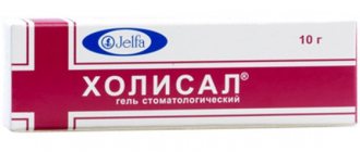
· To accelerate epithelization - keratoplasty preparations - cholisal gel, Vitaon oil, carotolin, rosehip and sea buckthorn oil
Ticket No. 8
The parents of a 5-year-old child noticed a change in the color and shape of the tongue. The child suffered from an acute respiratory viral infection, took antibiotics and multivitamins, and on the 3rd day pain appeared in the mouth when eating. Upon examination, swelling of the lips and tongue, teeth marks on the lateral surfaces of the tongue, bad breath, and a urticarial rash on the body were revealed.
Determine the diagnosis.
Make a treatment plan
Withdrawal of the drug
• Anesthesia - applications of 5% suspension of anesthesin in peach oil, trimecaine 1%
• antiseptic treatment (0.02-0.05% Chlorhexidine solution, Stomatidine, Givalex, Furagin)
· Keratoplasty applications - cholisal gel, Vitaon oil, carotolin, rosehip and sea buckthorn oil
· Antihistamines - Kestin - 1 tablet. 1 time per day, Zyrtec - 1 tablet. 1 time per day, Suprastin 0.025 g, diphenhydramine 0.05
Ticket No. 9
During treatment of pneumonia with antibiotics, a 2-year-old child developed dryness and burning of the oral mucosa, then a cheesy coating developed on the tongue. The mother cleaned off the plaque, but it appeared again. We applied for a consultation. Upon examination, hyperemia of the oral mucosa was revealed; the white coating on the tongue was not completely removed when scraped.
Determine the diagnosis.
Make a treatment plan
1. Acute pseudomembranous candidiasis, moderate form.
2. Laboratory:
Cytological MD - scrapings and washings from the affected areas are taken as research material. Plaques are removed with a Volkmann spoon, a spatula or a loop, and from erosive areas - with a sterile damp swab. The resulting material is used for microscopy of smears (unstained - using a mixture of alcohol and glycuerine and Gram-stained) and cytological preparations. There are 2 forms of fungal growth - budding and hyphae formation. The detection in one or more fields of view of many (more than 10) blastoconidia (daughter cells formed during budding), pseudohyphae (chains of yeast cells forming pseudomycelium) or true hyphae indicates a positive result. For hron f-m – predominance of filamentous fungi.
Iron supplements.
Ticket No. 10
Child 2 years old. Three days ago I felt unwell, T-38.1-39.2 C, my condition worsened, I don’t eat, I speak with difficulty. On the skin of the face, arms, and torso there are blisters from 0.2 to 0.5 cm in diameter. The mucous membrane of the oral cavity is eroded, covered almost all over with fragments of blisters, small erosions with clear contours.
Determine the diagnosis.
Make a treatment plan.
Treatment. In case of combined damage to the skin and oral mucosa, the patient should be treated in a hospital by a dermatologist. Even with isolated damage to the oral mucosa, general treatment is prescribed after consultation with a dermatologist.
Until recent years, pemphigus was considered an almost incurable disease, as it was fatal. Recognition of the autoimmune theory of the occurrence of the disease reduces all therapeutic measures to immunosuppressive effects on autoallergic processes through the use of corticosteroid drugs and cytostatics. Corticosteroids have a number of advantages - they have anti-inflammatory, desensitizing and antiallergic effects, and also have antishock and antitoxic properties. Their appointment is carried out according to vital indications. However, long-term use of these drugs causes the development of serious complications and side effects. If you abruptly stop taking the drug, a “withdrawal” syndrome occurs, and the disease recurs.
As a rule, treatment with corticosteroids (prednisolone, dexamethasone, polcortolone) begins with the administration of loading doses. Then the dose is slowly reduced to maintenance against the background of clinical remission. In case of resistance to corticosteroids, the antimalarial drug delagil (rezoquin, hingamine) is prescribed, 1 tablet. per day (0.25). Once the process has stabilized (cessation of new rashes), the dose is gradually and carefully reduced. Discontinuation of treatment leads to relapse, and therefore maintenance therapy is prescribed, i.e. The dose of the drug is prescribed at which new blisters do not form.
When taking glucocorticoids, it is necessary to take vitamins (tocopherol acetate, retinol palmitate, retinol acetate), calcium supplements (Calcium D3 Nycomed), and potassium. With increasing anemia - drip blood transfusion (150-200 ml), vitamin B12 intravenously, antianamine 2-4 ml intramuscularly every other day. Currently, extracorporeal detoxification methods (hemosorption, plasmapheresis) are successfully used to treat pemphigus. When a secondary infection occurs, the patient is prescribed antibacterial drugs.
Ticket No. 11
The child is 8 years old. Three days ago, the orthodontist took impressions and damaged the right corner of the mouth. A crack formed, covered with a yellow crust, and then pustules appeared on the skin around it, the temperature rose to 37.6 C, eating became painful.
Determine the diagnosis.
Make a treatment plan.
Infectious cheilitis, the most common form of which is the so-called jam - angular cheilitis. When seizures of streptococcal nature occur, erosion of the corner of the mouth is covered with a yellow crust, the skin is often affected, and lymphadenitis of the submandibular lymph nodes is pronounced.
SYMPTOMS
- painful crack;
- redness in the corner of the mouth;
- bubble;
- crusty ulcer
Outpatient at the clinic
TREATMENT It is necessary to eliminate the cause of the disease and reduce the impact of risk factors. Local therapy must be combined with the treatment of somatic diseases. Treatment of somatic pathology leads to a reduction in the symptoms of cheilitis. If the cause of cheilitis is a violation of the architectonics of the lips, caries or malocclusion, treatment by an orthodontist and myogymnastics is necessary. Local treatment depends on the results of bacteriological examination, and, first of all, includes oral hygiene. Antibacterial therapy is carried out with ointments containing antibiotics [1% tetracycline, 1% gentamicin, 1% erythromycin, chloramphenicol liniment (syntomycin)]. Tetracycline ointment of this concentration does not have an antibacterial effect. The use of tetracycline helps to reduce the production of lipase by S. epidermidis and S. aureus, reduce by-products of free fatty acid metabolism and, as a result, regression of clinical manifestations. It is advisable to locally prescribe agents that increase oral immunity: imudon 1 tablet 6 times a day, lycopid 50 mcg once a day. physiotherapy, the task of which is to provide bactericidal or antiparasitic, anti-inflammatory, trophic and resorption effects (UV irradiation, electrophoresis, phonophoresis)
· Physiotherapy. UV irradiation of the area of the corners of the mouth (UF), 2 biodoses, increasing the dose with each irradiation after 1-2 days by 1 biodose, bringing up to 4-5 biodoses; for a course of 4-5 irradiations.
Electrophoresis with nystatin on the affected area for candidiasis or galascorbine electrophoresis for vitamin deficiency, 15 minutes, daily; for a course of treatment 8-10 procedures.
Phonophoresis of nystatin or galascorbine on the affected area. Continuous mode, dose 0.2 W/cm2, procedure duration 5-6 minutes; for a course of treatment 8-10 procedures.
FORECAST The forecast is favorable. When exposed to the pathogen and eliminating the risk factor (normalizing the architectonics of the lips, treating caries and malocclusions), recovery occurs
Ticket No. 12
Teenager, 14 years old. He complains of pain when eating, malaise, body temperature 37.5-37.8 C. He has been sick for five days. Yesterday, white-gray dotted elements appeared on the projection of the lateral teeth, surrounded by a bright red rim, and enanthemas appeared on the palate in the form of small red-pink spots. On the scalp, face, and torso there are rashes in the form of maculopapular exanthema.
Determine the diagnosis.
Make a treatment plan.
Measles
When treating measles, the hygienic maintenance of the skin and mucous membranes is very important. Several times a day you need to rinse your eyes with warm boiled water or a 2% solution of sodium bicarbonate, then instill a solution of sodium sulfacyl or vitamin A 3-4 times a day. Clean the nose with cotton swabs moistened with warm Vaseline oil. Dry, cracked lips are lubricated with boric petroleum jelly or fat. In addition, drug therapy prescribed by a doctor must be carried out.
Patients should remain in bed during the entire period of fever; they must be provided with adequate, age-appropriate nutrition with a sufficient amount of liquid (tea, fruit juices, compotes, liquid jelly, fruit drinks, etc.). Patients who have undergone previous immunoprophylaxis tolerate measles more easily. Emergency immunoprophylaxis (in case of contact with a patient with measles) is carried out for children aged from 3 months to 4 years who have not previously been ill and have not been vaccinated using a single dose of intramuscular immunoglobulin (for older children, for special indications).
The most reliable way to prevent this disease is to vaccinate children aged 12-15 months with live measles vaccine, followed by revaccination at 6 years. Contraindications for vaccination are only acute infectious diseases, blood diseases, organic heart damage during the period of decompensation, acute nephritis, rheumatism in the acute and subacute stages.
Vaccination in most cases protects against measles disease or helps alleviate its course, and also prevents long-term persistence of the measles virus in the body (“slow” infection), which in recent years has been associated with the development of some severe chronic diseases of the central nervous system (multiple sclerosis).
Ticket No. 13
Child 9 years old. Complaints of sore throat. Small erosions were found on the highly hyperemic mucous membrane of the pharynx and soft palate. Regional lymph nodes are enlarged.
Determine the diagnosis.
Make a treatment plan
Herpangina
Treatment of herpetic sore throat is usually symptomatic. First of all, hyposensitizing drugs (antiallergic) are prescribed:
· Suprastin;
· Diazolin;
· Loratadine;
· Claritin;
· Telfast.
REMOVING TEMPERATURE
Antipyretics should be used to relieve fever. The most relevant for children are: paracetamol (Panadol) and ibuprofen (Ibufen D). Adults can take Aspirin, Ibuklin, Nimesulide.
Physiotherapy: needle, ultraviolet irradiation
Local:
• Local anesthesia - applications of 5% suspension of anesthesin in peach oil, trimecaine 1%
• antiseptic treatment (0.02-0.05% Chlorhexidine solution, Stomatidine, Givalex, Furagin)
• Applications of antiviral drugs (0.1% solution of glycyrrhizic acid in an aerosol, irrigation 6 times a day for 5 days), interferon ointment must be used repeatedly (4-5 times a day).
STRENGTHENING IMMUNITY
As with other viral diseases, special attention must be paid to strengthening the protective properties of the immune system. This is achieved by prescribing immunomodulators, such as Immunal, echinacea tincture, Imudon.
Ticket No. 14
Parents with a 3-year-old child came to the clinic. Body temperature up to 39 C; rashes on the lips, facial skin, and oral mucosa appeared in the evening of the second day of the disease. The child refuses to eat. Upon examination, multiple erosions are determined on the oral mucosa, on the tongue, along the line of closure of the teeth, the mucous part and the red border of the lips, extending to the skin of the face. Catarrhal gingivitis and lymphadenitis of the submandibular lymph nodes are noted.
Determine the diagnosis.
Make a treatment plan.
Acute herpetic stomatitis (moderate-severe form) catarrhal period
This disease, according to its etiology, is viral and is caused by the herpes simplex virus type 1. In the scraping, giant multinucleated cells can be found; they are distinguished by sharp polymorphism in shape, color and size. The center of the cell is occupied by a dense conglomerate of nuclei.
Antiviral therapy: 1. Treatment of a viral infection is carried out from the first day of the disease, that is, already in the prodromal period. 2. During the period of extinction of the disease, antiviral therapy is canceled. 3. Drugs: Zavirax, acyclovir, Poludanum, interferon, interferon ointment, must be used repeatedly (4-5 times a day).4. antiviral drugs are applied not only to the affected, but also to apparently healthy areas of the mucous membrane.
Ticket No. 15
The child is 3 years old. Referred for consultation regarding bleeding gums and tooth mobility. An external examination reveals sweating and hyperkeratosis of the palms and soles. Mobility of teeth and the presence of periodontal pockets more than 5 mm deep were revealed in the oral cavity.
Determine the diagnosis.
Make a treatment plan.
Papillon-Lefevre syndrome.
Differential diagnosis is carried out with rapidly progressing periodontitis and severe periodontitis. This syndrome has a characteristic radiological expression: resorption of periodontal bone tissue, significant loss of bone tissue of the interdental septa until they are completely reduced.
In a number of patients, clear manifestation of characteristic specific signs of the disease occurs relatively late. Periodontolysis in temporary and permanent dentition can be expressed to varying degrees. Anomaly of tissues - derivatives of the ectoderm: palmar-plantar dyskeratosis, damage to the knee and elbow joints, inferior hair, deformation of the nails, not all patients have clear clinical manifestations and can occur in an “erased” form.
Relatively late manifestation, vagueness of some clinical signs, and an incomplete set of symptoms complicate differential diagnosis, clarification of the diagnosis, and treatment. It is not possible to stop the destructive-inflammatory process in periodontal tissues. Proper organization of clinical trials, a comprehensive qualified approach to diagnosis, prescription of symptomatic treatment are required, and consultations with specialists are required: geneticists and dermatologists.
Comprehensive treatment plan:
Ticket No. 16
A 13-year-old teenager came to the clinic with complaints of gum growth, pain and bleeding when brushing teeth, and eating hard foods.
Determine the diagnosis.
Make a treatment plan.
Local status: the child’s general condition does not suffer, no facial asymmetry is detected, growth of gingival papillae is observed in the oral cavity, they are loose, false gingival pockets, but the dentogingival junction is not broken, bleeding occurs with light probing. In the cervical area of the teeth, in areas of gum hypertrophy, a large amount of soft or pigmented plaque is found, firmly associated with the hard tissues of the tooth.
Ticket No. 17
The girl is 8 years old. Complaints of bleeding gums when brushing teeth and when biting hard food. The interdental papillae in the area of the anterior group of teeth of the lower jaw are hyperemic with a bluish tint, slightly swollen, and bleed when touched. Such symptoms were previously noted, but disappeared after appropriate treatment.
Determine the diagnosis.
Make a treatment plan.
Ticket No. 18
Girl 4 years old. She complained of pain when eating. Objectively: body temperature 38°C, swelling and cyanosis of the soft palate and tonsils. The pharynx and the edges of the gums are covered with a gray-white coating, which, when removed, causes bleeding in the affected area. Lymphadenitis of the submandibular nodes.
Determine the diagnosis.
Make a treatment plan.
Diphtheria
Treatment is carried out in a hospital. The main treatment measure is the administration of anti-diphtheria antitoxic serum. The serum is administered according to the modified Bezredki method: first, 0.1 ml is injected subcutaneously, after 30 minutes - 0.2 ml and after 1-1.5 hours - the remaining dose is injected intramuscularly (into the upper outer quadrant of the buttock or into the anterior thigh muscles). In severe cases, the serum is administered intravenously. Serum is dosed in international units (IU). The amount of the administered drug depends on the severity of the disease and the period that has elapsed since its onset: from 10,000-20,000 IU for a localized form to 80,000-100,000 IU. As a rule, the serum is administered once. In case of a toxic form, detoxification therapy is indicated: intravenous administration of plasma, hemodez in combination with a 10% solution of glucose, vitamins, cocarboxylase, and corticosteroids. The use of plasmapheresis is effective. For diphtheria of the respiratory tract that threatens suffocation, tracheal intubation or tracheostomy is performed. Antibiotics are prescribed to children with croup complicated by pneumonia, otitis media, etc., caused by other bacterial flora.
Ticket No. 19
The child is 15 years old. He complained of changes in the position and mobility of teeth, pain in the gums when eating and brushing teeth. On external examination, the skin is pale, the child is lethargic, apathetic, and has tinnitus, headaches, palpitations, sleep disturbances, and decreased appetite.
Determine the diagnosis.
Make a treatment plan
Ticket No. 20
Child 10 years old. The examination revealed bright hyperemia with a cyanotic tint of the desquamated gingival margin in the area of all teeth, periodontal pockets with purulent-bloody contents, 1.6, 2.6, 3.1, 3.2, 3.6, 4.1, 4.2, 4.6 have mobility of 1 and 2 degrees. From the anamnesis it was found that the mother suffers from diabetes.
Determine the diagnosis.
Make a treatment plan.
1) Questioning, examination, palpation, probing to determine the depth of periodontal pockets, RMA, Schiller-Pisarev, orthopantomogram.
Ticket No. 21
In a 1-month-old artificially fed child, the mother noticed two “ulcers” on the palate. From the anamnesis it was revealed that the child was born premature and does not gain weight well. When eating, he cries and does not suck the pacifier well. Objectively: two rounded symmetrical erosions measuring about 0.9 cm in diameter were found at the border of the hard and soft palate.
Determine the diagnosis.
Make a treatment plan.
Afta Bednara
· Identification and elimination of traumatic factors
· relieving pain in the oral cavity - applications of 5% suspension of anesthesin in peach oil, trimecaine 1%
· applications with keratoplasty preparations - salcoseryl, sea buckthorn oil, rosehip oil, kyzyl may, fat-soluble vitamins A, E
Ticket No. 22
A 1.5 month old child erupted 7.1 teeth a week ago. In recent days, the child has become restless when eating and is crying. From the anamnesis it was found that the child was born at term and, according to the pediatrician, is healthy. Objectively: erosion was found on the lower surface of the tongue, the surrounding mucous membrane is hyperemic, slightly swollen, and painful on palpation.
Determine the diagnosis.
Make a treatment plan.
Ticket No. 23
A 6-year-old child received aspirin and ampicillin for 5 days due to acute respiratory infections, his health improved, but today he began to have pain in the mouth while eating and his body temperature increased. An objective examination revealed a pinpoint rash on the neck and chest. The lips are swollen, the tongue has teeth marks on the lateral surface, and there is diffuse erythema with clear contours on the soft and hard palate.
Determine the diagnosis.
Withdrawal of the drug
• Anesthesia - applications of 5% suspension of anesthesin in peach oil, trimecaine 1%
• antiseptic treatment (0.02-0.05% Chlorhexidine solution, Stomatidine, Givalex, Furagin)
· Keratoplasty applications - cholisal gel, Vitaon oil, carotolin, rosehip and sea buckthorn oil
· Antihistamines - Kestin - 1 tablet. 1 time per day, Zyrtec - 1 tablet. 1 time per day, Suprastin 0.025 g, diphenhydramine 0.05
Ticket No. 24
A 5-year-old child is practically healthy; his mother noticed bright pink spots on his tongue and an unusual appearance of his tongue, and went to the dentist. From the anamnesis it was found that pain occasionally occurs when eating spicy food, and the child previously had gastritis.
Determine the diagnosis.
Make a treatment plan.
· Desquamative glossitis
· Consultation with a pediatrician, gastroenterologist
· Anesthesia - 5% suspension of anesthesin in peach oil, trimecaine 1%
· Keratoplasty - cholisal gel, Vitaon oil, carotolin, rosehip and sea buckthorn oil
· Purpose of vitamin B5
Ticket No. 25
A mother with a 2-year-old child came to the clinic on the third day from the onset of the child’s illness. The rash appeared the day before, temperature up to 38.30 C, refusal to eat, poor sleep, restless behavior. Objectively: on the red border of the upper lip there are several bubbles with transparent contents. Catarrhal gingivitis, separate and merging erosions on the tongue, mucous membrane of the lips and cheeks.
Determine the diagnosis.
Make a treatment plan.
Ticket No. 26
The child is 10 days old, discharged from the maternity hospital the day before. According to the pediatrician, he is healthy. The delay in discharge was due to the mother's health condition. The mother noticed a white coating on the child’s lips and tongue and went to the dentist.
Determine the diagnosis.
Make a treatment plan.
Answer: Acute pseudomembranous candidiasis
The disease most often affects newborn children, to whom a fungal infection was transmitted during childbirth from a sick mother.
At the same time, newborns refuse to breastfeed and eat food, become lethargic and capricious. Infection of infants occurs from a mother with vulvovaginal candidiasis during childbirth. Therefore, a pregnant woman, in order to prevent acute pseudomembranous candidiasis in newborns, should treat fungal diseases of the genital organs. And after birth, maintain newborn hygiene, provide proper care for the nipples of the mammary glands, and maintain the sterility of baby care items.
Ticket No. 27
A 7-year-old child received ampicillin for 10 days due to pneumonia. The body temperature is normal, but the child is lethargic and pale. The mother noticed plaque in her mouth and went to the dentist. Upon examination, a yellow coating was found on the mucous membrane of the cheeks, palate, and back of the tongue, firmly welded together, rising above the level of the surface of the mucosa. The mucous membrane is hyperemic and painful when touched and forcibly removing plaque. Dry mouth. There is a thickening of the regional lymph nodes. From the anamnesis it was revealed that before the age of one year there was thrush.
Determine the diagnosis.
Compose
1Next ⇒
Mobile electrified feed dispenser: diagram and process of operation of the device...
General conditions for choosing a drainage system: The drainage system is selected depending on the nature of the protected…
Mechanical retention of earth masses: Mechanical retention of earth masses on a slope is provided by buttress structures of various designs...
Papillary patterns of the fingers are a marker of athletic ability: dermatoglyphic signs are formed at 3-5 months of pregnancy and do not change throughout life...
Severity of hypertrophic gingivitis
Chronic hypertrophic gingivitis has 3 degrees of development: mild, moderate and severe.
| Mild degree | Average degree | Severe degree |
| It is considered the initial stage of the disease. The papillary region of the gums is affected. The edge of the affected periodontal tissue covers the crown at 1/3 of its height. | The papillary marginal zone becomes dome-shaped. Observed: bleeding gums, pain when brushing teeth, poor oral hygiene. | Clear changes are observed throughout the periodontal area. In addition to discomfort, patients complain of a cosmetic defect in the form of gum enlargement up to 2/3 of the crown. |
Lightweight
The gum margin and gingival papillae thicken and become ridge-shaped. Periodontal pockets bleed and fill with purulent contents. Mineralized stones are found inside the pockets, and there is an unpleasant odor from the mouth.
As with the edematous form, patients complain of bleeding and sore gums.
Average
The gum relief is deformed, the gingival papillae increase in size, which leads to the overlap of the crown part of the tooth to half its height. The depth of the periodontal pocket reaches up to 5 mm. Carrying out hygiene measures becomes almost impossible due to sore gums.
Heavy
In the absence of treatment and non-compliance with hygiene rules, the disease progresses to a severe stage of development. The gum covers the tooth up to its cutting edge. Hypertrophy of periodontal tissue makes chewing and speaking more difficult. Multiple erosions and ulcers are observed on the gingival margin. In some cases, the erosive process is accompanied by suppuration.
Gingivitis and periodontitis: cause and effect relationships
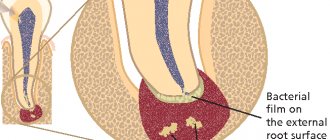
Hypertrophic gingivitis can manifest itself as an independent disease or occur during an exacerbation of periodontitis. Despite similar symptoms (pain, bleeding gums), the medical history has a completely different scenario: the mucous membrane becomes inflamed, a hypertrophy mechanism is triggered in its cells, and the volume of periodontal tissue increases. But! The integrity of the dental epithelial junction is not compromised and no pathological changes occur in the bone tissue. Inflammation does not penetrate deeper than the upper layer of the gum epithelium.
While periodontitis is characterized by a much more serious clinical picture: the inflammatory process first covers the mucous membrane, and then goes deeper - it affects the lower layers of periodontal tissue and reaches the bone.
The danger of hypertrophic gingivitis (like any other) is that it can develop into periodontitis, which is more difficult to treat and leads to serious consequences, including tooth loss. Why is this happening? The fact is that when periodontal tissue swells and false periodontal pockets form, the problem grows like a snowball. The gums become very sensitive and react with acute pain and burning to the slightest irritation. In this case, complete teeth cleaning is simply not possible and the number of bacteria in the oral cavity increases rapidly, worsening the disease every day. This is why it is so important to seek dental care in time to clear plaque from your gums and teeth. This is the only way to minimize the bacterial factor and reduce the likelihood of periodontitis.
Diagnostics
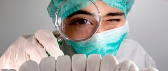
The diagnosis is made by the dentist based on diagnostic data. After collecting complaints, medical history and visual examination of the red border of the lips, the doctor conducts the following studies:
- Examination of the oral cavity with a thorough examination of periodontal tissue.
- Conducting oral hygiene indices (Fedorov-Volodkina, PMA, dental plaque index).
- Orthopantomography (to determine damage to the periodontium, root apexes, periosteum).
- Rheoparodontography (determination of blood supply to soft tissues).
- Biopsy from affected gum tissue.
Treatment of hypertrophic gingivitis
Before starting treatment for hypertrophic gingivitis, it is necessary to identify the cause of the disease. After determining the cause, the doctor chooses one of the types of treatment:
- medicinal;
- surgical.
Before any treatment method, professional cleaning of the oral cavity is carried out, including:
- removal of supragingival and subgingival calculus;
- removal of pigmented plaque;
- polishing teeth using abrasive pastes;
- treatment of wounds and erosions with antiseptic agents.
Since the key to a quick recovery is careful hygienic care of the oral cavity, the doctor teaches the patient proper dental technique, the use of additional hygiene items, and also gives recommendations for home care for inflamed gums.
Medication
For hypertrophic gingivitis, the patient is prescribed the following therapy:
- Injections into the inflamed papillae and marginal part of the gums with a 10% calcium chloride solution (done by a dentist).
- Sclerosing therapy in the form of injections and applications.
- Rinsing the mouth with anti-inflammatory herbal infusions (chamomile, sage, calendula).
- Oral baths with antiseptic drugs (miramistin, chlorhexidine).
- Carrying out applications with regenerating and antiseptic gels (methylprednisolone).
- Use of medicated toothpastes (Asepta).
After completing the prescribed course of treatment, you must visit the dentist for dynamic monitoring.
Surgical
Surgical treatment is indicated when it is impossible to get rid of the disease with drug therapy alone. As a rule, gingivectomy (removal of overgrown periodontal tissue) is performed for the fibrous form of hypertrophic gingivitis.
Under general anesthesia, the dental surgeon makes incisions on the oral and buccal sides. After excision of unnecessary tissue, the doctor applies a medicinal bandage to the wound site.
After surgery, drug therapy is prescribed. Brushing your teeth should be gentle; it is recommended to use a soft toothbrush for the first 2 days.
Treatment
Therapy for hypertrophic gingivitis always begins with a thorough diagnosis and establishment of the type and causes of the disorder. It is always carried out comprehensively, together with the elimination of the symptoms of the disease and supportive treatment.
Diagnostics includes:
- X-ray of the jaw;
- determination of the hygiene index;
- biopsy;
- detection of periodontal damage;
- orthopantomography;
Conservative
Before drug treatment, as a rule, a professional comprehensive cleaning of the oral cavity is performed. Next, the cause of the negative changes is eliminated (if possible). After this, the patient is prescribed medications to treat gingivitis. They include antibacterial, anti-inflammatory and immunostimulating medications. In addition, medications are also used to promote tissue restoration and renewal.
For severe swelling, the following are most often used:
- hormonal ointments for gums (Prednisolone, Hydrocortisone);
- vitamin therapy (including vitamins C and P);
- rinsing with herbal decoctions (chamomile, oak bark, sage);
- hot paraffin applications;
- calcium gluconate injections;
- Biocide compresses;
- heparin injections.
Physiotherapy using laser, ultrasound, and electrophoresis can also be used. The duration of treatment depends on the size and extent of the lesion.
Elimination of the fibrous form of gingivitis is carried out surgically. In this case, excision of enlarged gums or coagulation is performed.
Injections and the use of hormonal ointments are contraindicated for pregnant women. Vitamins also need to be taken with caution. Most often, rinsing with decoctions and Chlorhexidine solution, as well as applications and lotions are used for the treatment of pregnant women. Treatment using acetylsalicylic ointment is possible. Therapy is carried out under the close supervision of the attending physician.
Folk remedies
Medicinal herbs have an anti-inflammatory effect and can relieve swelling with regular rinsing and making lotions. The following traditional medicine recipes based on them are recommended:
- Pour two tablespoons of sage into a glass of water and boil. Rinse your mouth with warm mixture twice a day.
- Mix one tablespoon each of calendula, yarrow and chamomile flowers, pour into a thermos and pour half a liter of boiling water over the mixture. Leave for an hour. Rinse your mouth with the mixture for one minute three times a day.
- Moisten a cotton swab or piece of gauze with aloe juice and apply to the affected areas. It is also possible to periodically chew the leaves of the plant to relieve swelling and achieve an antibacterial effect.
Propolis tincture has a good effect on oral tissues. To prepare the medicine, you need to take a tablespoon of the product and dilute it with a glass of clean water. Rinse up to five times a day.
To speed up the healing process, it is recommended to follow a diet high in fresh fruits and vegetables.
Prognosis and prevention
Gingivitis in pregnant women, just like gingivitis in children, goes away on its own when hormonal levels normalize. There is no point in any treatment in this case. It is enough to just have your teeth cleaned by a dentist once every 6 months, and also carefully monitor your oral hygiene.
To protect yourself from the occurrence of hypertrophic gingivitis, you need to strengthen your body with the necessary vitamins (in particular group C) and minerals. An important step is timely treatment of concomitant diseases and giving up bad habits.
Prevention
First of all, prevention and timely treatment of pathologies that provoke the disease will help prevent the development of hypertrophic gingivitis. In addition, the following rules will help reduce the risk of violations:
- periodic professional teeth cleanings;
- regular oral hygiene;
- exclusion of traumatic situations and factors traumatic to the oral cavity;
- timely treatment of teeth and gums;
- periodic checking of the body's hormonal system;
- Carrying out preventive examinations at the dentist at least twice a year.
It is also recommended to use mouth rinses (preferably with an antibacterial effect), high-quality soft brushes and dental floss. During pregnancy, it is useful to include foods rich in vitamin C in your diet, as well as exclude harmful sugar-based sweets from the menu.
Consequences and complications of hypertrophic gingivitis
If you delay a visit to the dentist for a long time, or even begin independent treatment without the supervision of a doctor, you can cause the development of the following complications:
- atrophy of interdental spaces;
- change in bite;
- tooth mobility with subsequent loss;
- addition of an additional infection (periodontitis, candidiasis, stomatitis);
- speech dysfunction.
To protect yourself from the occurrence of hypertrophic gingivitis and prevent the development of complications, you must follow preventive recommendations.
To identify dental disease at an early stage of development, you should visit a dentist once a year. It must be remembered that timely treatment is the key to a successful and quick recovery, therefore self-medication and postponing a visit to the doctor for a long time are unacceptable.
Bibliography
- Maksimovsky Yu.M., Mitronin A.V. - Therapeutic dentistry - M.: Geotar-Media, 2012.
- Dmitrieva L.A. — Modern aspects of clinical periodontology, M., 2001.
- Barer G.M. – Periodontal diseases. Textbook, M.: GEOTAR-Media, 2008.
- Kuzmina E.M. et al. — Prevention of dental diseases in pregnant women and young children, M., MMSI. 1999.
- Afanasyev V.V. — Surgical dentistry: textbook, M.: Geotar—Media, 2011
Read related posts:
Diagnosis and treatment
First of all, an X-ray examination is performed to make a diagnosis. The image shows structural changes, which determine the true cause of swelling and other clinical symptoms.
For example, with periodontitis or periodontal disease, destructive changes occur in the deep layers of bone tissue, and with gingivitis, inflammation of the upper soft layers occurs. Sometimes gingivitis is characterized by osteoporosis.
If necessary, other diagnostic measures are prescribed:
- identification of pathological deposits on teeth - plaque, stones;
- assessment of the area of inflammatory changes;
- assessment of the severity and extent of pathological changes;
- Schiller-Pisarev test (helps determine the level of glycogen in periodontal tissues, which increases during the inflammatory process).
Differential diagnosis of the disease is carried out with epulis and fibromatosis.
Treatment of hypertrophic gingivitis is as follows:
- cleaning teeth and gums from pathological plaque and stones;
- conducting physiotherapy to resolve swelling and tissue regeneration;
- the use of antiseptic drugs to disinfect tissues, including natural remedies with this effect (herbal decoctions, infusions, etc.);
- performing gum massage;
- use of corticosteroids in the form of ointments if anti-inflammatory drugs are not effective;
- injections of hormones into the papillae in advanced cases.
If the disease develops in a fibrous form, as a rule, conservative treatment does not bring the desired recovery. In this case, other methods of therapy are used: gingivectomy, diathermocoagulation, cryodestruction.
In addition, all traumatic factors are excluded (prostheses are adjusted, the correct anatomical shape of damaged or normal teeth is restored, old fillings are replaced, etc.).