Tooth root resorption is one of the most mysterious dental phenomena. It occurs in people regardless of gender, age and condition of the oral cavity. This is a dental complication that can cause the patient to lose teeth. It occurs quite often in dental surgery.
Types of pathology
The following types of tooth root resorption are distinguished: physiological at the roots of temporary teeth and pathological resorption.
Depending on where resorption occurs, it is divided into internal and external.
Due to the appearance of the external one, it is classified conditionally into replacement, superficial and inflammatory.
Cervical resorption is also added to the types of external resorption.
Internal resorption may be complicated by perforation.
Concomitant factors in the appearance of pathology are trauma (chronic or acute) - constant mechanical impact, inflammation of the periodontal and pulp complex, tumors, cysts, unknown etiology.
The beginning of physiological root resorption of primary teeth is noted during the replacement of temporary teeth with permanent ones. It is based on the selective activity of osteoclasts using the nuclear factor kappa B activator receptor. However, studies provide vague explanations for the process of pulp resorption.
It has been established that pathological resorption of the roots of primary teeth occurs unevenly, uniformly, and also in the furcation area.
With uniform resorption, all dental roots are absorbed at the same time, the furcation zone is slightly affected.
Uneven flow occurs on the root closest to the follicle of the permanent tooth. Often the root of the tooth hurts.
The third type of resorption originates in the furcation zone, and then moves to the roots of temporary teeth. After bone resorption, the role of osteoclasts passes to odontoblast and pulp cells.
Internal root resorption is an asymptomatic phenomenon that is almost always determined by chance.
A common cause is trauma and inflammation of the pulp. Damage to dentinal tubules and predentin by inflammatory mediators (interleukins 1B) stimulates the RANKL system, which selectively activates osteoclasts in pulpal and/or periodontal tissues.
Since normal pulp is transformed into granulomatous tissue due to internal resorption, it is visible through the dental tissue. A “pink spot” appears. Subsequent pulp necrosis changes the color from pink to gray.
If resorption progresses, the patient complains of pain, periodontal damage leads to increasing dental mobility.
Classification of tooth root resorption
Depending on the reasons that led to the development of the disease, the following types of this pathological condition are distinguished:
| Type of resorption | Causes | Features of the course |
| Physiological | Occurs in children during the change of primary teeth to permanent ones due to the selective activity of osteoclasts. | More often it develops in healthy teeth, as well as teeth affected by caries, which has or has not been treated. X-rays can reveal the preservation of bone tissue. |
| Pathological | It develops as a complication of trauma, reimplantation, pressure from other teeth, cysts and other neoplasms, pulpal or periodontal infection. | The disease occurs against the background of chronic inflammatory processes in periodontal tissues resulting from tumors and cysts. X-rays reveal destruction and absence of bone tissue. |
Depending on the localization of pathological processes, external and internal tooth root resorption are distinguished.
| Type of resorption | Internal root resorption | External root resorption |
| Localization | Pathology develops on the inner surface of the tooth root in the area where it borders the pulp cavity. | The disease develops on the outer surface, in the area where ligaments connect the tooth root to the jaw. |
| Causes | Inflammatory processes of the dental nerve or trauma to dental tissues. | Penetration of pathogenic bacteria into the root canal of the tooth, tooth dislocation, death of the nerve, completed process of root formation. |
| Clinical manifestations | the presence of a “pink spot”, which appears due to the transformation of pulp tissue into granulomatous. It is visible because it shines through the tooth tissue, and after dying it changes its color to gray. As the disease progresses, the patient may experience pain, and the teeth become mobile. | It has virtually no symptoms and occurs against the background of penetration of blood vessels from surrounding tissues into the tooth root and connective tissue in different areas below the gum line. Due to pathological processes, the tooth may change color, but as a rule, the disease is detected by X-ray examination. |
| Features of the pathological process | Resorption occurs without clinical manifestations, so it is difficult to identify. As a rule, it is detected during a preventive dental examination. | It is considered the most aggressive and rapidly developing type of disease, characterized by extensive destructive processes of root tissue. |
Depending on the characteristics of the disease, three subtypes of external resorption are distinguished:
- Superficial is a physiological process, the development of which occurs as a reaction to orthodontic treatment or injury. The development of pathological processes rarely extends beyond the cement, so the defects are small in size and difficult to notice. The disease does not lead to impairment of the functionality of the affected tooth. After the trigger for its development is eliminated, the formation of new structures begins, which eliminates the need to treat root resorption;
- Inflammatory - characterized by very rapid development, since it occurs due to the penetration of pathogenic bacteria into the tooth canals or the death of the tooth nerve. In the absence of adequate treatment, the pathological condition causes extensive destruction of dental root tissue, which ultimately leads to disruption of its function and loss of the enamel-cementum junction.
- Replacement - develops as a result of injuries (especially when a tooth is dislocated) or there may be a complication of inflammatory resorption, even if it was treated. The disease is chronic, requires professional dental treatment and in the vast majority of cases ends in tooth loss. It is characterized by damage to the periodontal ligament and the formation of bone in its place, which leads to tooth immobility.
Superficial root resorption
This type of tooth root resorption is a physiological process, since it responds to damage during orthodontic treatment or during trauma - necrosis and ischemia of cementoblasts occur. It acts in the area of cement and rarely goes beyond it. Usually the defects are small and rarely identified, especially on the oral and vestibular sides. No functional impairment occurs.
After the trigger mechanism is eliminated, new structures are immediately built, so treatment is not necessary.
Diagnostics
In most cases, pathological tooth root resorption is asymptomatic and is detected during routine radiographic examination. At the present stage, the number of detected resorptions has increased due to the introduction of cone beam computed tomography (CBCT).
In his study, K. Aziz [9] proved that a two-dimensional image often does not reflect the true nature or extent of the resorptive process.
K. Vasconcelos [67] in his report indicates that CBCT helps to determine the presence and topography of the resorptive area of the root, which determines treatment tactics.
Ankylosis, or replacement resorption
This type of external resorption of tooth roots, however, is not inferior to inflammatory destruction in terms of the severity of tissue destruction. Appears during injuries, especially with complete or impacted dental dislocation. Usually represents the outcome of inflammatory resorption, even with its therapy. Replacement resorption is a chronic process that appears in the area of damage to the periodontal ligament on the outer root surface, never stops on its own and in almost all cases ends in tooth loss.
A strong pathological influence damages the periodontal ligament, resulting in the inevitable formation of bone at this site. The tooth becomes immobile.
When replacement resorption stops, they speak of the transient type. If it comes to tooth loss, then resorption is called progressive.
Types of pathology and basic classification
Insufficient knowledge of pathology allows us to use only the following factors as criteria for classification:
- The presence of physiological destruction in the root structure of temporary elements;
- Manifestation of multifactorial pathology arising due to negative stimulation.
From the point of view of external signs, inflammatory, superficial and replacement resorption are distinguished. In addition, external forms include cervical destruction, characteristic of pulpless units. In turn, internal resorption is characterized by perforation that occurs in the area of connection between the pulp chamber and the root walls.
Cervical resorption
Whatever this type of resorption is called - invasive, cervical external or cervical peripheral, it is an idiopathic type of external. Its development is caused by trauma, scaling, bruxism and orthodontics. In the course of some studies, scientists presented cases of the impact of intracanal bleaching on the appearance of external resorption. It turned out that hydrogen peroxide (30%) is able to penetrate the cement surface through the tubules and destroy both it and the periodontium.
It does not always appear in the cervical area, which is determined by the depth of the pathological pocket. Resorption is supported by infection in the gingival sulcus and gradually goes around the pulp chamber. It is not damaged. Cervical resorption occurs without symptoms until pulpal and periodontal infections occur. If the defect is deep, sensitivity to temperature appears, the walls of the cavity become hard, bleed slightly during probing and creak. Dental treatment in dentistry will be discussed below. First, let's find out how pathology is diagnosed.
Diagnosis of invasive cervical resorption
For many practicing doctors, dental diseases of a resorptive nature remain pathologies shrouded in mystery, not fully understood and mysterious. Historically, when studying these lesions, they were most often presented as such, which arose as a result of the action of certain factors of the external or internal environment.
Classically, it was assumed that internal resorption of vital teeth develops due to traumatic damage to the pulp (Figure 1), and external resorption occurs strictly in non-vital teeth (Figure 2). The latter can be caused by injury to the periodontal ligament due to external factors, pressure that occurs as a result of teething, or developmental pathology (photo 3). Sometimes external resorption is observed after orthodontic treatment, which provokes an increase in the activity of osteoclasts in the area of cement and dentin at the root apex. Most doctors, of course, rely on the knowledge gained during preliminary training, but some are still interested in the dynamics of development of evidence-based facts regarding certain dental pathologies.
Photo 1: Radiograph showing an example of internal root resorption.
Figure 2: Radiograph showing an example of extrinsic inflammatory root resorption.
Figure 3: Radiograph showing an example of pressure-induced resorption.
Over the past two decades, a significant number of publications have been presented in the dental literature describing unique cases of external resorption of completely vital teeth in the cervical region. Consequently, this type of pathology had to be added to the two already known (photo 4). Sometimes the terms used to describe this type of lesion are only subjective, but for a clear understanding, this article will use the name invasive cervical root resorption (IPR), since this is the name given to this pathology by Dr. Geoffrey S. Heithersay, who initially described it. This article will review both the clinical and radiological features of resorption, as well as aspects related to its etiology, pathophysiology and treatment.
Figure 4: Radiograph showing an example of invasive cervical resorption.
Relevance of the topic
Given the unique nature of the IPPC, it is important to ensure a thorough understanding of the basic principles of development of this process. Regardless of its manifestation, resorption is the loss of dental hard tissue caused by the action of immune-mediated cells with clastic activity. Under normal conditions, the activity of these cells is prevented by the presence of non-mineralized tissue layers present in both cementum and dentin. When the integrity of these layers is violated through injury, infection or other factors, conditions are created under which clastic cells can begin to show their activity. The cells responsible for IPCC originate from the attached or junctional epithelium at the base of the gingival sulcus. Considering this origin, it becomes clear why pathology most often affects the area of the neck of the tooth. Exceptions, however, are noted in areas of loss of periodontal attachment or in areas of teeth that are in the process of eruption - in such cases the level of the gingival sulcus decreases in the apical direction (photos 5 - 6).
Photo 5. In this case, the focus of IPCC developed in the apical area of the tooth (in most cases it develops in the area of the neck of the tooth), which provoked the migration of the periodontal junction. The pathology developed a year after the tooth was treated due to dislocation.
Photo 6. In this case, the focus of IPCC developed on the apical part of the tooth (in most cases it develops in the area of the neck of the tooth), which provoked the migration of the periodontal junction. The pathology developed a year after the tooth was treated due to dislocation.
The cells adhere to the tooth structure in areas of cementum loss, thereby allowing invasion of fibrovascular tissue, and thus deterioration of mineralized tooth root structures develops. If epithelial invasion or bacterial contamination of the defect is not prevented, then there is no hope for restoration of bone tissue from adjacent structures. Although similar histologically identified replacement resorption can also develop during the restoration of areas of IPRC after loss of periodontal attachment. Predentin, which surrounds the pulp, like the precement, serves as a protective layer that limits the penetration of resorptive tissue into the pulp space. Of course, such protection is not eternal, and sooner or later, without treatment, the pulp is still affected by the pathological process. Therefore, the spread of IPCC most often occurs around the pulp without its direct involvement, in the coronal or apical direction of the relative primary focus.
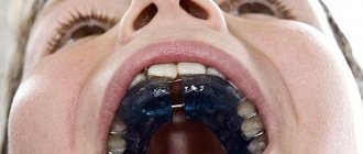
Etiology
A number of etiological factors are associated with the development of IPCC, the direct cause of which has yet to be proven. The most expected of these are pre-orthodontic treatment, traumatic lesions, orthognathic surgery, intracoronal restorations and whitening of non-vital teeth. Most of these factors provoke damage to the cement in the neck of the tooth, thus causing physical or chemical injury. Other habits of the patient, such as playing wind instruments with a wooden mouthpiece, can also create conditions that are not much different in nature from the condition after orthodontic intervention. Such habits can cause damage and pathological abrasion in the area of the front teeth. Taking systemic bisphosphonates is also associated with the development of IPCC, since these drugs have been shown to influence changes in bone metabolism, and therefore the activity of bone cells. In addition, a viral etiology for the development of ICC has also been proposed. Odontoclastic resorption is common in cats after infection with Feline Herpesvirus 1 (FeHV-1), and the signs of this lesion are very similar to IPCC, which is diagnosed in humans. A small study of patients with multiple ICC lesions found that 100% of subjects tested positive for FeHV-1, supporting the validity of the above theory. The mechanism of development of IPCC may also be genetic in nature, since it is often observed in members of the same family. In many cases, the connection between IPCC and any factors could not be established; therefore, the disease in such situations can be diagnosed as idiopathic.
Diagnostics
In most cases, IPCC is an asymptomatic pathology and is difficult to diagnose. Lesions are often identified during routine radiographic examinations during the treatment of other dental conditions. Occasionally, the physician may be able to locate the area of ICC during soft tissue retraction to perform restoration. Probing can often reveal the presence of a cavity, or a chalky surface of the lesion below the gingival margin. More advanced lesions may appear as a fairly wide and clearly visible cavity of dark pigmentation. Such defects are often filled with soft surrounding tissue that bleeds easily when probing. Since the predentin layer protects the pulp for a certain time, it remains undisturbed for a long time, and the development of pulpitis or necrosis is extremely rare, even in the most advanced stages of IPCC. Symptoms of inflammation of the periodontal ligament, such as a reaction to percussion or sensitivity to palpation, as well as the formation of periodontal pockets, are usually absent.
Sometimes the lesion manifests itself in a clinically obvious way. Photos 7-10 show a case where the patient himself noted pink discoloration in the cervical region of the right lateral incisor. Although pulp sensitivity was normal, a perforating lesion was found along the distobuccal aspect of the tooth. Although it was previously believed that the presence of pink discolorations indicates internal tooth resorption, in fact, in most such cases, IPCC is clinically diagnosed.
Photo 7. A case of IPCC in the area of the 10th tooth with the formation of pink discoloration.
Photo 8. Confirmation of the diagnosis of IPCC on an x-ray.
Photo 9. Confirmation of the diagnosis on a CT scan.
Photo 10. Extension of IPCT in the parapulpal apical direction (CBCT slice).
Due to the principles of radiological imaging, the two-dimensional nature of radiographs and the significant overlap of multiple anatomical structures, periapical or bitewing images are often not sensitive enough to diagnose iCC. Also, the information content of these methods for IPCC is often associated with the amount of loss of hard dental tissue. Cone beam computed tomography (CBCT) is particularly useful in the early stages of identifying a lesion, although given that this method is rarely used for screening purposes, ICC is again often diagnosed as an additional finding in the diagnosis of some other disease or in the planning of complex treatment algorithms. Periodontal bone loss is extremely rare in the area of external cervical resorption.
When radiologically identified, IPCC most often resembles a class V carious defect. When lesions are localized on the buccal or lingual sides, due to the graphical overlay on the area of the dental pulp, they can cause some blurring of its contour. As the pathology develops, it progresses towards the pulp chamber without causing direct impact on the pulp. In most cases, even among extensive lesions, the hard tissue surrounding the pulp often appears almost intact, which appears on radiographs as a thin radiopaque line between the IPC defect and the dental pulp.
IPCC lesions are classified according to their degree of invasion and extension, with class 1 lesions being small and well-defined defects localized only to the cervical dentin; Class 2 lesions are slightly larger, but still well-defined lesions localized in the cervical region and approaching the pulp; Class 3 lesions are even larger and less clearly defined lesions that extend into the coronal third of the root structure of the tooth; Class 4 lesions are foci of pathology that extend beyond the coronal third of the root. Classes 1 and 2 lesions may have more pronounced radioluminescence, especially when present on the mesial or distal sides of the tooth, while class 3 and 4 lesions are characterized by blurred edges and a thin projection contour around the endodontic space. The IPKR classification scheme is shown in photo 11.
Photo 11. Clinical classification of IPCC according to Heithersay.
Treatment
Research shows that treatment of early ICC lesions provides a highly predictable outcome; while extensive forms of pathology are increasingly difficult to control. Treatment of the defects requires surgery, complete cleansing of the affected area with trichloroacetic acid (TCA), and further repair. As with other interventions, the doctor needs to consider access to the defect. For example, lesions located on the buccal side of the root are more accessible, while the proximal location of the defect significantly complicates access. The use of TCA for 1-4 minutes allows you to completely get rid of residual cells that exhibit clastic activity - thus, the doctor provides a kind of prevention of relapse of IPCC. TCA is an extremely caustic and reactive substance that can damage surrounding soft tissue, so to ensure proper sealing, clinicians are advised to use glycerin gel to protect the surrounding periodontal tissue. After application of TCA, the area should be rinsed with sterile water or saline, then dried, and restored with a suitable restorative material (eg, glass ionomer modified composite resin). Depending on the location of the defect, appropriate surgical interventions may be required to provide access and create an appropriate flap. Class 1 lesions are typically treated without the need for endodontic intervention, whereas all other types of ICC may require a concurrent root canal procedure. Class 4 lesions are generally characterized by a very unfavorable prognosis, given the volume of such a defect. From a clinical point of view, such teeth cannot be treated. If they are asymptomatic, they can be monitored for a certain period of time, since there is a chance of bone replacement of the existing defect to some extent. On the other hand, it is always possible to remove such a tooth, and the risk of it compromising the local periodontal condition is quite small.
Considering the localization and similarity of IPCC to class V carious defects or internal type of resorption, it is necessary to ensure adequate differential diagnosis of this process. The astute clinician must consider the entire clinical and radiographic picture, including potential etiologic factors, location, and the relationship of clinical findings to CBCT findings. IPCR is distinguished by the preservation of the intact state of the walls of the defect, their solid consistency, until the carious process is connected to the IPCR for the second time. Bleeding of tissue during cavity preparation in the absence of signs of perforation of the pulp chamber should also suggest possible external resorption. In this case, the doctor must ensure complete physical and chemical clearance of the defect from clastic cells with the additional use of IPC. Areas of external resorption are visualized radiographically as lesions around the dental pulp, which are often cervical in location. Perforations in cases of IPCC cause expansion of the periodontal ligament area and loss of surrounding bone tissue, which, in principle, is not typical for classical cases of IPCC.
conclusions
IPCR is a unique diagnosis that differs from classic cases of internal or external root resorption. The peculiar pathophysiology of this process clearly determines its differences, and also helps to select an adequate algorithm of interventions for treatment. Early forms of lesions can be successfully treated with chemomechanical procedures, while late forms of pathologies require monitoring in the hope of developing the process of bone remodeling. Modern research and technology have improved not only our understanding of IPCC, but also the clinical and radiographic diagnostic tools at our disposal, allowing physicians to recognize IPCC as an independent clinical pathology.
Source: stomatologclub.ru
Diagnosis of root resorption
The difficulty in diagnosing root internal resorption lies in the absence of patient complaints about health. This can go on for years. Therefore, we must pay tribute to X-ray diagnostics.
X-ray examination also helps in determining external resorption. Typically, defects have uneven edges, can penetrate into the depths of dentin, and are detected on any root surface. But there are situations when the focus of clearing has clear boundaries, as with internal resorption. In this case, you need to make several projections of the radiograph and make sure that the contour of the root canal overlaps the contours of the pathology. Even better is a CBCT scan.
It is more difficult to distinguish inflammatory resorption from replacement resorption. With replacement x-ray, it is difficult to identify foci of periodontal fissures and radiolucencies, since bone is being formed. Due to the chronic nature of the process, ankylosis occurs more easily.
The diagnosis of invasive cervical resorption is made based on x-ray diagnosis and clinical presentation.
Treatment of root resorption
When it comes to root resorption treatment, it is difficult to come up with a universal plan. The choice of method is determined by the type of resorption, its magnitude, involvement of the pulp, as well as ensuring adequate access.
In case of internal root resorption, root canal treatment is performed. They are filled with composite sealants, MTA and gutta-percha. This requires the dentist to be highly qualified and have good knowledge of the anatomy of the root canals.
In case of internal resorption with perforation from the oral cavity, treatment should be carried out after creating competent surgical access by cutting out and further peeling off the flap. At the same time, high-quality antiseptic treatment is performed.
With the correct approach in dental surgery to the treatment of inflammatory external resorption, it is necessary to eliminate the cause, scrupulous medicinal and mechanical treatment of the root canals and filling in the future (there may be an option to install a temporary filling for up to three weeks) if the tooth is functionally significant.
With replacement resorption, root canal treatment is carried out by removing pulp with necrosis. For some time, enamel matrix proteins were used, but their positive effects were not proven. Replacement resorption in most cases leads to tooth loss. The patient should be warned about this.
Dental treatment in dentistry must be comprehensive and timely.
Idiopathic cervical resorption is thought to be associated with bacterial trauma, bleaching, systemic pathology, periodontal ligament irritation, and urolithiasis. The results of using MTV simultaneously with the sandwich technique composite and GIC have proven themselves to be excellent.
Treatment of extensive external and internal root resorption with MTA
Internal root resorption is usually asymptomatic and very rarely occurs in permanent teeth. Such lesions are usually discovered incidentally on targeted x-rays in the form of a single round or oval radiolucent enlargement of the canals and pulp space. The edges are smooth and clearly defined with a violation of the length of the canal. This resorption is characterized by a progressive loss of tooth tissue, starting from the root canal wall, sometimes reaching perforation.
There are many etiological factors, but the most common causes are infection, trauma, tooth fracture or treatment. Resorption processes can develop when the pH level shifts to the acidic side, during infection of the pulp, which leads to the dissolution of dentin and enamel structures by chelates. In cases of trauma, intrapulpal bleeding with the formation of clots is replaced by granulation tissue with giant multinucleated cells that resorb dentin. Very often, the cause of internal resorption is unified and classified as “idiopathic internal resorption.”
Untreated internal resorption can progress to external resorption or cause tooth fracture. Perforations are also a consequence of internal resorption. However, early diagnosis and treatment of such pathology is still possible. Complete removal of resorbed tissue from the root canal is essential to prevent long-term structural loss. However, endodontic treatment with cleaning, shaping and obturation of the canal with suitable material in such cases remains difficult, especially if the tooth damage is extensive. In many cases, removal is the only solution.
MTA was originally developed for such purposes, since conventional material did not have the necessary characteristics. MTA is also recommended for pulp capping, pulpotomy, open apex apexification, perforation treatment, and root canal filling. MTA has satisfactory characteristics including biocompatibility, radiopacity, difficulty in impregnation, high biological properties due to hydroxyapatite, high fracture resistance, resistance to compressive forces and microhardness.
Clinical case 1
A targeted radiograph taken in 2009 of the asymptomatic upper incisors of a 14-year-old girl with a history of trauma revealed extensive internal and external resorption involving both upper central incisors (Figure 1).
Figure 1: Radiograph showing extensive external and internal resorption of the maxillary central incisors in a 14-year-old girl.
Initially, both teeth were planned to be removed due to extensive tissue loss. However, given the good periodontal condition and the patient's young age, the possibility of retaining 11 and 21 until the age of 18 was considered in order to replace the affected teeth with implants.
The teeth are isolated using a rubber dam without applying a clamp to avoid the possibility of horizontal fracture. Pulp tissue was removed through coronal access. The large communication between the resorbed cavity and the periodontium looked like an area of hemorrhage. After determining the working length, the canals are prepared with a size 70 H-file.
The apical stop was created using a 140 H-file using the step-back technique. All procedures were performed under copious irrigation with fresh 5.25% sodium hypochlorite solution (Farmacia Amazon, Brazil). Calcium hydroxide paste (Calen, SSW, Brazil) was applied to create an alkaline environment, dissolve remaining pulp fragments and control bleeding at the perforation.
After 7 days, the liner material was removed using irrigation with a solution of 5.25% sodium hypochlorite. The root canals were washed for 3 minutes with an EDTA buffer solution pH 7.4 (Odahcam, Dentsply, Brazil) with agitation with an instrument. Then MTA is mixed with sterile water until a grainy, sandy mixture is obtained. The resulting material is condensed into an open apex using a hand plugger, Hu Friedy #11. MTA, being hydrophilic, requires moisture, so absolute dryness of the field is not only not necessary, but also contraindicated. After this, a transparent plastic pin of size 3 (Luminex post, Dentatus, USA) coated with Vaseline was inserted into the canal lumen. After 24 hours, the pin is removed and the opened space is filled with gutta-percha (Dentsply Maillefer Switzerland). The filling was performed within 3 mm of the apex using a Pexit Plus sealer (Ivoclar Vivadent AG, Liechtenstein). All excess filling material was removed from the pulp chamber, which was then obturated with cement. The crown was finally restored with composite material.
Immediately after filling in February 2009, an x-ray was taken (Photo 2), confirming satisfactory filling of the root canals and resorptive defects. Clinical and radiological monitoring was carried out 24 months after treatment (Photo 3), demonstrating the functionality of the teeth without endodontic pathology. Despite the stability of the teeth and their functionality, discoloration has appeared to a gray tint. Composite veneers were made for the patient.
Photo 2: X-ray after treatment
Photo 3: After 24 months
Clinical case 2
A targeted x-ray of an asymptomatic maxillary right central incisor in a 26-year-old man with a history of trauma performed in June 2009 revealed extensive internal root resorption (Figure 4).
Photo 4: Pre-intervention X-ray: extensive resorptive process in the upper right central incisor in a 26-year-old man
The tooth is isolated with a rubber dam without a clamp. Pulp tissue was removed through coronal access. The small communication between the resorbed cavity and the periodontium looked like an area of hemorrhage. After determining the working length, the canal is prepared and obturated similarly to the case described above.
Ultimately, the crown was restored with composite. After treatment, an x-ray was taken to confirm satisfactory filling of the root canal and resorptive defect (Photo 5). After 2 weeks, the crown was made (Photo 6).
Photo 5: X-ray immediately after treatment
Photo 6: Vestibular view 1 month after crown fixation
Clinical and radiological examination after 24 months in February 2001 (Photo 7) and demonstrated the functionality of the tooth without endodontic pathology.
Photo 7: X-ray after 24 months
Photo 8: View 24 months after treatment
Clinical case 3
A targeted x-ray of an asymptomatic maxillary right central incisor in a 23-year-old woman with a history of trauma revealed extensive internal root resorption (Figure 9). The tooth is isolated with a rubber dam without a clamp. Pulp tissue was removed through coronal access. The large communication between the resorbed cavity and the periodontium looked like an area of hemorrhage.
Figure 9: Pre-treatment X-ray showing extensive internal root resorption in the upper right central incisor
After determining the working length, the canal is passed through a 25 size H-file. The apical stop was created using a 40 H-file using the step-back technique. All procedures were performed under copious irrigation with fresh 5.25% sodium hypochlorite and piezo ultrasound. Calcium hydroxide paste (Calen, SSW, Brazil) was applied to create an alkaline environment, dissolve remaining pulp fragments and control bleeding at the perforation.
After 7 days, obturation was carried out exactly according to the scheme with the previous two cases. The crown was restored with composite. An x-ray was taken (Photo 10), confirming satisfactory filling of the root canal and resorptive defect. After 2 weeks, the crown was made. A clinical and x-ray examination was carried out 38 months later in February 2011 (Photo 11), demonstrating the functionality of the tooth and the absence of pathology in the endodontic tissues.
Photo 10: X-ray immediately after filling
Photo 11: 38 months after treatment
Discussion
The prognosis for internal resorption is very favorable in cases where resorption has not affected the majority of the tooth volume. However, with the use of high-quality reinforcing materials, even a tooth with a large defect can be saved.
Treatment consists of complete removal of all affected tissue. Several materials have been used to fill resorptive defects: hydrophilic polymers, zinc oxide eugenol paste, zinc acetate cement, amalgam alloy, and thermoplastic gutta-percha injected or condensed. Some of these materials are not able to impart the required strength to the tooth. For the cases described, MTA was used due to its obstructive and reparative qualities, as well as mechanical strength. It is made of hydrophilic particles that harden in the presence of liquid and blood. The only disadvantage of MTA is the difficulty of its removal during repeated endodontic treatment, as well as the placement of a pin. To prevent these undesirable effects, the central segment is obturated with gutta-percha.
The exact mechanism of root enhancement by MTA remains unclear. There is very little material available to study the qualities of MTA.
There is no reliable data on the issue of MTA fillings and crowns. Further research is needed to clarify this.
Teeth change their shade after using MTA, so after working with this material, as a rule, a crown is required. Further research is required to overcome this shortcoming.
Conclusion
MTA can be successfully used to mechanically strengthen weakened teeth and treat internal root resorption.
Author: Salma BA Abdo
Reviews about resection of the apex of the tooth root
This pathology occurs quite often. Reviews confirm this. This is usually due to injuries and inflammation of the pulp. When treating, it is important to identify the possible cause and then eliminate it.
There are many opinions regarding temporary calcium hydroxide fillings. Some specialists successfully use calcium, placing it in the canals for six months or more. But there are studies that prove the progression and development of resorption that did not appear before. In addition, temporary filling with calcium (more than three months) reduces the elasticity of dentin, and there is a risk of fractures.