The presence of milk teeth in adults cannot be attributed to the realm of fantasy or humor. This phenomenon can sometimes be encountered in dental practice. What contributes to the fact that a baby tooth can be preserved in adults and what to do with such a childhood “relic”?
Echo of childhood
Most people believe that baby teeth are closely associated with a touching and carefree childhood. It’s not for nothing that James Barry, a Scottish writer, in his fairy tale about Peter Pan - a boy who wants to always be young and not grow up - specifically describes that there were many milk pearl teeth in his mouth, and none of them had fallen out yet. Replacing baby teeth with permanent ones can be considered the same step into adulthood as the first room at school and the first two.
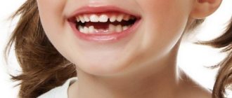
Teeth begin to change at about 5-6 years of age, and this process ends at about 12-14 years. Currently, according to the observations of many doctors, the replacement of baby teeth with permanent ones occurs in younger children than happened in past decades. But it also happens that baby teeth can be preserved in adults. Such cases can be encountered at 20, 30, and sometimes at 50 years of age. Why does this happen and what to do in such a situation?
Milk teeth and their features
Temporary and permanent teeth have certain differences in structure. The shape of baby teeth is the same as in molars (permanent), but their size is much smaller, the roots are shorter, and the number is different - there are only 20 of them versus 32 permanent teeth (including wisdom teeth). “Children’s” teeth are characterized by a short service life: their roots dissolve over time (as dentists say, “resorb”) approximately 2-3 years after they are fully formed. This process begins with the area touched by the crowns of the permanent teeth growing underneath them.
But it happens that for one reason or another the formation of the rudiments of permanent teeth does not occur. In this case, the roots of baby teeth are more often resorbed as a result of the influence of the rudiments of permanent adjacent teeth. But it happens that this does not happen and “children’s” teeth can then remain in adults - dentists call them persistent (translated from Latin persistere - to remain, to remain).
Why may there be no rudiments of permanent teeth?
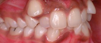
There are a variety of reasons that contribute to the absence of permanent tooth buds. These include hereditary characteristics, osteomyelitis of the jaws and their traumatic lesions, metabolic disorders, pathology of the endocrine glands. In addition, acute and chronic inflammation of baby teeth, in particular, untreated periodontitis in a timely manner, can also damage the rudiments of permanent teeth and contribute to their death.
Sometimes it happens that the rudiments of permanent teeth are formed, but can lie quite deep without touching the roots of milk teeth. This may occur due to the incorrect position of the permanent tooth or lack of space. In such cases, baby teeth can also be found in adults.
Do adults need to have their baby teeth removed?
Undoubtedly, baby teeth can often cause problems in adults. Firstly, their service life is short, which is why their resistance to caries is much lower compared to permanent ones. Secondly, teeth that do not fall out on time can interfere with the growth of permanent teeth and also cause them to be incorrectly positioned. But this does not mean that if a baby tooth is found in an adult, it must be removed. Everything is individual and depends on the specific situation. Very often, it is recommended to leave baby teeth in adults that are well preserved so that they last as long as they can. After all, sometimes it happens that permanent teeth may never appear in their place.
In any case, what to do with a baby tooth in adults needs to be decided after an X-ray examination is carried out, which will help determine the presence or absence of the rudiments of an unerupted permanent tooth, as well as whether the process of resorption of the baby tooth root is ongoing. If the rudiments are not found and the roots of a baby tooth have not been reabsorbed, and it looks aesthetically pleasing and immobile, then such a baby tooth should not be removed. The same applies to cases where it is X-ray proven that the position of the permanent tooth is such that even after removing the milk tooth, it will not be able to erupt. However, in this case it is better to consult an orthodontist.
What is edentia
Most likely, having heard such a term as “edentia,” the average patient of a dental clinic will not understand what we are talking about, because this disease, unlike caries, is not at all well-known. However, in dental practice, adentia is diagnosed quite often.
Of course, we are not talking about a congenital anomaly, when teeth are completely absent - an extremely rare pathology. But dentists encounter partial edentia (when several teeth are missing) almost every day.
Is it worth treating the disease or should it be classified as an aesthetic problem?
Adentia is a type of pathology characterized by a lack of teeth of varying degrees of distribution. The disease is usually classified into subtypes, depending on:
- nature of appearance (congenital or acquired);
- degree of severity (full or partial).
To date, it has not been possible to collect irrefutable facts to explain the cause of the disease.
Explaining the reasons for the absence of permanent teeth, doctors are inclined to believe that it appears as a consequence of poorly healed baby teeth.
Also, factors that increase the chances of this anomaly occurring include hereditary predisposition and metabolic disruptions, which prevents the proper formation of tooth buds.
Often, patients have concomitant abnormalities in the form of impaired formation of nails or hair.
The absence of some teeth, for example, lateral incisors or wisdom teeth, is subject to a certain pattern. It has been statistically confirmed that a lack of second incisors is observed in 0.9% of cases, and the rudiments of the second premolars in the lower row in childhood are absent in 0.5%.
This developmental anomaly is explained by a general decrease in the load when chewing on the jaw bone of modern humans, compared to our distant ancestors.
During evolution, the size of the jaw bone changed, which caused a situation of a banal lack of space for the development of the rudiments of molars.
The influence of heredity is also great in the case of symmetrical incomplete edentia. There have been cases where teeth, even in the presence of rudiments, never erupted, remaining at the level of the alveolar bone, which is clearly visible on x-rays.
As a result, such a tooth creates many problems, starting with the appearance of neuralgic pain and ending with deformation of the adjacent roots. It is important to note that in primary occlusion such a developmental anomaly is diagnosed extremely rarely.
Regarding children, one should take into account the fact that some teeth tend to erupt later than the period that falls within the limits of physiological norms. The reason for this is the lack of space in the dentition.
True adentia should be distinguished from retention. Retention is a time delay in the development (growth) of permanent teeth. Causes of retention: metabolic disorders, heredity.
Most often, displacement of impacted teeth is observed. Impacted teeth often do erupt, and sometimes the waiting period can be as long as decades.
You can speed up eruption with the help of orthopedic intervention. Consequences of retention: displacement of adjacent teeth, jaw deformation, and the addition of inflammatory processes.
Therefore, we should not forget about the importance of control over this process.
Congenital (complete)
It is noted in isolated cases. The patient is diagnosed with a total absence of molar rudiments. This naturally affects the symmetry of the face.
Diagnosis of this type is not difficult - adentia is noticeable even with normal palpation of the jaw. X-rays clearly demonstrate the total absence of all tooth germs. The jaw bone is not developed, so there is a significant decrease in the size of the lower areas of the face.
It is often possible to detect edentulous molars during the period of physiological change of teeth. X-rays clearly visualize the absence of rudiments and the characteristic disproportion of the face.
Congenital (partial)
This type of pathology is much more common. The dentition reveals single or multiple missing teeth. The X-ray image shows the total absence of all rudiments and the presence of three - the distance between the existing teeth. If a noticeable part of the teeth in a row is missing, a general underdevelopment of the jaw is observed.
There are symmetrical and asymmetrical congenital partial edentia. Based on the name, it becomes clear:
- symmetrical – lack of teeth of the same name on the left and right sides of the row;
- asymmetrical – lack of opposite teeth on different sides
Purchased (full)
This type of adentia is usually called secondary. The lack of teeth in this pathology can be observed in both jaws. Acquired adentia can be diagnosed in the primary and permanent dentition. The main causes: tooth loss due to destruction or surgical removal.
The advanced form of acquired adentia is characterized by a total lack of teeth in the oral cavity; as a result, the lower jaw shifts to the nose area, and the tissue surface of the gums begins to significantly recede. Atrophy of the alveolar processes occurs, as well as the entire jawbone as a whole. The patient loses the ability to bite off and chew food thoroughly, and diction also deteriorates significantly.
Acquired (partial)
The most common type of adentia in dental practice. Characterized by single or multiple lack of teeth in a row. The picture is usually aggravated by too thin tooth enamel, which entails abrasion of hard dental tissues, which means that it becomes difficult for the patient to take solid, as well as hot and cold food.
Clinical picture
A characteristic symptom is lack of teeth of any severity.
Other symptoms:
- reduction in jaw size,
- atrophy of the alveolar processes,
- the appearance of retraction of soft tissues in the perioral area of the face,
- the appearance of small wrinkles around the mouth,
- The angle of the jaw smooths out and becomes more obtuse.
When edentulism is incomplete, a distortion of the bite occurs due to the displacement of the teeth towards the site of the lost teeth.
Diagnostics
Does not pose any particular difficulties for any experienced doctor. Already during the initial examination, the doctor diagnoses a lack of teeth of varying severity.
Diagnosis of any form of adentia is carried out accompanied by an x-ray examination of both jaws.
This rule especially applies to the treatment of congenital anomalies, since only an image can show the total absence of tooth germs.
In pediatric dental practice, when diagnosing adentia, the method of panoramic radiography is used. This type of study fully reflects the overall picture: the absence of rudiments, the structural features of the roots and the entire jaw bone tissue.
At the time of developing the upcoming treatment plan, phenomena that interfere with prosthetics should be completely eliminated. These include:
- the presence of roots from previously destroyed teeth,
- the presence of exostoses,
- the presence of inflammation and neoplasms,
- diseases of the oral mucosa.
Treatment of adentia in adults
It has been practically established that the orthopedic method of eliminating the disease is the most effective method.
The preparatory stage includes the development of a treatment plan. First, the degree of atrophy of the alveolar tubercles and processes should be determined. Next, the patient is fitted with a pre-orthodontic trainer.
Treatment of acquired severe adentia consists of restoring the overall performance of the entire dental system while simultaneously eliminating all possible complications. Restoration is carried out by installing plate prostheses.
An important aspect in eliminating secondary adentia is the complete elimination of factors influencing the development of the pathological process that causes adentia.
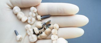
Treatment of complete edentia requires the use of implant prosthetics.
Treatment of edentulism: pediatric dentistry
Congenital unexpressed adentia in children involves taking measures aimed at stimulating the correct process of teething, which creates optimal conditions to eliminate the likelihood of deformation of the jaw bones. After the last teeth in the row have erupted, the dentist analyzes all available prosthetic options using:
- metal-ceramic crowns and inlays;
- adhesive bridge;
- method of implanting dental implants.
Prosthetics, as a method of treating congenital adentia in childhood, is allowed when the child reaches 3 years of age. A mandatory condition is constant monitoring and monitoring of the progress of treatment due to possible risks of disruption of the normal development of the child’s jaw caused by the pressure of the installed prosthesis on the jaw bone.
Possible complications when treating adentia using prosthetics:
- inability to sufficiently fix the prosthesis due to jaw atrophy;
- the occurrence of inflammatory phenomena;
- allergic reactions;
- bedsores.
It is important not to forget that tooth loss is always a traumatic factor for the patient’s psyche, therefore, providing the patient with psychological assistance is a necessary and mandatory condition.
What does lack of treatment lead to?
Adentia is a serious dental pathology that requires mandatory treatment. In the absence of adequate medical intervention, the patient’s quality of life decreases markedly:
- loss of the ability to chew food thoroughly, causing disruption of the gastrointestinal tract, metabolic failure and vitamin deficiency;
- complete adentia leads to disruption of the entire temporomandibular joint, which in the future often causes inflammatory processes;
- problems with diction, leading to constant psychological discomfort, which in turn can provoke depression;
The danger of ignoring such a disease is too great, so do not waste time on unnecessary thoughts - consult a dentist. Remember, edentia is a serious disease that entails a number of negative consequences that can significantly worsen a person’s quality of life!
Source: https://clinicpartner.ru/articles/CHto-takoe-adentija/
| Some clinicians distinguish between acquired (as a result of disease or injury) and congenital or hereditary adentia. “Partial secondary adentia” as an independent nosological form of damage to the dental system is a disease characterized by a violation of the integrity of the dentition. In the definition of this nosological form, the term “edentia” is supplemented with the word “secondary”, which indicates that a tooth (teeth) is lost after its eruption as a result of disease or injury. This definition, according to the author, contains a differential diagnostic feature that makes it possible to distinguish this disease from primary, congenital adentia and tooth retention. To summarize, it should be noted that it is more convenient to use the terms “defect” instead of “secondary adentia”; “true adentia,” when there is no tooth in the dentition and its rudiment in the jaw, and “retention or false adentia,” that is, a tooth that has not erupted. The causes of edentia can be heredity, disorders of the function of the endocrine glands, disorders of mineral metabolism in the prenatal period due to diseases of the mother and after the birth of the child due to diseases of early childhood. Death of tooth germs occurs with ichthyosis and endocrinopathies, hypothyroidism and cerebral dwarfism. Disorders of the embryogenesis of dental tissues, acute inflammatory processes that developed during the period of primary occlusion also lead to the death of the rudiments of permanent teeth and subsequently to underdevelopment of the jaw. The same processes can cause partial or complete retention. Stanton Capdepont syndrome is described in the literature under various names: “dentinogenesis imperfecta”, “transparent teeth”, “opalescent dentin”, etc. This disease is hereditary and affects milk and permanent teeth. With normally formed enamel, the dentin structure may be disrupted (less mineral salts, fewer tubules and they are wider, their direction is changed). The radiograph reveals a reduction in size or complete obliteration of the dental cavity and root canals due to the formation of replacement dentin. Due to the thinness of the roots, there is a higher risk of fractures due to injury. The color of the teeth is blue-brown, violet or amber. Due to less mineralization of the teeth, early progressive wear occurs, right down to the gums. The treatment is prosthetic, without removing teeth, that is, the production of overlapping removable dentures (partial or complete). Partial adentia (hypodontia) can occur without obvious systemic diseases.
Canine edentia is rare. The most common missing teeth are the upper lateral incisors, wisdom teeth on both jaws, and the lower second premolars. Sometimes very severe forms of partial adentia occur, when almost all milk teeth are missing or only 6 permanent teeth are present. YES. Kalvelis observed such a patient for 6 years (9-15 years): the cause of adentia could not be determined, the general development and growth of the jaws were normal, despite the absence of permanent teeth. Adentia in primary occlusion should be considered congenital, since the formation of the crowns of almost all primary teeth ends in the prenatal period. As for adentia in the permanent dentition, the conclusion about its congenitality should be made with caution, since the death of the tooth germ is possible due to exposure to infection or dysfunction of the endocrine glands, accompanied by a violation of calcification in the phase of calcification of the crowns of the teeth. The dental follicle, without calcification, loses its viability and dies. The clinical picture of complete edentia is characterized by a disturbance in appearance, a reduction in the lower third of the face, recession of the lips and cheeks, and impaired chewing and speech. According to the literature, multiple adentia occurs in 0.3% of people. Dr. L.E. Davidson reports on an 8-year-old boy who was born from healthy parents and had no abnormalities in pediatric status, and in the oral cavity there were conical deciduous frontal teeth, up to 4.0 mm wide; the roots of the molars had a rounded shape; X-ray examination revealed a complete absence of permanent tooth buds on both jaws, mobility of milk teeth due to the absence of roots; The child received removable dentures. With partial edentia, clinical manifestations are more varied and depend on the number of missing teeth and their former location. In the absence of one or two symmetrical teeth of the same name, there may not be a free space in their place, because the jaw in this area developed poorly, and the teeth behind them erupted next to the ones in front. When a tooth is retained, there usually remains a free space, albeit narrowed, in the place where it should have erupted. This is one of the differential signs of adentia and retention. In addition, edentulism is characterized by a thinned toothless alveolar process or the presence of milk teeth in a discrepancy with the timing of the eruption of permanent teeth.
In the absence of a lateral incisor, its place is taken by a mesially moving primary canine, and then a permanent one and the entire dental arch is shortened. In the absence of both lateral incisors, the central ones are displaced distally, and a diastema appears between them. Edentia, as a rule, leads to underdevelopment of the jaws, which is more noticeable the more teeth are missing. Thus, having a complete set of teeth is important not only for chewing and aesthetics, but also for preventing the movement of the lateral teeth. TreatmentWhen treating adentia, the following activities can be planned depending on the clinical situation:
Implantation may be the treatment of choice if space is available or can be pre-created through orthodontic tooth movement. The width of the narrowest implant is 4.0-5.0 mm, and the distance between it and the adjacent tooth should be at least 1.0 mm. In total, a gap of ~7.0 mm is required. It should be borne in mind that in adolescents, before planning implantation, it is necessary to obtain information about the growth period. The latter ends at different times for boys and girls (~ 14-16 years old, To illustrate the treatment of crowded teeth, in combination with a defect in the dentition, we present the clinical observation of Dr. JB Romero et al. The patient, aged 11 years, came to the clinic to eliminate aesthetic and functional disorders caused by the loss of the 11th tooth (sports injury). In both jaws there was pronounced crowding of the anterior teeth, which progressed significantly after the loss of the tooth so that the gap was practically closed by the adjacent teeth. After a consultation (surgeon, orthodontist, implantologist), it was decided to carry out orthodontic treatment, and when the patient reaches adulthood, implant-supported prosthetics. For successful orthodontic treatment, one premolar was removed from each quadrant of the upper and lower jaws, and at the final stage, a removable retention polymer splint with an artificial tooth was made to replace the 11th missing tooth. All subsequent stages of therapy were carried out after the patient turned 21 years old. During a preliminary x-ray examination, it was found that the 38th tooth is located almost horizontally. Therefore, when drawing up a treatment plan, it was decided to perform two surgical operations at once: removal of the 38th tooth and taking a block bone autograft for subsequent augmentation (building up) of the severely atrophied vestibular cortical plate of the alveolar part in the area of the 11th tooth.
The surgeon placed the autograft in the area with maximum atrophy of the cortical plate of the alveolar process and secured it with two titanium screws with a diameter of 1.2 mm. The period of engraftment of the autograft was 4 months, during which the tooth was corrected, an implantation operation was performed, the duration of osteoregeneration was 6 months. After this, an all-ceramic structure was made with fixation to the implant in the area of the 11th tooth. In conclusion, it should be noted that the possibilities for eliminating partial edentia and dental defects when treating children are significantly limited, since implants cannot be used before reaching a certain age. Therefore, as a rule, temporary structures such as adhesive prostheses are used. If implantation is planned for true edentia, then it is necessary to preserve baby teeth longer until a suitable age is reached, and in adults with a defect in the dentition, on the contrary, implantation should be carried out faster in order to reduce atrophy of the alveolar process. Most often, we are talking about the possibility of implantation in the absence of lateral incisors of the upper jaw. |
What to do if a baby tooth can cause problems?
If you are not satisfied with the aesthetics of a baby tooth or it is mobile, you still need to start with an x-ray examination. Having discovered on an x-ray that there are no rudiments of a permanent tooth, and the roots of a baby tooth have been reabsorbed, and mobility of the 3rd - 4th degree is observed (the tooth is very mobile), then it needs to be removed and then a decision must be made about what type of prosthetics to use in this case. case.
If you are not satisfied with the appearance of the tooth, you also need to use an x-ray to determine the condition of the roots of the baby tooth and the rudiments of the permanent tooth. Further, the decision must be made individually in a specific case. It depends on the age of the patient, as well as on the place of the baby tooth in the dentition. If there are no rudiments and the roots of a baby tooth are present, a veneer can be installed on it or the tooth can be restored, making it invisible in the dentition. Those who want to completely transform their smile will benefit from Lumineers.
If there are rudiments of a permanent tooth, you need to estimate how long it will take before erupting and decide to remove the baby tooth and pull out the permanent one.
Although the presence of baby teeth in adults can be considered an anomaly, this is not a reason to part with them - after all, very often they can serve you for many years. But sometimes it happens that such “greetings from childhood” can become an obstacle to the eruption of a permanent tooth. Therefore, if you discover that you have a baby tooth, be sure to take an x-ray and consult a specialist.
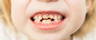
Edentia: lack of formation of one or more molars in children
It is very important at the age of 5-7 years before changing baby teeth in children to take a panoramic photograph of the dentition to make sure that all the molars are present in the bookmark. Because
The absence of molars in children is becoming more and more common, and then the milk teeth are retained as long as possible.
If you don’t know about the absence of molars and remove the baby teeth, you will have to use a more radical method to restore the dentition, for example, implantology.
Permanent teeth are formed in a child in the first year of life; it is during this period that taking antibiotics by children is not advisable and should be prescribed by doctors with great caution.
What is edentia
The term “ edentia ” is not the most common in dentistry, so not every patient understands what we are talking about on the first try. The phenomenon of edentia - congenital or acquired absence of teeth - is not so rare. Complete edentia (the absence of all teeth) is rare, but partial (with the loss of several) is a common occurrence.
Adentia is the complete or partial absence of permanent or baby teeth. There are several types of edentia:
- full;
- partial;
- primary;
- secondary.
If you analyze this list, you can see the pattern of classification according to the principle of appearance - primary (the second name is congenital) and secondary (in another way - acquired) and according to the type of prevalence (complete or partial). The causes of adentia are not fully understood. It is believed that it occurs after the resorption of the follicle, which occurs under the influence of general diseases or inflammation.
Edentia of permanent teeth can appear as a complication for milk teeth, especially if the latter were not treated on time and with poor quality.
Doctors do not rule out a hereditary factor, problems in the endocrine system, as a result of which deviations occur during the formation of tooth germs.
In most cases, in the presence of edentia, patients may experience improper formation of nails, hair and other organs of actodermal origin.
There is a pattern in the absence of some permanent teeth - lateral incisors, lower premolars, wisdom teeth. According to statistics, dentists do not observe second incisors in 0.9%. The rudiments of the second lower premolar are absent in 0.5% of children.
The reasons for this phenomenon are explained by the fact that the masticatory apparatus in modern conditions does not have such a serious load as in its distant ancestors.
Evolution has changed the size of the jaw, the number of rudiments of permanent teeth, since there is no place for them in the changed jaw - reduction of the jaw leads to reduction of teeth.
With a symmetrical incomplete number of teeth, the role of hereditary factors is great. There are cases when all the tooth germs are present, but some of them do not erupt, remaining retained in the alveolar bone. This fact is confirmed by radiography.
In primary occlusion this phenomenon is rare. An impacted tooth can create many problems for the jaw: displacement of adjacent teeth, deformation of adjacent roots.
Often such a tooth causes neuralgic pain and can serve as a source of focal infection.
In childhood, it is necessary to take into account the likelihood of teeth erupting late, sometimes beyond the physiological period. A tooth may be delayed due to lack of space in the dentition. Timely orthodontic intervention is important here.
Primary full
Complete primary adentia is a very serious anomaly, which, fortunately, is very rare. It occurs in the bite of baby or permanent teeth.
The patient is completely deprived of the rudiments of all permanent teeth. This condition inevitably provokes violations of facial symmetry. At the same time, the alveolar processes of both jaws develop incorrectly.
The mucous membrane of the oral cavity is pale and dry.
When baby teeth are edentulous, their rudiments are completely absent; by palpating the jaw, this is easy to diagnose. On the radiograph, the rudiments of baby teeth are completely absent, and the jaws are underdeveloped, which causes a severe reduction in the lower part of the face.
Edentia of permanent teeth is usually detected when milk teeth are replaced by permanent teeth. On the x-ray, the doctor observes the absence of the rudiments of permanent teeth, the pulling of the lower jaw towards the upper, with subsequent asymmetry of the face.
Primary partial
Primary partial adentia is much more common than complete adentia. In the dentition with this form, several or one primary or permanent teeth are missing. On the radiograph there are no rudiments of missing teeth, but gaps appear between the erupted teeth - trema. If a significant part of the teeth is missing in the dentition, then the jaw is formed underdeveloped.
Partial edentia can be symmetrical or asymmetrical. With symmetrical edentia, there are no teeth of the same name on the right and left in the dentition - for example, the right and left incisors. With asymmetrical – there are no teeth of different names on different sides.
Secondary full
Secondary adentia has another name - acquired. In the secondary form, teeth in the dentition are completely absent, both on the upper and lower jaws. Secondary adentia occurs in both permanent and baby teeth. This phenomenon occurs after tooth loss or extraction.
With complete secondary edentia, the patient’s mouth has no teeth at all, so the lower jaw moves closer to the nose, and the soft tissues of the mouth area noticeably recede. With complete secondary adentia, the alveolar processes and the body of the jaw atrophy. The patient is unable to bite or chew food and is unable to clearly pronounce sounds.
Secondary partial
Partial secondary adentia is a more common form. With this disease, several (or one) primary or permanent teeth are missing from the dentition.
If there is insufficient tooth enamel, the hard tissues of the tooth are worn away, causing hyperesthesia. The disease makes it difficult to eat hot or cold food, creating a habit of liquid food that does not need to be chewed.
In the photo - complete and partial edentia, edentia in children.
Symptoms of edentia
The symptoms of edentulism are simple - complete or partial absence of teeth. In addition to direct symptoms, there are also indirect ones:
- reduction of one or both jaws;
- retraction of the soft tissues of the oral part of the face;
- atrophy of the alveolar processes;
- formation of a network of wrinkles around the mouth;
- atrophied muscles in the mouth area;
- dullness of the angle of the jaw.
With partial edentia, a deep (distorted) bite is formed. The teeth gradually shift towards the missing ones. In the area where there are no antagonist teeth, the dentoalveolar processes of healthy teeth lengthen.
Diagnosis of edentia
Diagnosing edentia is not difficult. When examining the patient’s oral cavity, the dentist notes the complete or partial absence of teeth in a row. An X-ray examination of both jaws is required, especially in cases of primary adentia, since only in the image can one see the absence of the rudiments of permanent or baby teeth.
When diagnosing adentia in children, a panoramic X-ray of the jaw is taken - it is this that allows one to determine the absence of tooth germs, the structure of the roots of the teeth and the bone tissue of the alveolar process.
When diagnosing, it is necessary to exclude factors that do not allow urgent prosthetics. The dentist highlights the following points:
- the presence of unremoved roots covered with mucous membrane;
- the presence of exostoses;
- the presence of tumors and inflammation;
- the presence of diseases of the oral mucosa.
After the final elimination of all provoking factors, prosthetics can begin.
Treatment of adentia
The most effective method of treating adentia is orthopedic. The doctor draws up a treatment regimen based on the degree of atrophy of the alveolar processes and tubercles. When treating primary adentia, depending on the patient’s age, the patient is registered at a dispensary and a pre-orthodontic trainer is installed.
In case of partial primary adentia in children, it is necessary to stimulate proper teething to prevent jaw deformation. When the seventh permanent teeth erupt, the dentist explores options for prosthetics for missing teeth:
- prosthetics with metal-ceramic crowns and inlays;
- production of an adhesive bridge;
- implantation of missing teeth.
Treatment of primary adentia in children with the help of prosthetics is carried out by prosthetics from the age of 3 years. Such children should be under the constant supervision of a specialist - due to the pressure of the prosthesis, there is a danger of impaired jaw growth in the baby.
When treating secondary complete adentia, the dentist restores the functionality of the dentofacial system, preventing the development of complications and pathologies, and after restoration, performs prosthetics using removable plate dentures. When treating secondary adentia, it is important to eliminate the cause of the pathological process that provokes adentia.
In case of complete edentia, preliminary dental implantation is performed.
When treating adentia with prosthetics, complications are possible
- disruption of normal fixation of the prosthesis due to jaw atrophy;
- allergic reaction to denture material;
- inflammatory process;
- formation of bedsores.
An important point is psychological assistance to patients experiencing psychological discomfort from tooth loss.
Consequences of edentia
- Edentia is a complex dental disease, and without proper treatment, the patient’s quality of life can significantly suffer. With complete edentia, speech impairment occurs and it becomes inarticulate. The patient is unable to chew or bite off solid food. Poor nutrition leads to gastrointestinal problems and vitamin deficiency.
- In the absence of teeth, the temporomandibular joint does not function properly, which often leads to the development of inflammatory processes.
- It is impossible not to take into account psychological discomfort, a decrease in the patient’s social status, and self-esteem. All this provokes regular stress and the occurrence of nervous disorders.
Edentia must be treated without fail, and without much thought.
Source: https://sandora.in.ua/adentiya-otsutstvie-zakladki-odnogo-ili-neskolkih-korennyh-zubov-u-detej/