Danger of jaw cancer
Jaw cancer is a malignant neoplasm originating from bone tissue. The disease has its own clinical severity, and treatment is prescribed depending on the degree of damage.
Jaw cancer is a disease that spares no one. It can occur both in infancy and in old age.
It is noteworthy that, according to statistics, malignant neoplasms occur in 1-2% of all visits to the dentist, while cancer of the maxillofacial area in general affects people in 15% of cases. Due to the fact that the maxillofacial area has a complex structure with many nearby nerves and vessels, treatment of this pathology can be extremely difficult and requires the intervention of many highly specialized doctors.
Damage to the jaw by a malignant tumor: generally accepted classification
The appearance of a malignant neoplasm on the jaw begins with bone tissue.
Cancer cells themselves can develop from various areas, for example:
- periosteum;
- ondotogenic structures;
- spongy bone marrow;
- neurogenic cells;
- blood vessels;
- elements of unknown origin.
In this case, there is a classification of malignant neoplasm on the upper or lower jaw:
- chondrosarcoma, sarcoma (connective tissue tumor);
- cylindroma, carcinoma (epithelial tumor);
- schwannoma, melanoblastoma.
Depending on how advanced the disease is and how quickly it progresses, another classification can be distinguished according to the TNM system:
- At the first stage, only one anatomical region (T1) is affected by a malignant neoplasm.
- At the second stage, the disease affects no more than two anatomical areas (T2).
- At the third stage, the pathology has progressed so much that more than two areas are affected (T3).
- At the fourth stage, a large organ or some part of it is affected, and the pathological process itself begins to spread to distant tissues (T4).
Metastases can appear early, and their rapid occurrence is characterized by pronounced pain. Metastasis is most often detected in the lungs.
According to the form of the disease
Depending on what caused the tumor on the jaw, its symptoms, etc., forms of the disease can be distinguished.
In this case, there are only two of them:
- Primary form. Formed exclusively on the jaw bone. These include the following malignant neoplasms:
- osteosarcoma;
- Young's sarcoma;
- giant cell tumor.
- Secondary form. In this case, metastases from tumors in other areas spread to the jaws, mainly the head and neck.
The size of the tumor-like neoplasm is most often small, but since rapid growth can be observed, contacting a specialist in the early stages will avoid many complications. Only a doctor will make the correct diagnosis and prescribe the necessary treatment.
Lower jaw cancer
Causes of tumor
The etiology of the disease is still a very difficult issue for doctors. There are many theories, but there is no definitive conclusion on what exactly provokes the development of a malignant tumor on the jaw.
However, we can identify some factors that become the “culprits” for the occurrence of cancer:
- mechanical damage to the upper or lower jaw (trauma);
- advanced caries process in teeth, pulpitis, etc.;
- exposure to external adverse factors, for example, radioactive or ionizing radiation;
- damage to the mucous membrane in the oral cavity;
- incorrectly selected dentures, crowns;
- the occurrence of an acute, sluggish inflammatory process or frequent relapses in the chronic stage;
- abuse of bad habits, in particular smoking, etc.
As already mentioned, in the secondary form of the disease the cause is the formation of a tumor-like process in another anatomical area, and it is here that the following etiology can be distinguished:
- The presence of a cancer tumor localized on other organs with the appearance of metastases in the jaw:
- kidney cancer;
- mammary cancer;
- prostate cancer;
- lungs' cancer;
- throat cancer, etc.
- Precancerous diseases of the maxillofacial area, for example:
- oral papillomas;
- leukoplakia;
- leukokeratosis, etc.
You can learn more about the causes, symptoms and the disease itself by simply watching the video in this article.
How does oral squamous cell carcinoma develop?
Squamous cell carcinoma of the oral cavity is a malignant neoplasm that forms from degenerated epithelial tissue. The degeneration of epithelial cells begins gradually - under the influence of negative factors, metabolic and biochemical processes are disrupted, leading to mutation of the genes of one or more cells.
Mutated cells have only two functions left - growth and uncontrolled division. When the number of degenerated cells increases, they form a tumor-like formation characterized by invasive growth.
Squamous cell carcinoma of the oral mucosa is most common in men, but recently there are more and more female cancer patients due to alcohol and tobacco smoke. But statistics show that modern treatments have significantly reduced the mortality rate from oral squamous cell carcinoma.
By what signs can the disease be recognized?
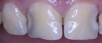
Most often, the patient begins to suspect a deterioration in health based on the onset of symptoms. Although the formation of a cancerous tumor on the lower or upper jaw has some differences in clinical severity, it is still possible to identify the general symptoms of the disease:
- teeth become mobile, shift and begin to fall out;
- There is asymmetry of the face due to edema and a growing tumor;
- pronounced pain in the upper or lower jaw;
- contracture of the lower jaw and upper jaw is observed (limited mobility);
- difficulty swallowing;
- the jawbone is deformed;
- there is an unpleasant odor coming from the mouth;
- there may be discharge of purulent contents from the nose;
- frequent headaches occur;
- there is numbness in the skin in the facial area, etc.
However, more detailed clinical characteristics are described in the table below:
| Form of the disease | Upper jaw lesion | Lower jaw lesion |
| Primary | Clinical severity of a malignant lesion of the upper jaw will have the following symptoms:
| The primary form is characterized by the following symptoms:
|
| Secondary | The secondary form of the disease will be characterized by the manifestation of symptoms that directly depend on the location of the malignant neoplasm. The location of the tumor in the anterioinferior region carries with it the following symptoms:
The postero-superior location of the tumor-like neoplasm is manifested by the following symptoms:
| If cancer of the lower jaw appears, symptoms in its secondary form may be as follows:
|
Symptoms of cancer of the upper or lower jaw
Attention: In the initial stages of the disease, signs of jaw cancer may be completely absent, which significantly complicates timely diagnosis and diagnosis. In this case, it is advisable to conduct an annual preventive examination with a doctor in order to identify the pathology at the very beginning.
Maxillary cancer symptoms
Recovery after treatment
Methods to combat jaw cancer are aggressive, and after them the patient needs rehabilitation. In addition to complex prosthetics, a person requires updated operations, speech correction, and health improvement in general sanatoriums. Three-stage prosthetics are usually used:
- Before the operation, an individual prosthetic plate is made,
- production of a formative prosthesis within 2 weeks after surgery,
- creation of the final prosthesis, compensation of soft tissue defects using splints and bone plates.
For cancer of the upper jaw, a commission is carried out (disability group II). Bone grafting is recommended 10-12 months after tumor removal. Radical intervention leads to disability and decreased ability to work, but over time, patients can return to mental work and other activities.
What is the danger of such a malignant formation?
A malignant neoplasm is already dangerous just by its name. This disease provokes many complications, and after treatment the immune system becomes thinner. This contributes to the development of various infectious and inflammatory processes.
Also, the most common consequences of cancer of the lower or upper jaw are the following pathologies:
- osteomyelitis;
- development of bleeding;
- pathological fractures of the jaw bones;
- visual impairment;
- relapses of the disease, etc.
What kind of diagnostics are performed?
Diagnosis of the disease is based on an initial examination of the patient, collecting anamnesis and prescribing the necessary research methods.
These include the following diagnostic methods:
- X-ray. This is the most accessible way to detect a tumor. It is carried out in lateral and direct projection.
- CT scan. Allows the doctor to visually assess the location of the tumor-like neoplasm, its spread and size.
- Lab tests. A general blood and urine test is required, as well as more detailed tests if necessary.
- Fluorography. Allows you to detect the presence of metastases in the lungs.
- Lymph node biopsy. Most often, the submandibular lymph nodes are examined to determine metastasis to this area.
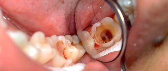
- Histological examination. If the tumor-like neoplasm originates from the alveolar substance, then the material for research can be taken from the socket of the extracted tooth. In case of bone formation, trepanation of the jaw is required. Such diagnostics allow us to identify the type of cancer cells.
Also, do not forget that this disease is fraught with various complications, so consultation with other doctors will be required. For example, if the orbit is affected, an examination by an ophthalmologist is necessary. If cancer of the maxillary sinus is detected, you cannot do without an otolaryngologist.
Treatment tactics
Therapeutic measures to eliminate pathology are carried out only after a thorough diagnosis, where the stage of the disease, the size of the tumor, the patient’s condition, etc. are determined.
Depending on these factors, the following common surgical methods are performed:
- Resection is partial. It is carried out only if the lesion is superficial.
- Segmental resection. This method of surgical intervention is effective in the absence of deep lesions, and also provided that the alveolar process is not involved in the pathological process.
- Removal of half the jaw. This method is indicated for damage to the angle of the jaw.
- Complete removal of the jaw and nearby soft tissues. Indicated for extensive damage, as well as if the tumor is observed in the chin area.
Nowadays, distraction of the lower jaw is carried out using special devices - distractors. Such a device provides gradual stretching of the lower jaw with the formation of replacement new tissue.
The lower jaw is dissected using an ultrasonic scalpel. This allows you to avoid affecting the roots of the teeth and mucous membranes during the operation. The price for such an operation varies depending on the clinic, its location and the availability of highly specialized doctors on staff.
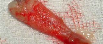
Photo of surgery
Do not forget about chemotherapy or radiation therapy to prevent tumor growth or metastases.
Radiation therapy
This method is carried out after surgery (when the wounds have healed) or before surgery to prevent the risk of metastasis. In some cases, surgical treatment is unacceptable for jaw cancer, so in this case, ionizing radiation is a “way out” of the current situation.
The occurrence of side effects during radiation therapy is inevitable, and their occurrence is characterized by the following symptoms:
- severe dry mouth;
- teeth begin to fall out;
- the taste and smell of food changes;
- the timbre of the voice becomes different;
- Infectious processes with characteristic clinical severity develop.
The severity of side effects depends on the prescribed course of radiation therapy and the extent of the surgical intervention.
Chemotherapy
Chemotherapy may be used as the primary treatment. Side effects after such treatment are more aggressive and are expressed in the same symptoms as with radiation therapy. However, the use of toxic drugs can cause bleeding, severe pain, baldness, etc.
With extensive removal of the jaw, incl. and teeth require long-term recovery in the future. After eliminating the pathological focus, plastic surgery with the insertion of special prostheses can be used.
Treatment of oral squamous cell carcinoma
Treatment of squamous cell carcinoma of the oral cavity occurs in two stages: initially, treatment of the primary tumor is carried out, then prevention or elimination of metastases. Therefore, the treatment regimen includes several methods at once - surgical, antitumor, radiological.
Surgical treatment
The tactics of surgery for squamous cell carcinoma of the oral cavity are selected taking into account the stage and extent of the tumor. A small tumor of the oral cavity is excised, including 2-3 cm of healthy tissue, to prevent the development of relapse.
In case of extensive neoplasms, squamous cell carcinoma of the oral cavity is removed with partial or complete resection of structures - palate, tongue, cheek, often involving adjacent anatomical areas. After extensive operations, important functions such as speech, breathing, chewing and swallowing are disrupted.
If possible, during surgery to remove oral squamous cell carcinoma, reconstruction is performed to restore the lost organs and their functions. But more often than not, surgeons wait until after surgical recovery, carry out preparations, and only if the patient’s condition is satisfactory, perform plastic surgery.
Chemotherapy
Chemotherapy for squamous cell carcinoma of the oral cavity is carried out to prevent relapse and destroy metastases. This treatment method involves the use of drugs that have a detrimental effect on cancer cells. For squamous cell carcinoma of the oral cavity, intravenous infusion of antitumor drugs is required, since less effect is observed when taken orally.
What drugs are used:
- Cisplatin.
- Methotrexate.
- 5-fluorouracil.
- Paclitaxel.
- Gemcitabine.
- Taxotere.
Chemotherapy for squamous cell carcinoma of the oral cavity requires a combination of two or three antitumor drugs - this way malignant tissues are destroyed more quickly, reducing the likelihood of metastases spreading throughout the body.
Chemotherapy is prescribed as palliative treatment for late-stage cancer. It allows you to stop the progression of the tumor and relieve body pain caused by the spread of metastases.
Radiation therapy
Radiation therapy for squamous cell carcinoma of the oral cavity is an effective method of reducing the size of the tumor and preventing the formation of secondary tumors. At the first stage of squamous cell carcinoma of the oral mucosa, it allows you to destroy the tumor without surgery, and at later stages of development it allows you to achieve a longer remission.
Irradiation is carried out using two methods:
- External irradiation is the effect of waves directly on the mouth and neck. Not only malignant but also healthy tissues are exposed to rays, which leads to the development of severe complications.
- Brachytherapy – radioactive substances are injected directly into the tumor tissue, which avoids damage to healthy tissue.
Radiation therapy for oral squamous cell carcinoma is often combined with chemotherapy to improve the chances of killing all cancer cells.
Disease prognosis
The prognosis of the disease directly depends on the time of visiting a doctor and identifying the pathology. In the early stages, tumor removal is more successful, and the effectiveness of the treatment will depend only on how the patient follows the medical instructions for behavior after the operation. In any case, at the first and second stages the prognosis is quite favorable, and a person lives after the disease for another dozen years.
At the third and fourth stages, various complications appear, and getting rid of the tumor is much more difficult. In this case, doctors carry out replacement therapy and improve the patient’s quality of life. The life prognosis in this case is 5-6 years.
Attention: the main cause of death from jaw cancer is late diagnosis and untimely treatment. Also, the first relapse after therapy occurs in the first 1-2 years, so you need to be very careful.
To avoid progression of the disease, it is necessary to undergo mandatory preventive diagnostics. It is also worth paying attention to the peculiar “signals” of the body. If the first symptoms of jaw cancer appear, you should immediately consult a specialist. Monitor your health and follow all recommendations regarding a healthy lifestyle.
Life forecast
The prognosis for life with squamous cell carcinoma of the oral cavity depends on the stage of the disease and the location of the tumor. If the tumor of the oral cavity is localized at the bottom or root of the tongue, the first metastases appear very early, so the prognosis is worse than for cancer of other localizations.
The most favorable course is for highly differentiated squamous cell keratinizing cancer of the oral cavity - it slowly progresses in size, does not penetrate into deep tissues and metastasizes only in the late stages of development. Five-year relapse-free period of tongue cancer at stages 1-2 in 75-90% of patients treated. For tumors of the cheek and floor of the mouth, the course of the disease is favorable in 46-65% of patients. At stage 3 of the disease, regardless of the location of the tumor, the absence of relapses is observed in 25-35% of patients.
Poorly differentiated tumors grow rapidly, cover deep layers and metastasize even at a small size. Frequent relapses are observed, and it is difficult to predict the course of the disease.