Cyst or tumor of the jaw in a child: when is it time to sound the alarm
A dental cyst in a child is by no means a rare occurrence among young patients. The most common reasons for its appearance, experts include maxillofacial injuries, poor quality of dental treatment and infection. The trouble is that a milk tooth cyst can have a detrimental effect on the formation of the rudiments of permanent teeth. What it is and how the disease can be cured will be discussed further.
Causes of dental cysts in children
Most parents do not seriously care about the health of their children’s teeth until they replace their baby teeth with permanent ones, arguing that the baby teeth will fall out anyway. This opinion is wrong - it is quite possible for a cyst to appear on a child’s baby tooth.
If caries has progressed to pulpitis, and then to periodontitis, then such a tooth becomes a source of infection, and the body, in order to stop its spread, turns on self-defense. To do this, it forms, most often at the root of the tooth, a protective bubble, or granuloma, which then transforms into a jaw cyst.
But not only advanced dental diseases can cause the formation of a tooth root cyst. Any injury, from cracking nuts to a blow to the jaw, can trigger the appearance of a dental cyst in a child. Those parents whose children are young hockey players or boxers need to be especially careful.
Also, if the treatment of pulpitis is poorly carried out, the infection will reach the roots through the canals of the tooth, as a result of which a rare type of dental cyst in children may occur - follicular, which occurs only in 6% of cases. The only thing you can wish for in such circumstances is to approach the choice of clinic and doctor competently and with all seriousness, otherwise your teeth will have to be constantly re-treated.
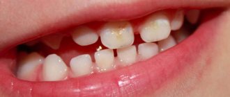
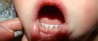
In addition, there is such a thing as an eruption cyst. It occurs several weeks before the appearance of a new baby or molar tooth. Teething cysts in children can be caused by infection, trauma, caries, or dental abnormalities.
Photo of eruption cyst in children
Types of dental cysts
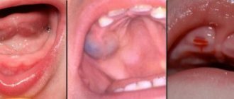
In dentistry, a cyst is a phenomenon in which a new formation appears in the form of a round cavity near the apex of the tooth root. It is localized in bone tissue, so it replaces its cells. Gradually it increases in size and is filled with purulent masses. In some cases, it manifests itself in the form of a purulent lump that appears directly on the gum.
It is customary to distinguish the following types of this unpleasant phenomenon:
- periodontal cyst: appears near unerupted teeth as a result of damage to the gum tissue. Usually occurs when wisdom teeth erupt,
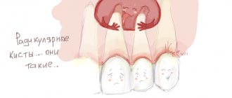
- radicular: formed near a tooth with pulpitis, untreated caries or poorly filled canals,
- follicular cyst: a separate type, which is characterized by the appearance of a cavity in the buds of damaged teeth. Often leads to the formation of large fluxes and fistulas, and can provoke the spread of the inflammatory process into deeper layers of tissue,
- keratocyst: this type is congenital - the cavity appears near defective rudiments,
- eruption cyst: a common type of pathology among children, which usually occurs during the eruption of permanent teeth between the ages of 6 and 14 years,
- residual: occurs against the background of injury to dental roots, unsuccessful tooth extraction and infection penetration into the open socket,
- eye tooth cyst: a special type of complication of inflammation of the maxillary sinuses.
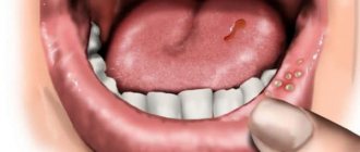
In the photo - a tooth cyst in a child
Causes
The prognosis of the disease depends on the type, size and location of the cyst.
Most cysts are benign and some do not require treatment at all. Sometimes a cystic lesion can provoke a local aggressive tumor, which, if not treated correctly, can cause destruction of surrounding tissue. This type is usually removed surgically to prevent new cysts from forming. If the cyst reaches a large size, the lower jaw may be weakened so that a pathological crack appears. Source of information: Wikipedia.
The most common cause is infection of the root canal. About 450 species of bacteria live in the oral cavity of a healthy person, which enter there along with food and feed on its remains on the teeth. If the dentist does not follow safety rules when treating a tooth (namely, does not use antiseptic baths during endodontic treatment), small particles of the tooth may get inside the canal.
Also of great importance:
- Poor oral hygiene.
- Weak immunity.
- Mechanical damage to teeth and gums.
- Gum diseases.
- Damage to the tissue of the jaws themselves.
The most common causes of cyst formation in childhood
- advanced caries: quite often young children are diagnosed with periodontitis. This disease usually results from an advanced form of caries or untreated pulpitis. According to statistics, in 6% of cases the result of advanced pulpitis is a follicular cyst1. As a result, a characteristic purulent lump appears on the gum of the baby tooth,
- medical errors made when filling dental canals: here we are talking about damage to molars and permanent teeth in older children. If the doctor has not completely sealed one of the canals, there is a risk of a source of infection. Due to the fact that the purulent masses come out, a pronounced lump forms on the gum tissue,
- accidental perforation of a tooth element: another common medical error is perforation of the wall of the dental canal, which can also lead to the appearance of a purulent cavity near the root,
- periodontitis: often a cyst becomes a symptom of inflammation of periodontal tissue, which, in turn, occurs as a consequence of an advanced stage of pulpitis. Often, against the background of impaired outflow of purulent masses, a purulent abscess is formed.
On a note! A fairly common occurrence among children is an eruption cyst. In this case, we are talking about the appearance of a purulent lump on an unerupted tooth. Among the common causes, experts identify caries, anomalies in the structure of the dentition and injury to the gums, for example, after the removal of a baby tooth.
Potential Complications
As a rule, at first the cyst does not cause the child any significant discomfort. However, the cavity gradually increases in size, after which suppuration occurs near the causative tooth. If for some reason the integrity of the neoplasm is disrupted, purulent masses will fall on the bone tissue and can provoke infection. As a result, the child's gums and cheek may become swollen.
World experts in the field of periodontology and pediatric dentistry argue that if the risk of spread of pus is not eliminated in time, odontogenic periostitis may appear - a serious pathology that leads to the gradual destruction of the jaw bone. In addition, the infection can spread to the baby’s internal organs and cause the development of even more serious problems for the body as a whole.
Features of treatment in children
As for the treatment of dental cysts in children, surgical methods are usually used. Based on the initial clinical picture, the specialist can choose one of the following options:
- cystectomy: cutting the gum and completely removing the cystic formation. If the cavity is larger than one and a half centimeters, resection of the root apex may be necessary. Among the disadvantages, it is worth highlighting the high traumatic nature of the method, but, on the other hand, the operation is characterized by minimal recovery time,
“My 7-year-old son was recently diagnosed with whale tooth. The doctor explained that it appeared as a result of improperly sealed canals, and then I remembered that before that we had once turned to an outside dentist, since the case was an emergency. Apparently, the hasty treatment took its toll. Many people here are concerned about the operation itself, so I’ll tell you: we were given sedation, so the child was not in pain, and he was not capricious at all. By the way, this is an excellent option for pain relief - by the evening my son fully recovered, there were no side effects. The doctor cut his gum and quickly removed the purulent cyst. Everything took no more than half an hour...”
Elena F., Moscow, fragment of message from correspondence on the forum
- cystotomy: surgical intervention involves removing only the anterior wall of the tumor. This method allows treatment with minimal risk of damage to the buds of permanent teeth, therefore this operation is most often prescribed for children.
- laser dialysis: cyst removal is carried out using a special light guide, which is inserted into the cystic cavity. This method is not suitable for large tumors, but is the most preferable when it comes to treating cysts in children. True, this is rare - the clinic must have the appropriate equipment installed, plus the specialist must be able to work with it, i.e. undergo training.
We should not forget about the importance of therapeutic treatment, which involves, first of all, thorough treatment of the canals with antiseptics. Treatment should be performed exclusively in a doctor's office under the direction of a pediatric dentist.
The best defense is prevention
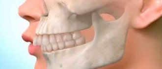
An x-ray examination will help detect a cyst in the early stages, which is recommended every six months, especially for those children who have already encountered pulpitis and periodontitis. Most often, the cyst occurs in the area of the first molars on the lower jaw.
X-ray of a child's teeth
It is important to accustom your child to good and regular oral hygiene from an early age. You should definitely attend all scheduled preventive examinations at the dentist’s office - only such a responsible approach will allow you to recognize the problem in time and take timely measures to eliminate it.
- Nikitin A.A., Titova N.V. Surgical treatment of cystic formations of the jaws in children using biocomposite materials, 2005.
Odontogenic cysts of the jaws
Odontogenic cysts of the jaws are a very common pathology. Currently, surgical treatment of this pathology is the most effective, since all odontogenic perihilar cysts are chronic foci of infection that have an adverse effect on the body. Odontogenic cysts are intraosseous cavity formations, the appearance of which is caused either by a violation of the development of the dental follicle, or by a chronic inflammatory process in the periodontium. The cyst consists of a shell, a cavity in which there is an opalescent liquid with cholesterol crystals.
According to morpho- and pathogenesis, as well as localization, the following types of cysts are distinguished: 1. Cysts formed from the epithelium of the tooth-forming plate (radicular): - periodontal - lateral periodontal - residual (residual) 2. Cysts developing from the enamel organ or follicle: - follicular -primordial -gingival 3. Cysts developing from the enamel organ or islands of Malasse: -keratocyst Clinical picture: -complaints may be absent, and the cyst may be an accidental finding during an X-ray examination -as the size of the cyst increases, deformation of the jaw may occur and bulging of the mucous membrane may occur - with an increase in the size of the cyst in the projection of the masticatory group of teeth in the upper jaw, the cyst can adhere to, push aside, or penetrate into the maxillary sinus. In this connection, headaches, a feeling of heaviness in the middle zone of the face on the affected side, difficulty in nasal breathing (including when the cyst grows into the lower nasal passage) may be disturbing - if the cyst is localized on the lower jaw, as it grows, compression of the lower alveolus may occur nerve, which can lead to numbness in the skin and mucous membrane of the lower lip and chin, possibly teeth, on the affected side (Vincent's symptom). With a significant increase in the size of the cyst, a pathological fracture may occur in the lower jaw. -exacerbation of the process can lead to suppuration of the cyst. As a rule, swelling of the soft tissues appears, in some cases difficulty opening the mouth (if the cyst is located in the projection of the third molars); during an intraoral examination, hyperemia of the mucous membrane, a symptom of fluctuation (swelling), pain when touched, and pathological mobility of the causative tooth are possible. Diagnosis: 1. intraoral contact radiograph 2. orthopantomogram 3. radiograph of the lower jaw in lateral projection 4. computed tomogram Treatment There are two methods of treating odontogenic cysts: 1. Cystotomy - removal of part of the cyst wall and creation of conditions for long-term communication (with the oral cavity, cavity nose, maxillary sinus), eliminating the main mechanism of cyst growth - an increase in hydrostatic pressure. Indications for cystotomy: - a cyst into the cavity of which 3 or more intact teeth are projected, the periodontal fissure is not identified on the radiograph at the roots of the latter - associated diseases - large cysts of the upper jaw with destruction of the bone floor of the nasal cavity and palatal plate - extensive cysts of the lower jaw with sharp thinning of the base of the jaw (bone thickness less than 1-0.5 cm) 2. Cystectomy - removal of the entire cyst shell of the bone cavity Indications for cystectomy: - cyst, as a consequence of a malformation of the odontogenic epithelium - small-sized cyst located within 1-2 intact teeth - an extensive cyst, in which there are no teeth in its area and a sufficient volume of bone tissue is preserved (for the upper jaw - cysts adjacent to or pushing aside the maxillary sinus without inflammation in the sinus) During cystectomy, the causative teeth are either removed or preserved. In this case, before surgical intervention, the root canal is hermetically sealed, after which an operation is usually performed with resection of the root apex.
These methods for treating odontogenic cysts are effective, but it must be remembered that the disease is easier to prevent than to treat!
The best prevention of diseases is an annual visit to the dentist for a preventive examination and periodic X-ray observations of previously treated teeth.