Acid necrosis of tooth tissue
Dental enamel necrosis is a pathology in which damage and destruction of hard dental tissues occurs.
The appearance and development of the pathological process is influenced by external and internal factors. Acid necrosis of teeth in the initial stage manifests itself as chalky spots, which over time become cavities with jagged edges. The enamel of the teeth is gradually destroyed, the patient feels pain from temperature changes in the oral cavity. In therapeutic dentistry, radiography and clinical examination data are used to diagnose necrosis of hard dental tissues. Treatment is used both therapeutic and orthopedic.
Acid necrosis of teeth - complete destruction of tissue
The concept of necrosis of hard dental tissues and the causes of its occurrence
Necrosis of hard dental tissues means a serious pathology, during the development of which enamel and dentin cells slowly die. As a result, this affects a person’s chewing function, which in the future leads to problems in the functioning of the entire digestive system. Diction problems are also possible.
At the initial stage, the disease affects an area characteristic of a specific type of tooth necrosis. Subsequently, necrosis begins to affect the entire surface of the enamel. Without timely treatment, the dental crown is completely destroyed, and after tooth loss, necrosis in some cases can spread to the gums.
There are a number of reasons that can provoke the development of necrosis of hard dental tissues. They are usually divided into external and internal factors. Internal include:
- disruptions in the functioning of the central nervous system,
- pregnancy, especially several cases in a row,
- dysfunction of the thyroid gland, for example, hypothyroidism,
- unbalanced hormone production, which is often observed in adolescents,
- constant intoxication of the human body,
- predisposition.
External factors negatively affect the dental tissue itself and come down to:
- Excessive or prolonged exposure to substances rich in acids or aggressive chemicals. These include: medications, food products, production means.
- Exposure to high doses. Occurs in cancer patients with an appropriate course of therapy.
- Constant exposure to electromagnetic radiation.
Characteristics of acid necrosis of teeth. Causes of appearance, symptoms. Stages of damage
Necrosis of hard dental tissues is multiple damage to teeth, leading to the appearance of defective areas on the smooth surface of the enamel and under it (dentin layer). The destruction of tooth enamel occurs under the influence of harmful factors and is non-carious in nature. Pathology is observed equally in men and women.
People at increased risk of developing necrotic lesions in the oral cavity:
- Workers in hazardous chemical industries.
- Cancer patients.
- Suffering from hormonal imbalances.
- With disorders of the digestive tract.
If left untreated, the disease leads to loss of chewing function and complete tooth destruction. The sooner a doctor diagnoses the disease and identifies the cause, the better for the patient.
Initial necrosis of dental tissues
Pathogenesis of acid necrosis
With acid necrosis of teeth, the formation of decalcified zones of enamel is observed under chronic exposure to acids. Further, these areas soften and gradually lose the enamel layer.
Occupational acid necrosis is usually observed in factories where there is a high concentration of acid fumes and hydrogen chloride gas in the air. These substances enter the oral cavity and dissolve in saliva, as a result of which the saliva oxidizes and demineralizes the hard tissues of the tooth. It should be noted that excessive consumption of citrus fruits and acidic drinks affects the enamel in the same way as acid fumes in factories.
Reasons for appearance
The reasons for the formation of enamel necrosis are divided into:
- Internal processes of the body (endogenous).
- Exogenous (externally influencing).
Internal factors contributing to the destruction of enamel are based on changes in hormonal levels. With endocrine disorders, a qualitative change in the composition of the enamel occurs, it becomes thinner. And when the gastrointestinal tract is disrupted, tooth enamel is destroyed due to the release of hydrochloric acid into the esophagus and oral cavity.
Wedge-shaped necrosis on teeth has a different nature
External factors contributing to necrosis are based on the use of drugs toxic to enamel, or on inhalation of toxic fumes from the air. When exposed to exogenous factors, central teeth and canines are often destroyed.
When toxic chemicals enter the oral cavity, the qualitative composition of saliva is disrupted and trophism in the neurovascular apparatus of the teeth decreases. The nutrition of hard tissues with nutrients decreases, enamel demineralization occurs, and tooth tissue becomes thinner.
Symptoms of radiation necrosis of dental tissues
Radiation (post-radiation) necrotic damage occurs in people exposed to ionizing radiation.
- This pathology is recorded in patients after radiation therapy for oncology. That is why, as a preventative measure, the patient is put on a lead mouthguard over his teeth during irradiation, and in preparation for irradiation, a course of remineralizing therapy is prescribed.
- Radiation necrosis of teeth also occurs, like an occupational disease , in people engaged in production in the presence of various radioactive substances.
Symptoms of radiation necrosis of teeth:
- After radiation exposure, a person experiences unpleasant sensations in the oral cavity - dry mucous membranes and tongue, a change or complete loss of taste. Radiomucoside appears in the mucous membranes of the oral cavity.
- After some time (2-3 months), the tooth enamel becomes dull and acquires a yellowish-grayish tint.
- There is fragility of the enamel and rapid abrasion of the chewing surfaces of the teeth.
- First, single, then multiple areas of dark or black color appear on the surface of the teeth, filled with loose contents.
- As a rule, there is no toothache - this is an important indicator of radiation necrosis of teeth.
- Gradually, the areas of necrosis increase and merge with each other. Removing necrotic tissue from them is painless.
After a year or two, all teeth may be affected.
Classification of stages of destruction
Doctors divide the stages of necrosis according to the changes occurring in hard tissues. The following stages are characteristic of necrosis:
- Initial. Under the influence of negative factors, demineralization of areas of the teeth occurs, and a chalky stain forms in the cervical part.
- Demineralized areas expose light dentin.
- In the cervical area, in place of exposed dentin, a pigmented funnel appears. Cariogenic microorganisms multiply in the funnel.
- The patient has individually multiple lesions of the crowns of the teeth at different stages, or single necrotic pathological processes.
Multiple lesions of tooth crowns due to acid necrosis
Necrotic lesions of hard dental tissues are accompanied by the progression of caries, which leads to rapid destruction of the deep layers of crowns.
Necrotic lesions of hard tissues of teeth of an endogenous nature
Internal pathological factors can lead to dental necrosis in the same way as those listed above.
Internal or endogenous factors include:
- Disruption of the endocrine glands (the most common cause is hypothyroidism or other thyroid diseases).
- Chronic intoxication of the human body.
- Disorders of the central nervous system.
- Pregnancy (or many pregnancies, one after another).
- Hormonal imbalances (for example, during puberty).
- Hereditary factors.
As a rule, dental necrosis, which occurs as a complication of any internal diseases or disorders in the body, has a cervical form, primarily affecting the cervical areas of the teeth.
Symptoms of the disease
Most patients with necrotic lesions of dental crowns note severe pigmentation of tooth enamel. When exposed to various acids, the color of dental crowns differs from the original. Hydrochloric acid turns crowns yellow, and sulfuric acid turns them black. When nitric acid is inhaled, dentin turns an unnatural white color. Under the influence of acids, crowns lose their natural shine, their surface becomes rough and dull.
After aggressive exposure, chalky spots are visible on the crowns. Sometimes replacement dentin forms in their place, and the patient experiences discomfort when closing the jaw. The progression of necrosis causes pain. Weak enamel reacts to temperature changes with painful sensations. Pain syndrome is often present when organic acids (lemon, strawberries, sweet or sticky foods) enter the oral cavity.
Chemical necrosis of dental tissues
The clinical manifestation of the disease is multiple, and rapid development of necrotic processes is observed. The non-carious cervical lesion has an uneven edge; no pain is noted on probing. The chewing efficiency of teeth decreases, and horizontal abrasion of the enamel is noted.
Acid necrosis clinic
The clinical picture of acid necrosis of teeth develops slowly. With chemical necrosis, fangs and incisors are more often affected: in the area of the cutting edges, the enamel disappears, resulting in the formation of sharp edges. In the initial stage, patients have a feeling of teeth on edge, and there may also be a feeling of teeth sticking, which is associated with the “washing out” of minerals from the tooth enamel, resulting in a feeling of softness of the teeth when they are closed. In later stages of the disease, pain from temperature and chemical stimuli appears. However, it should be borne in mind that pain may not be observed, due to the fact that tertiary dentin is being produced. As necrosis progresses, the enamel loses its shine, becomes dull and rough. And after complete loss of enamel, dentin becomes pigmented and becomes dark. In advanced cases, complete dissolution and abrasion of the tooth crown may occur.
Diagnosis of the disease and treatment methods
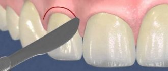
Diagnostic measures consist of a clinical examination of the oral cavity and medical history. Differential diagnosis consists of distinguishing between the wedge-shaped effect, enamel erosion and necrosis. The differential difference lies in the absence of the natural shine of the enamel and the determination of a different shape of the lesion.
To exclude damage to the upper layers of the tooth root, doctors use radiography (orthopantomogram). And to find the main cause of necrotic changes in the oral cavity, the patient is prescribed a study by a gastroenterologist and endocrinologist.
Clinical picture and pathogenesis
The cause of acid necrosis is most often an increased concentration of vapors in the air. This usually takes place in the production areas of chemical plants.
Once in the oral cavity, hydrogen chloride gas and vapors of other substances dissolve in saliva, giving an acidic reaction.
The main effect on dental tissues comes down to decalcification - the process of dissolution and loss of calcium salts by dental tissues. The loss of calcium leads to a decrease in the strength of enamel and dentin, the appearance of microcracks, chips and breaks on them.
Teeth become sensitive to temperature, chemical and mechanical influences and quickly wear out. Over time, the pulp also dies.
Reference . Normally, saliva has a pH of 6.8–7.4 units. The pH of a particular person's saliva depends on its composition and the rate of salivation. With abundant salivation, the acidity of saliva can reach 7.8 pH. A medium with a pH of 7.0 is considered neutral.
Saliva has buffering properties - the ability to neutralize a certain amount of acid entering the mouth. However, if its volume and concentration in the oral area exceeds a certain value, the buffering properties to neutralize the acid are not enough.
With the development of pathology, it is also possible not only a direct effect of acid on dental tissues, but also an indirect one, when the vapors negatively affect the entire body, reducing immunity and disrupting the traffic (nutrition) of dental tissues, which ultimately also leads to their decalcification and loss strength.
Symptomatically, the disease manifests itself by the appearance of a sore throat, sticking of the upper and lower teeth to each other due to softening of tissues, sensitivity to cold and hot, mechanical and chemical irritants, changes in the color and structure of dental tissues.
The enamel becomes rough and matte. The defects have uneven contours, the cutting edge becomes thinner and breaks off. The teeth shorten and narrow, taking on a wedge shape. The gaps between them increase, the bite decreases.
The dentin exposed when the enamel is broken and abraded has a dark tint. Destruction occurs in the direction from the cutting edge to the neck of the tooth. First, the outer (vestibular) surface is affected, followed by the occlusal and lingual surfaces.
Due to the formation of replacement dentin and the complete death of the pulp, over time, the initial edge and sensitivity of the teeth may become dull or disappear altogether.
Histologically, acid necrosis manifests itself by the destruction of enamel prisms, the formation of replacement dentin, and the closure (obliteration) of root cavities. Electron microscopy shows demineralized amorphous areas of dentin and enamel.
What do Turner's teeth look like, and is there a chance to correct the situation?
Visit here to learn more about dental enamel implants.
At this address https://www.vash-dentist.ru/lechenie/zubyi/emal/chem-opasna-eroziya.html all the most important things about the treatment of tooth enamel erosion.
Treatment of acid necrosis of teeth
The goal of treatment of necrotic changes in tooth enamel is aimed at restoring the integrity of hard dental tissues and increasing their density. Initial treatment is aimed at eliminating the pathological processes in the body that provoked necrosis. After eliminating the cause of necrotic changes and prescribing a treatment regimen, the doctor treats the crowns with minerals. Such treatment allows you to saturate dental tissues with nutrients and restore trophism.
At more advanced stages, prosthetics or crown restoration with polymer materials are used.
Treatment regimen
An effective treatment regimen is prescribed by a dentist and includes:
- Oral use of calcium glycerophosphate. The dose of the drug is calculated individually.
- Supplementing the diet with multivitamin complexes. The course is calculated individually.
- The drug Klamin. The dosage is calculated individually.
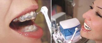
- Daily use of applications based on fluoridated pastes.
- Use of the drug phytonol for oral administration.
Computer necrosis of teeth
Over the past three years, dentists have encountered a new, completely unexpected and practically unexplored pathological process in teeth, characterized by systemic damage, more intense than that observed with post-radiation damage.
First of all, the following was unexpected for everyone:
- all applicants had worked with computers of different models for more than 3-5 years;
- all patients did not comply with the work schedule, professional protection and occupational hygiene (the working day lasted at least 8-10 hours, often including Saturdays);
- the side of the jaws facing the monitor was more intensely affected;
- all those who applied were previously practically healthy;
- all patients did not complain of pain in the teeth;
- none of the patients were exposed to any ionizing radiation or toxic or chemical substances;
- all applicants were aged 25-35 years.
A thorough examination of the patients was carried out with a detailed survey, including an analysis of working conditions with a computer and the length of the working day, familiarization with the brand of equipment and the year of its manufacture, etc. Along with this, when examining the patients, a clinical picture of the lesion was revealed, characterized by systemic, extensive damage to the teeth, which should be characterized as necrosis of enamel and dentin, somewhat reminiscent of their post-radiation damage, but at the same time different from it. This condition was designated as computer necrosis of teeth.
The computer itself is far from the main culprit in the identified dental pathology. Judging by the time spent by patients at the computer, the same dental lesions would probably appear in fans of TV series sitting in front of household televisions. The only question is how much time you need to stay at the screen, how far or close to it and in determining a number of other conditions.
How to identify?
- foci of necrosis cover a significant or even most of the crowns of the teeth, primarily the caries-immune surface, their cervical part, roots and alveolar bone;
- the lesions are mostly pigmented - dark brown, almost black, filled with a softened mass of tooth tissue of the same or dirty brown color;
- they are usually painless;
- the teeth have no vibrant shine.
How to avoid dangerous defeats?
You should follow sanitary standards when working with a computer:
- the area of the workplace should not be less than 6 m2 with a minimum volume of the entire room of 20-24 m2;
- natural light should be on the left;
- when there are 2 or more computers in the workroom, the distances between video monitors must be maintained (2 m when the screens are directed in one direction);
- the employee should be at a distance of 0.6-0.7 m from the screen;
- every 2 hours of work it is necessary to take breaks of 15-20 minutes to ventilate the room;
- The total duration of work with a computer should not exceed 6 hours for adults and no more than 3-4 hours for children and adolescents; for women during pregnancy and breastfeeding, work with a computer is contraindicated.
Some preventative measures should be taken:
- taking multivitamins 2-3 tablets every other day in winter and spring;
- taking Klamin or Phytolon every other day 15 minutes before meals;
- use of phosphate-containing pastes on all teeth 3 times a week for 15 minutes;
- daily dental care using phosphate-containing toothpastes.
SECTION DENTISTRY
All articles Addresses and telephone numbers of clinics
Tooth necrosis - from a harmless defect to serious problems
Exposure to negative factors is often the main cause of the development of most dental pathologies.
All of them require timely treatment, otherwise further progression of the disease is fraught with loss of the affected organ.
One of these diseases, necrosis, will be discussed in this article.
Clinical picture
This pathology accounts for 10% of all cases of non-carious enamel lesions. And these statistics are trending upward. The range of its varieties and forms, which have not yet been thoroughly studied, is expanding.
In most cases, the localization zone of the anomaly is the cervical part of the frontal jaw segment of the teeth. Damage to the indigenous organs of this disease is diagnosed extremely rarely.
The main danger posed by the diagnosis is a gradual impairment of chewing function .
What it is?
Dental tissue necrosis is the gradual death of enamel and dentin cells. This pathology is a complex dental disease that is difficult to treat.
As a rule, the chewing function of a person suffers first . As the disease progresses, diction disorders may occur.
Each type of pathology is characterized by its own specific area of localization at the initial stages of development. But as the course of the disease progresses, necrosis gradually spreads to the entire surface of the enamel.
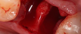
Which, if left untreated, leads to complete destruction of the crown of the tooth and its loss. In this case, necrosis of the gums is sometimes observed after tooth extraction.
Photo: what tooth necrosis looks like
Statistics of clinical cases show that in recent years the frequency of diagnosis of necrosis of dental tissue has been constantly increasing .
Reasons for the development of pathology
The first group includes:
- dysfunction of endocrine activity - against the background of the disease, the structural mineral content of hard tissues changes qualitatively, necrosis develops;
- lesions of the central nervous system of a chronic nature - they inhibit the processes of tissue regeneration and lead to their rapid destruction;
- severe and long-term intoxication - toxic processes affect the most vulnerable tissues of the body, and with thin and sensitive enamel they can provoke disease;
- bearing a fetus - pregnancy, is an additional load on the main organs and vital systems, teeth are no exception;
- genetic factor - it has been proven that a predisposition to this disease has a genetic code that can be transmitted during gestation and not manifest itself for a long time;
- problems with the gastrointestinal tract - a malfunction of the intestines causes destruction of the organ through the action of stomach acid, which is released in greater quantities than required by the norm.
To the second group:
- unbalanced diet - excessive consumption of food acids negatively affects the condition of the hard tissues of the oral cavity;
- harmful industries - interaction with acidic components provokes the development of necrosis;
- radiation exposure - irradiating flows in contact with air produce toxins that have a negative effect on organs located in the open area of the oral cavity;
- electromagnetic rays – people whose occupation is related to computer equipment are at risk.
Classification
There are several stages of destruction:
- chalky spot - this is how focal demineralization of an organ fragment in its cervical part manifests itself. In this case, the patient does not experience any discomfort;
- exposure of dentin - the phenomenon is facilitated by chips on the surface, at the site of demineralization, which, not fully completed, lightens and exposes dentin;
- pulp damage - externally manifests itself as an anomaly with a funnel-shaped cavity. This sharply reduces the size of the pulp chamber, the tissues become fragile, and pain occurs upon contact with them. The tooth takes on a grayish tint.
Read here about effective drugs used to remineralize teeth.
The following types of disease are distinguished:
- Cervical necrosis – affects the dental neck. The enamel becomes dull, whitish focal manifestations appear, hard tissue loses strength, crumbles and becomes loose. Progressing rapidly, the disease can completely destroy hard tissue. The main reason is hormonal imbalance.
- Acidic, where the provoking factor of development is acidic vapors. They actively wash away the calcium component responsible for the strength of the enamel. Clinical manifestations are surface roughness and a change in color pigment to dull gray. The organ becomes extremely susceptible to temperature changes.
- Post-radiation – considered a consequence of exposure to radiation. In advanced stages, the tooth can become almost black. In this case, pain syndrome is completely absent. The insidiousness of the disease is that if treatment is not started immediately, you can lose an entire fragment of the jaw row.
- Computer necrosis - the disease progresses against the background of the negative influence of ionizing radiation flows coming from the monitor. This form of pathology is characterized by dark brown pigmentation of the enamel and a matte, rough surface.
Necrosis of hard dental tissues
It is generally accepted that necrotic damage (necrosis) of hard tissues of teeth is a rather serious disease, in advanced stages leading to a significant (from a physiological point of view) deterioration of the usual chewing function. As sad as it is to realize this, with each new year, the overall incidence of this disease (necrotic damage to hard dental tissues) increases noticeably.
Statistics say that today, the total number of people suffering from necrotic lesions of hard dental tissues is becoming significantly larger than what was recorded 10 or 15 years ago. In addition, today doctors are faced with the emergence of ever new (unknown ten years ago) forms of necrotic lesions of teeth, which creates certain practical difficulties in the treatment of such diseases.
The causes of the development of necrotic lesions (necrosis) of hard tissues of teeth can be a variety of factors. Moreover, the reasons for the development of this disease can relate to both external and internal influences.
If we talk about external factors in the development of the problem, then the cause of some disruption of the physiologically normal integrity of dense tooth enamel may be excessively frequent exposure of teeth to various food acids. In such cases, doctors diagnose the so-called acid necrotic lesion (Necrosis) of hard dental tissues. Often, this pathology can develop in people who work for a long time at large industrial enterprises, whose work is directly related to the use of certain aggressive acids, etc.
Another factor that obviously contributes to the partial or complete destruction of enamel may be powerful radioactive radiation, for example, we are talking about undergoing treatment courses of powerful radiation therapy. In addition, doctors identify another very special form of so-called computer necrosis. This is a condition when the problem develops primarily due to the negative impact of powerful electromagnetic radiation from computer monitors directly on the enamel of our teeth.
But let’s say that endogenous (or internal) factors of the active development of necrosis include certain disorders of the full functioning of the endocrine glands, numerous diseases of the central nervous system (CNS). Chronic intoxication of the body with certain substances, normal pregnancy, and even just heredity can contribute to the development of necrosis.
Quite often, when the functionality of the thyroid gland decreases (with a disease such as hypothyroidism), a cervical form of necrotic damage to hard dental tissues can develop. No less often, cervical necrosis can occur in women carrying a baby.
It must be said that necrotic damage to hard dental tissues can be different in its varieties. This could be, for example, cervical necrosis, which is characterized by the formation of some necrotic foci directly in the area of the necks of certain teeth - incisors, premolars or canines. Note that in extremely rare cases, necrotic lesions can form on the necks of molars.
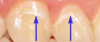
This disease usually debuts with the primary formation of very small chalky spots, which differ from other problems by their shiny surface. Quite quickly over a short period of time, the area of such whitish lesions increases. After a while, the spots lose their shine, and in return, on the contrary, a certain roughness appears.
A little later, the enamel with this disease becomes matte and changes its color to darker shades. Subsequently, directly on the areas previously affected by stains, the tooth enamel simply disappears and the dentin is almost completely exposed. Further, if the disease progresses, the boundaries of such dental lesions only increase noticeably over time.
At the most advanced stages, the enamel of teeth affected by necrosis becomes so weak and friable that it becomes possible to simply scrape it off with the simplest dental instruments. In addition, in patients suffering from cervical necrosis, as a rule, everything becomes as sensitive as possible.
Moreover, increased sensitivity extends to literally all types of existing irritants: it can be cold, too hot, salty or excessively sour food. It is believed that with a timely (usually urgent) visit to an experienced dentist, necrotic lesions of tooth enamel can be eliminated, thereby significantly inhibiting the further progress of such an unpleasant and dangerous disease.
Secondly, it could of course be acid necrosis. Such damage to the hard tissues of certain teeth can occur as a result of some extremely aggressive effects on tooth enamel of certain acids and sometimes alkalis. It is generally accepted that at certain enterprises associated with the production of acids (or in other places where certain acids are used daily on a large industrial scale) a huge amount of so-called acid vapors can accumulate directly in the air.
Actually, such vapors can quite easily penetrate the oral cavity of any person who works daily at such an enterprise. Further, after the initial entry into the oral cavity, acidic vapors mix (and quite easily) with saliva, thereby making it acutely acidic. As a result, acidic saliva gradually begins to destroy tooth enamel, washing out calcium from them every day.
The most dangerous for tooth enamel are considered to be nitric, sulfuric, as well as acetic, lactic and, of course, phosphoric acids. Some alkalis can be no less dangerous in this sense.
Most often, the acid variant of the disease is characterized by patient complaints of excessive sensitivity of tooth enamel. Unbearable pain when exposed to even minimal temperature or mechanical external stimuli. In some cases, the disease is accompanied by a feeling of the teeth themselves sticking together immediately when they are habitually closed.
Often, as this disease progresses, the enamel can become downright rough; in some cases, it can acquire an unpleasant grayish tint. As a rule, in the future, fairly rapid abrasion of the surface of the teeth can occur. After which some shortening and even destruction of the dental crowns is observed, and the destruction can occur right down to the nearest gingival margin.
Symptoms
The first thing a person suffering from this diagnosis begins to complain about is a change in color pigment. When affected by hydrochloric acid, the shade changes to yellowish-gray; against the background of the influence of sulfuric acid vapors, the pigment becomes almost black; when toxic with nitrogen, it becomes snow-white.
At the same time, the enamel itself becomes dull, and when palpated, its roughness is felt. This is the initial stage of the disease.
Hard tissues lose strength, focal destructive fragments appear. When eating cold or too acidic food, pain occurs, which, however, quickly passes.
As necrosis progresses, discomfort occurs more and more often, and it becomes difficult to brush the organs with a toothbrush. Any mechanical pressure causes severe discomfort.
The damaging processes, growing rapidly, begin to have an increasingly brighter color - the outlines of the formation are uneven, the cavity bottom is matte, its probing is too painful . The teeth begin to wear down in all directions, which causes partial chewing dysfunction.
Kinds
The following types of disease are distinguished:
- Cervical necrosis – affects the dental neck. The enamel becomes dull, whitish focal manifestations appear, hard tissue loses strength, crumbles and becomes loose. Progressing rapidly, the disease can completely destroy hard tissue. The main reason is hormonal imbalance.
- Acidic, where the provoking factor of development is acidic vapors. They actively wash away the calcium component responsible for the strength of the enamel. Clinical manifestations are surface roughness and a change in color pigment to dull gray. The organ becomes extremely susceptible to temperature changes.
- Post-radiation – considered a consequence of exposure to radiation. In advanced stages, the tooth can become almost black. In this case, pain syndrome is completely absent. The insidiousness of the disease is that if treatment is not started immediately, you can lose an entire fragment of the jaw row.
- Computer necrosis - the disease progresses against the background of the negative influence of ionizing radiation flows coming from the monitor. This form of pathology is characterized by dark brown pigmentation of the enamel and a matte, rough surface.
Cervical
The type of necrosis received its name due to the fact that tooth decay begins in the cervical area. From a small, outwardly almost imperceptible spot, the lesion grows, covering the tooth enamel.
This type is often caused by endocrine pathologies and hormonal changes during pregnancy. The main symptoms of this type of hard tissue necrosis include increased sensitivity of teeth to temperature and acidity of foods and drinks. Patients experience an unpleasant odor from the oral cavity.
This is interesting: Treatment methods for loose teeth in adults
Acid
Acid necrosis of teeth occurs when enamel comes into contact with various acids, both organic and inorganic.
Common among chemical production workers, with frequent vomiting during pregnancy, poisoning, and gastrointestinal diseases. Acids or their vapors destroy the mineral composition of the teeth, in particular calcium, in the damaged areas and acid necrosis begins.
Unlike the cervical type, the symptoms of the acidic type are not pronounced; thinning of the enamel and exposure of dentin can be completely painless.
Radiation
Quite common among cancer patients during radiation therapy, as well as people working with radio wave radiation. The extent of the damage and the general condition of the tissues depend primarily on the degree of irradiation. The localization of the lesion in radiation necrosis most often occurs in the enamel area adjacent to the gum. The symptoms are quite extensive.
The main features include:
- swelling and redness of the gums in the area adjacent to the tooth;
- general malaise;
- decreased saliva production;
- discomfort in the gum area;
- development of iron deficiency in the blood.
Computer
Computer necrosis is observed in patients who spend a long time at the monitor, on average several hours a day for 2–4 years. Radiation leads to damage.
With computer necrosis of teeth, pain is almost completely absent, and the damage begins not from a tiny area of enamel, as in other cases, but from a fairly large area of hard tooth tissue. In some cases, a change in the pulp is observed; it acquires a bluish-gray tint, while the tooth enamel, without being completely destroyed, becomes rough. The root and even the bones of the jaw are also affected, which makes treatment quite labor-intensive and time-consuming.
Diagnostics
High-quality and timely diagnosis is the most important condition for preventing damaging processes from acting globally, leading to irreversible consequences.
Their main symptoms are massive tooth decay, up to complete loss. To prevent this from happening, use the following methods to identify the defect.
Visual inspection and equipment
When conducting a clinical examination of the patient, the doctor will certainly notice the external manifestations of the diagnosis, and after collecting a complete history and listening to the patient’s complaints, he will quickly give the correct conclusion.
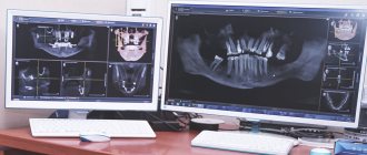
If the information obtained is not enough, an x-ray examination is performed. The problem area is specifically studied for the structural condition of hard tissues.
In the image, organs affected by necrotic lesions will have blurred boundaries, which are a direct consequence of demineralization.
Differential diagnosis
And although their localization is the same, there are still differences:
- with necrosis, the surface will always be matte and there will be no shine at all;
- another focal formation is observed.
Determining the type of pathology
In order to accurately understand the type of pathology, in addition to the signs described above, it is differentiated according to the following indications:
- computer necrosis has an immediate damaging effect on the pulp, which is unusual for other types of disease. This difference can be easily seen in the X-ray results. This same form of anomaly is characterized by a complete loss of shine of the entire tooth, and not the affected area;
- the acid type progresses several times more slowly; it is practically latent for a long time. This is uncharacteristic of the radiation form, the main difference of which is the sharp edge parts. In addition, this type of anomaly is associated with a general deterioration in the patient’s health and has more pronounced symptoms.
Consultation with specialists of other profiles
In some cases, it is simply necessary to listen to the opinion of specialized specialists, such as a gastroenterologist or endocrinologist.
Their consultation will help shed light on the true cause of the progression of the disease and develop the correct therapeutic treatment regimen.
Diagnosis of dental necrosis
If necrosis of hard dental tissues is suspected, radiography is performed. The image shows transparent and unclear teeth, which means they are hypomineralized. Given the large area affected, diagnosing the disease is easy. The task is complicated by the need to classify something that the doctor has not encountered before.
Modern PCs and color TVs have little radiation and create a special field that has an electrostatic effect, calling into question the stability of the body.
Computer necrosis of teeth involves partial destruction of odontoblasts and deterioration of the functioning of these cells, as well as other components of the pulp, including the response to the reaction of radiation on the structure of dentin and enamel. An unpleasant consequence of the disease is the problem of the functioning of the salivary glands with further remineralization of the enamel at the physiological level.
In order not to lose oxidative homeostasis, antioxidant reserves are used, but often they are not enough if there is a lack of antioxidants in the body.
Treatment regimen
Depending on the type of disease, various schemes for its elimination are used. In any case, this is a complex event consisting of a number of sequential manipulations.
At the stage of formation of the disease, the principle of treatment is to eliminate the factors that provoked it, as well as to return the tissues to their former hardness. At the progression stage, the emphasis is on restoring pronounced organ defects.
The following treatment regimen can be considered the most effective:
- Calcium glycerophosphate is a product for internal use. The daily dose is 1.5 g at a time. Strengthens tissues and prevents their further loosening;
- Klamin is a drug for systemic use and has a cumulative effect. Drink 2 tablets for 14 days;
- Phytonol - suspension is dissolved in water and consumed 15 minutes before meals. The course of treatment is 60 days. Single dosage 30 drops. Restores dentin at the cellular level;
- Multivitamins are a drug with a general strengthening spectrum of action. Drink 3 tablets throughout the month;
- Pastes - applied by application. Phosphate-saturated strips are applied to the affected area for several minutes. The procedure is repeated daily until the result improves.
After 90 days, the course of treatment is repeated.
Therapy for necrosis is strictly sequential and includes the following steps:
- gentle cleansing of the affected fragments;
- complex treatment according to the scheme discussed above;
- in a difficult clinical situation - orthopedic measures, including dissection of the affected fragments and the application of special mixtures with a strengthening effect. A temporary filling is placed on them;
- After 40-45 days, the cavity will be opened, re-cleaned, freed from filling material and a permanent glass ionomer will be installed.
In this article we will talk about the differential diagnosis of deep caries.
Treatment methods
Each type requires long-term treatment and the selection of an individual therapy regimen, which can only be prescribed by a dentist after a detailed examination.
As a rule, treatment is complex and includes local remineralization procedures, fluoridation and the use of general medications.
What is it aimed at?
The main goal of treatment is to restore the quality and integrity of dental tissues.
At the initial stages of pathology development, therapy is aimed at eliminating the causes that caused it, as well as restoring the density of the dental tissue structure by saturating it with minerals.
In more advanced cases, therapy will additionally solve the problem of eliminating tooth surface defects.
Our next publication will discuss methods of treating dental hyperesthesia.
In a separate article we will talk about how to get rid of yellow plaque between teeth.
Scheme
The main treatment can only be prescribed by the attending physician.
Of the publicly available methods, the following treatment regimen is used :
- Calcium glycerophosphate. Take 1.5 g per day orally for a month.
- Klamin . For at least 2 weeks, take 2 tablets per day;
- Phytonol . Dilute 30 drops of the drug in a small amount of warm water and drink at least 15 minutes before meals. The product must be taken for about 2 months.
- Multivitamin complex . Complivit or Kvadevit are recommended, taking 3 tablets per day for a month.
- Applications using pastes that contain large amounts of phosphates. Applications are applied to the enamel every day, leaving for 5 to 15 minutes.
The course of treatment according to this regimen must be carried out every 3 months .
How diverse the causes and treatment methods for dental necrosis can be, look at the video:
General rules
Treatment of any type of necrosis has a certain sequence:
- First, the affected tissues are cleansed.
- Then complex remineralizing therapy is used.
- In case of severe destruction, orthopedic treatment is carried out with preparation of the affected area and the application of strengthening pastes, which are covered with a temporary filling.
- After 1.5 months, the defect area is again opened, cleaned and filled with a permanent glass ionomer filling.
Prevention
The main prevention of the disease is to avoid or minimize the impact of factors that provoke its development, causing partial death of solid fragments.
This can often be achieved by adjusting your diet correctly, eliminating sweets and too acidic foods.
Oral hygiene and regular visits to the dentist will also reduce the risk of developing the disease , especially when the initial stages of its formation are already present. If you go to the clinic at this stage, the progression can be stopped.
The approximate cost of treatment, taking into account the regional average, is as follows: