In some cases, a pathology such as necrosis develops in the human body. It represents the death of tissue structure, which is irreversible. Quite often the pathology is localized in the gingival area. However, necrosis can also affect the tissue of the heart muscle and occur against the background of bedsores. An important characteristic is that the symptoms in the initial stages of the development of the pathology are blurred. When the symptoms manifest themselves in full, the process already becomes irreversible, it is practically impossible to stop it, and cell regeneration becomes impossible.
The causes of this pathology
The death of gum tissue can occur for a number of different reasons, the main one of which is poor circulation in the gums. Quite often, necrosis of the gums is accompanied by necrotic lesions of the teeth. The main causes of disruption of the structure of gum tissue are:
- Inadequate oral hygiene, which leads to the development of gingivitis, which in turn provokes an inflammatory process that affects the teeth and gums.
- Traumatic damage to the gums resulting from dental treatment due to malocclusion, burns, acid exposure, lightning strikes, constant compression of blood vessels, prolonged exposure to cold.
- Necrosis of the gums often occurs after anesthesia.
- Decreased immunity, vitamin deficiency, anemia.
- Regular use of narcotic or toxic substances.
- Heavy exposure to radioactive isotopes, radiation therapy.
- Endocrine pathologies, hormonal imbalances.
- Violation of blood composition and metabolism.
- Infection, advanced form of gingivitis or periodontal disease.
Preventive actions
Prevention includes activities carried out by the state or agency and individual measures taken by individuals.
State/departmental measures include:
- reducing the concentration of acid vapors in work areas to a level that is harmless to health using technical means (ventilation, filters, absorbers);
- fluoridation of drinking water (increasing the fluorine content to 2 mg/l;
- medical examination : regular (at least once every 3 months) dental examination by workers at chemical enterprises;
- if necessary, providing workers with personal protective equipment for the respiratory system and oral cavity;
- clinical examination of persons at risk for acid necrosis with preventive remineralization therapy.
Individual measures include:
- Mandatory wearing of PPE (if prescribed by safety regulations).
- Workers at chemical plants rinse their mouths every 2 hours with alkaline solutions (1% borax, 2-3% bicarbonate of soda).
- Applications or rubbing of fluoride-containing toothpastes into teeth.
- Using glass straws when drinking acidic drinks.
- A healthy diet that includes eating foods rich in calcium and fluoride - vegetables, fruits, dairy products.
Prophylaxis is an effective means of preventing acid necrosis. With its help, you can significantly reduce or completely eliminate damage to teeth from acid solutions.
The video provides additional information on the topic of the article.
Arsenic
There is also another reason that is toxic in nature, which should be highlighted separately. In some cases, gum necrosis develops from arsenic.
When devitalizing the pulp cavity, dentists still use a paste that is an arsenic compound. This poison has a destructive effect on the skin, bone marrow, gastrointestinal tract, lungs and kidneys. It has an extremely destructive effect on body tissue. Over time, the arsenic filling can become depressurized and cause gum necrosis. The incubation period takes several years, so the patient notices the appearance of negative symptoms too late.
Signs
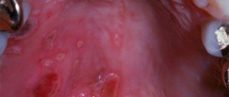
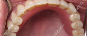
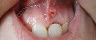
Gum necrosis goes through several stages in its development. At the initial stages, fairly harmless symptoms appear - the surface of the gum slightly peels off from the tooth, bleeding of the gums increases, pigmentation is disrupted, and the enamel loses its shine. Another symptom of gum necrosis is discoloration of the tooth surface and the appearance of roughness on it.
At the middle stage of development, there is the appearance of an unpleasant odor from the oral cavity, swelling, redness of the gums, and enlarged lymph nodes. In the absence of the necessary therapeutic measures, the condition begins to deteriorate: the temperature rises to borderline values, a gray coating appears, and bleeding of the gingival papillae increases. The tissue areas gradually begin to turn black, become covered with ulcers, and the teeth become unstable. As a result of constant fever, the patient's appetite and sleep patterns are disrupted, and headaches develop.
At the last stage, the destructive process and symptoms of gum necrosis worsen. Foci of inflammation become visible visually, severe swelling appears. The gum tissue begins to die, causing a foul smell, and the bone structures are exposed. Dyspeptic disorders and fever become permanent. The process develops until the chewing function is completely lost.
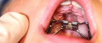
Post-injection necrosis of the hard palate and other anesthetized areas
Many cases of necrosis of injected tissues, especially the mucous membrane of the hard palate, have been described after infiltration anesthesia with a solution of novocaine-adrenaline [V. S. Belkovich, P. K. Broshnikovsky, I. M. Vainberg, G. I. Vidensky, V. M. Mikhelson, G. F. Nikolaev, E. A. Rabovskaya, A. B. Raiz, I. X. Safinsky, Hoffmann, Villiger, etc.].
To explain the predominant damage to post-injection necrosis of the hard palate, the following etiological points are put forward: 1) the presence of dense, inextensible and inflexible tissue in the palate area; 2) damage by the anesthetic solution to the only vascularizing this area of the anterior palatine artery or its branches, as well as the palatine vein, or their injury, followed by hemorrhage, entry into the vessel of the anesthetic solution, the occurrence of thrombosis and the onset of a severe circulatory disorder as a result; 3) the presence of bad exchange in this area.
Post-injection necrosis of other areas of the mucous membrane of the oral cavity and jaw is observed much less frequently. Here, when these areas are affected, the etiology is as follows: 1) the influence of the sharply anemic effect of adrenaline; 2) infection; 3) use of a highly concentrated anesthetic solution for infiltration anesthesia; 4) use of a non-isotonic anesthetic solution; 5) injecting an anesthetic solution too close to the edge of the gum into dense and stubborn tissue; 6) too rapid administration of fluid; 7) damage to nerve branches, leading to disruption of trophism; the formation of a hematoma, which increases pressure and disrupts tissue nutrition; 9) the presence of harmful and highly irritating impurities in the tissue in the anesthetic solution, syringe or needle, which can happen when the syringe and needle are stored in solutions of phenol, Lysol, etc. and used for anesthetic injection without prior rinsing; 10) erroneous replacement of the anesthetic solution with a caustic or highly irritating liquid (for example, ether, alcohol, lapis, formalin, tincture of iodine, ammonia, gasoline, various acids, etc.).
We observed cases of post-injection necrotic lesions of the mucomaxillary areas in patients referred to us regarding this complication for consultation and treatment. One can also note the predominant, but not
the exclusive localization of this complication in the palatal region.
Regarding the etiology of this complication, when it is not associated with an erroneous replacement of the anesthetic solution, we do not attach exclusive importance to any one of the above-mentioned points, but see the cause of this complication in a combination of a number of points or in any individual of them under appropriate conditions. Wrong, in our opinion, are those who see the only cause of the complication in question in the anemic effect of adrenaline and, moreover, do not quite correctly attribute a stronger anemic effect to the stale solution of the drug; those who see all the evil in a disorder of the blood circulation are also not entirely right.
with possible detachment of the periosteum, completely ignoring the influence of infection and other issues.
Among the cases we observed, where small necrotic areas of the mucous membrane and small bone sequesters several millimeters in size with rapid complete healing were predominantly separated, there were a number of cases where large ones, several millimeters in size, were isolated
centimeters, sequesters.
The release of sequestra due to postoperative necrosis in the palate in four of our cases led to the formation of a communication of significant size between the oral and maxillary cavities, which required plastic closure of the defect.
It must be assumed that various diseases can create favorable conditions for such complications.
In the most severe cases we observed, we identified malaria, tuberculosis, syphilis and
other diseases.
Necrosis of injected tissue most often occurs as a result of injecting an anesthetic solution into tissue that is difficult to stretch, especially when the injection is performed quickly and under high pressure.
One of the most unpleasant complications, leading to sequestration of the injected areas of the jaw and adjacent soft tissues, and often also to a severe general condition, during local injection anesthesia, of course, is the injection of another, mistakenly taken substance instead of an anesthetic. This can happen during mass anesthesia and, as a rule, is associated with a certain negligence of the doctor and his assistants.
Naturally, in the event of an erroneous injection of irritating and toxic substances, the course of the process in the affected areas of the injected tissues and the disease of the body depends on the nature of the injected substance and its concentration.
The literature provides a number of cases of these errors.
Villiger observed a case where soapy alcohol was injected instead of novocaine. The consequence was sequestration of the corresponding area of the jaw and adjacent soft tissues. The same author described a case where iodine tincture was injected instead of an anesthetic liquid. Here the matter was limited only to necrosis of soft tissues in the injection area.
E. M. Gofung observed a case where, to remove an upper molar, instead of novocaine, alcohol, which had previously been used to wash the syringe, was injected. Outcome - after several weeks of severe pain and necrotic phenomena, rejection of the entire palatal plate of the corresponding upper jaw adjacent to the injection site,
It should be noted that sequestration of the palatal plate in this case may have been associated not only with the injection of alcohol into the periodontal tissues, but also with the introduction of infection as a result of using a syringe that was not boiled, but only washed with alcohol.
N.I. Agapov describes a case when formalin was mistakenly injected instead of novocaine for pain relief during the extraction of a lower tooth on the right. Fortunately, due to the severe pain during the injection, very little of this substance was injected. However, by the morning of the second day, the patient’s right half of his face was swollen, radiating pain appeared in the right half of his head and significant swelling in the area of the lower jaw on the right, near the molars where the liquid was injected. The mouth opened only the width of a finger, and then it was extremely painful. These pains and all accompanying phenomena continued for 10 days; subsequently, sequestration appeared along all three molars and then the wound gradually healed. According to the author, such a relatively favorable outcome occurred only because the doctor managed to introduce only 0.5 ml of formalin and only under the gum, but not under the periosteum.
This author describes 2 more cases where ammonia and a solution of 10% lapis were injected. In both cases, severe pain appeared during injection; the patients appeared on the second day with large swelling and severe radiating pain. If these two cases resulted only in the sequestration of larger and smaller sections of bone, it is because immediately after the mistake was made, wide incisions were made in the soft tissue in the area where the injection was made.
In 1952, we observed a patient admitted to our clinic from the region, in whom, to produce intraoral mandibular conduction anesthesia, three days earlier, instead of a 2% novocaine solution, a 5% formaldehyde solution was mistakenly injected. As a result, the patient developed a general serious condition with severe damage to the maxillofacial area: severe general weakness, fainting, headaches, dizziness, cardiac weakness, an extensive and very painful necrotic ulcer in the area of the anterior edge of the lower jaw branch, cheeks, gums and the floor of the oral cavity within the molars, difficulty opening the mouth and subsequently perimaxillary phlegmon. The following activities were carried out in the clinic: opening of perimaxillary phlegmons, multiple blood transfusions, penicillin therapy, diet therapy, cardiac, painkillers, bromides, etc., as well as irrigation and rinsing of the mouth with antiseptic and antibiotic agents, mechanotherapy.
After a six-week stay in the clinic, the patient was discharged in satisfactory condition.
The cause of the misfortune in this case was that on the day of this unpleasant incident, 2% novocaine and a 5% formaldehyde solution were simultaneously received from the pharmacy in the same container. The sister mistakenly gave the doctor a bottle of formalin instead of novocaine. The doctor, without checking the contents of the bottle, performed a mandibular conductive injection of 4 ml of formalin solution.
In 1955, under our supervision was patient G., 46 years old, who was treated by a dentist in one of the clinics to remove b| under local injection anesthesia, instead of a solution of novocaine, 4 ml of hydrochloric acid were mistakenly injected (approximately 3 ml from the buccal and 1 ml from the palatal side of the tooth being removed).
In the dental office of the said clinic, in the medicine cabinet, on the same shelf and in the same containers, were placed a solution of novocaine and hydrochloric acid, which the office nurse sometimes used to clean various objects. One day, after a doctor suggested boiling a solution of novocaine to numb a tooth extraction, a sister boiled 5 ml of hydrochloric acid in a test tube. Despite the fact that hydrochloric acid smoked when boiled and injecting it into the patient’s gums was accompanied by severe pain, neither the nurse nor the doctor suspected the possibility of a fatal mistake. Only after the tooth extraction, during which the patient experienced very severe pain, did the doctor notice that a very unpleasant mistake had occurred. The doctor, confused, did not make any incisions in the tissues injected with hydrochloric acid and sent the patient home in order to visit him soon.
The swelling that appeared immediately after the tooth was removed began to quickly increase in all directions and spread to the temporal, parotid, submandibular and orbital areas, as well as to the upper lip. Paresis of the facial nerve of the corresponding side also occurred. Body temperature increased to 38.5°. The next morning we were called to the patient. He spent the night very restlessly, almost without sleep. He complained of pain in the entire half of his face and head, general weakness and lack of appetite. Body temperature is 38.5°, the pulse is rapid, uneven and somewhat weak. Significant swelling of the perimandibular tissues on the right; the eyelids of the right eye are swollen and closed. Mouth opening is limited. The gums and transitional fold of the mucous membrane in the wound area after tooth extraction are swollen, and the injection site is ulcerated. The inside of the gums and the mucous membrane of the palate are also swollen in this area, and the injection site is ulcerated. The extraction wound is covered with a dirty gray foul-smelling coating. There was stool, urination was somewhat delayed. Re-
39S
recommended: bed rest, intramuscular injection of penicillin 70,000 every three hours, drinking plenty of fluids, appropriate non-solid food, rinsing and irrigating the mouth with a soda solution (1 teaspoon per glass of digested water), painkillers powders (pyramidon 0.3, dionine 0. 01) 1 powder 2-3 times a day, subcutaneous injection of camphor oil 3 times a day.
Over the next two days, the temperature did not drop, the pulse improved somewhat, the patient slept a little, but his sleep was restless; the patient was sweating, urination was insufficient, and there was no stool. The swelling of the face on the right did not decrease, the eyelids of the right eye were closed. During these days, the opening of the mouth has become even more limited—the mouth opens by 1.5 cm. The same treatment was prescribed, as well as washing the right eye with a penicillin solution, bromide mixture, and a cleansing enema.
On the fourth day, the patient continued to complain of general weakness and pain in the affected area and in the head. Body temperature decreased - 37.8° in the morning, 38° in the evening. The pulse has leveled out somewhat. Appetite has improved slightly. Urination is normal. Stool - after an enema. Mouth opening - 1.5 cm. There is an unpleasant odor from the mouth. Disintegration of submucosal tissues began to be noted in the places where injections were made, especially pronounced on the buccal side. The extraction wound has cleaned up slightly.
Recommended: continue penicillin therapy and bromide mixture, injection of camphor - 2 times a day, removal of sequestering tissues at the site of former injections with subsequent iodization of the affected areas, rinsing the mouth with a soda solution, food rich in vitamins, mandatory intake of vitamins C, b] 3 times a day , taking painkillers as needed. Continue rinsing your eyes with penicillin solution and try to actively open your mouth. Outside - a dry cotton bandage.
On the sixth day, the temperature dropped to normal. The pulse became even more even and only slightly faster. The headaches have calmed down significantly. Sleep began to normalize. The swelling of the surrounding areas decreased significantly and remained only in the affected area. The facial nerve palsy continued. The eyelids of the right eye opened, the eye was watering, but there was no pus. The patient became more cheerful.
Therapeutic prescriptions: bed rest and camphor intake were canceled, penicillin therapy was recommended to continue for two more days - 2 times a day, 250,000 units, active opening of the mouth.
On the eighth day, the patient's general condition improved significantly. The headaches had almost stopped, but the dizziness still remained. Sleep and appetite have returned. Tempe-
Body temperature and pulse returned to normal. The bad breath did not disappear. The disintegration of tissues on the side of the palate decreased, and on the buccal side it increased - significant rags of sequestered tissue began to stand out, mainly due to the disintegration of Bisha's lump. The mouth opened 1.8 cm. The extraction wound healed. Penicillin therapy was canceled. Care for the affected area is the same: removal of sequestered areas, followed by iodization of this area; rinsing your mouth with a soda solution. Strengthening nutrition. Vitamin therapy - rosehip infusion 2 times a day, half a glass, lemons, black currants and other fruit juices.
To improve mouth opening and prevent scar contracture of the lower jaw, it is recommended to intensively engage in passive (using an apparatus) mechanotherapy - alternately inserting clothespins with the reverse side and rubber plugs between the teeth.
On the 13th day after the injection of hydrochloric acid, the disintegration of tissue on the palatal side stopped, and on the buccal side it began to decrease and continued for about another week. Over the following days, the mouth gradually began to open more. The unpleasant odor became less pungent. Swelling of the soft tissues in the area of the right upper jaw gradually decreased. The general condition gradually improved. The patient was prescribed physiotherapeutic procedures for paresis of the facial nerve and to eliminate residual inflammatory phenomena.
Only 5 weeks after the onset of the disease, the patient with traces of paresis of the facial nerve and swelling of the maxillary region on the right (the mouth opened almost to normal) began to work.
It is interesting to note that, according to the observations of many authors, as well as our own, the introduction of another erroneously taken substance instead of an anesthetic solution often occurs during an additional injection, due to the lack of analgesic effect from the first injection, carried out either with an anesthetic solution, or even more often with the same erroneously taken substance.
Prevention. General preventive measures boil down mainly to the following.
1. We should once again remind you of the need to always boil the syringe and needle before an anesthetic injection, and after the injection, rinse them with saline, alcohol or ether, then dry and store not in antiseptic solutions, but dry and in a dry place.
2. A minimum amount of anesthetic solution with the weakest possible concentration of adrenaline should be injected into the mucous membrane of the palate. When pale
mucous membrane of the palate during an anesthetic injection
you should either stop injection or reduce the intended dose.
3. For infiltration anesthesia in the palate, an anesthetic solution with a weak concentration of adrenaline is used (no more than 1 drop per 10 ml of solution).
4. The anesthetic injection should be given slowly and without excessive pressure on the piston. To ensure the latter circumstance, you should not use syringes with screw-on needles and with transverse projections for fixing fingers. When using Record type syringes, the needle usually jumps off the syringe as anesthetic liquid is injected into tight tissue under strong pressure, which prevents tissue ruptures and injury
cells released under such pressure by an anesthetic liquid.
5. It should also be recognized in relation to the prevention of this complication the advantage of conduction anesthesia over
infiltration, in particular in the area of the mucous membrane of the palate.
6. The doctor before drawing into the syringe,
solution for injection anesthesia, you must personally verify that the container contains exactly the indicated anesthetic solution. To avoid possible unfortunate errors, the anesthetizing physician should not rely on
only on the instructions of auxiliary personnel or doctors working together with them.
In the extraction room, on the table near the doctor during anesthesia, there should be no other liquids except solutions of novocaine and adrenaline.
The appearance of severe pain at the injection site of the “anesthetic” liquid should raise suspicion of an accidental mistaken injection of some other harmful liquid instead of the anesthetic solution. In these cases, you should immediately stop further injection and continue it again only after carefully checking the liquid contained in the syringe and being completely convinced that there is no such error. But this is possible if you follow the rules of very slow production of anesthetic injections and
carefully monitoring the patient’s reaction to the injection of an anesthetic solution.
It is strictly forbidden to store other liquids, especially poisonous ones, in the medicine cabinet together with injection solutions, in particular with anesthetic solutions.
'Toxic liquids must be stored in a separate cabinet, in special containers, in accordance with the instructions of the Pharmacopoeia. V. M. Mikhelson, having analyzed the reasons known to him
OCW
cases of complications during anesthesia in dental practice, in particular the erroneous administration of toxic substances instead of an anesthetic solution, comes to the following conclusion.
These errors are primarily the result of organizational problems, and the author points out that such errors predominantly occur in outpatient settings rather than inpatient settings. Errors are more often observed in those dental institutions where there are no special not only surgical days, but even hours, and the doctor often switches without any break from conservative therapy to surgery. Errors occur more often in clinics without a special surgical room and without regular checks of medication storage. Mistakes occur where the doctor does not pay attention to the patient’s restless behavior during the injection and attributes it to “the patient’s hysteria and nervousness.”
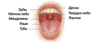
Diagnosis of gum pathologies
During a routine examination, a competent dentist will definitely identify gum diseases, detect the presence of bedsores under dental processes, and listen to the patient’s complaints. The main criteria for assessing a patient’s condition for a doctor are:
- The smell that comes from the mouth.
- Condition of gingival tissues.
- Gum color.
- Bleeding and pain that occur.
- Symptoms of intoxication (insomnia, upset stomach, headache, loss of appetite, hyperthermia).
Therapeutic effect
Therapy of necrotic changes involves, first of all, restoring blood circulation in the damaged tissue and stopping further destruction. Tissues that have already died should be removed, since their structure cannot be restored. Among other things, therapy for gum necrosis involves strengthening the general immune system. Specialists will prescribe a number of measures aimed at improving vascular and nerve patency and returning to the natural regenerative functions of cells.
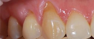
Gum necrosis (photos can be found on specialized resources) is divided into two types:
- Coagulation (dry). With such necrosis, the inflammatory process is replaced by the process of mummification of the affected tissues.
- Colliquation (wet). This form is characterized, on the contrary, by the presence of putrefactive inclusions, purulent foci, edema, and extensive inflammation.
Therapy for liquefaction necrosis is not aimed at its transformation into coagulation necrosis. For this purpose, the abscesses are opened, the inflamed areas are drained, and special bandages are applied. Damaged surfaces are treated using hydrogen peroxide. If such actions are not effective, removal of dead tissue is prescribed. It is also necessary to stop the poisoning of the body - the patient is prescribed antibiotic drugs and vascular measures. A negative characteristic of wet necrosis is its rapid spread, resulting in irreparable harm to the body.
Treatment of coagulation-type gum necrosis involves treating the affected gums with antiseptics, removing dead areas that have lost functionality, and conducting therapy aimed at restoring blood circulation.
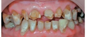
Necrotization of the gums can also be caused by a burn. Immediately after the incident, you should remove the product that caused it and seek help from a dentist. Regardless of the type of burn (chemical or thermal), it is necessary to take into account the depth of the injury. When tissue is rejected, initial treatment for a gum burn involves surgery to remove the area that is dying. If the burn is mild, the dentist will prescribe rinsing the mouth using aseptic medications and treating with medicated ointments and gels.
Treatment tactics
The method of treatment depends on the extent of the damage , but in any case, as a first step, measures should be taken to stop or at least reduce exposure to acids.
In the initial stage of the pathology, systemic and local remineralizing therapy is carried out.
Drugs containing calcium gluconate or glycerophosphate are prescribed internally. The usual daily dose is 1.5 g in 3 divided doses. The course of treatment lasts a month.
Additional medications - phytolon (30 drops twice or thrice a day, 15-20 minutes before meals, duration of treatment - 60 days), klamin (1 tablet per day). Multivitamins are also prescribed, in particular, Kvadevit - 3 tablets per day for a month.
Local remineralizing therapy involves applications with a solution of 2% sodium fluoride. Teeth isolated with cotton wool rolls are covered with a swab moistened with sodium fluoride, closed for 5 minutes, after which the swab and cotton rolls are removed, and the mouth is rinsed with water.
When caring for the oral cavity, fluoride-containing toothpastes are used for 5-6 months.
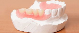
In case of severe tooth decay, 3-6 months after remineralization, the affected areas are filled with glass ionomer cement and, if necessary, prosthetics are performed.