Features of the maxillary sinus
The maxillary sinus (its other name is the maxillary sinus) is located in the thickness of the bone tissue of the upper jaw. It is separated from the oral cavity by the alveolar process of the upper jaw, which forms its bottom. The volume of such a sinus is quite large, and in adults it can reach 10 cubic centimeters.
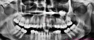
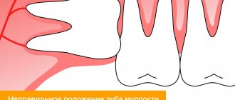
In the photo: the roots of the teeth at the bottom of the maxillary sinus
This sinus, or sinus, is not airtight. It communicates with the nasal cavity through a narrow slit.
Typically, perforation of the maxillary sinus occurs in the area of its bottom. Some of its features contribute to this:
- Close proximity of the roots of molars and premolars. In some cases, the thickness of the bone layer between the tooth roots and the bottom of the maxillary sinus can be relatively large - up to 1 cm, but in some people the bone border between these formations is very thin - no more than 1 mm.
- Sometimes the roots of the first and second molars are located in the sinus cavity itself, separated from it by just a layer of mucous membrane.
- Rapid thinning of the bone layer in the presence of acute or chronic inflammatory diseases: periodontitis, periodontitis, cysts.
- Relatively thin bony trabeculae in the tissue of the upper jaw.
All this predisposes to the occurrence of perforation during dental procedures, even if the treatment technique was not violated and the doctor did not apply significant traumatic force.
Causes of perforations of the maxillary sinus floor
The etiology of perforations of the maxillary sinus is always associated with any dental procedures. Perforation can occur:
- when removing teeth;
- during endodontic treatment;
- during dental implantation;
- during root resection.
When teeth are removed, damage to the bottom of the maxillary sinus can be a consequence of either rough actions by the dentist or failure to comply with treatment tactics, or as a result of the anatomical characteristics of the patient himself (for example, when the tooth roots are located directly in the sinus cavity).
In the photo: the tooth root is located close to the bottom of the maxillary sinus, which increases the likelihood of perforation during removal
When endodontic treatment is carried out, one of the complications is perforation of the tooth root, which is often combined with damage and perforation of the bottom of the maxillary sinus. This happens when the root canals are excessively expanded, when brute force is used when inserting pin elements or compacting filling cement. With this type of perforation of the maxillary sinus, filling material or root fragments almost always penetrate into its cavity.
If perforation occurs at the time of insertion of a dental implant (it can be an implant of any brand, for example, Mis, Nobel, Xive, etc.) or during root canal filling or insertion of pins into the tooth root, then it is always a therapeutic error doctor's tactics.
Damage to the bottom of the maxillary sinus is a serious complication when implanting artificial roots into bone tissue during prosthetics. This is explained by the fact that after tooth extraction, bone tissue very quickly undergoes degeneration processes. As a result, the height of the alveolar process of the jaw decreases. If the doctor does not take this point into account and incorrectly carries out preparations before implantation, and also incorrectly selects the size of the implant, then the risk of sinus perforation is very high.
Tooth root resection is a treatment method for the presence of cysts in the area of its apex. If the patient is underexamined, when the doctor does not know the exact size of the bone plate separating the bottom of the sinus from the wall of the cyst, and also if it is necessary to remove a large volume of jaw bone, then perforation of the maxillary sinus is not a rare phenomenon.
Tooth in the maxillary sinus consequences
The maxillary sinus is known to many thanks to such a common disease as sinusitis. A person has 2 maxillary sinuses, located in the cavity of the bone above the upper jaw on either side of the nose.
They are quite extensive in volume; in an adult they can be up to 10 cubic centimeters in volume. The danger of perforation of the maxillary sinus occurs when teeth are removed in this part of the upper jaw.
Features and causes of pathology development
Most often, perforation of the maxillary sinus is discussed in connection with a visit to the dentist. It is dental procedures to remove the upper teeth that most often lead to this phenomenon. In this case, we can talk about perforation of the sinus floor.
The upper teeth are separated from the maxillary sinuses by thin bone plates, which in some places are no thicker than a millimeter. It's quite easy to damage them.
Perforation of the maxillary sinus is not always the doctor’s fault. Sometimes the doctor acts correctly and quite carefully, but perforation of the bottom still occurs.
We can identify the main reasons for such a phenomenon as perforation of the maxillary sinuses:
- Tooth extraction, which was already mentioned above. The bottom of the sinus can be damaged if the tooth is removed too quickly and forcefully using forceps. However, there are also anatomical features that make the sinuses especially vulnerable.
- Endodontic treatment. These are quite complex dental procedures that involve manipulation of the tooth tissues, gums, and roots. That is, with such treatment it is necessary to penetrate deep into the gum and under the tooth itself, which can also lead to perforation of the maxillary sinus. This treatment is used in difficult cases when the tooth is infected or almost destroyed, but it can still be saved.
- Dental implantation. This is the replacement of a missing tooth with an artificial implant, which is implanted using a special mechanism made of a durable and safe alloy. This is a difficult and expensive operation in which the root of the tooth is replaced with a device similar to a screw. If implanted incorrectly, there is a possibility of damaging the bone plate.
- Chronic periodontitis. This is an inflammation of the tissue around the tooth. With this disease, the bone plate that separates the upper molar from the maxillary sinus becomes thinner and flakes off. As a result, the tooth must be removed, and perforation cannot be avoided even with the most careful removal.
- Root resection. This is one of the methods of treating chronic periodontitis, when part of the root (its apex) is removed along with a purulent formation (cyst). With such manipulations with the upper teeth, there is always a possibility of perforation of the maxillary sinuses.
We suggest you read: Why a splinter remains after tooth extraction and how to correct the situation
Symptoms
What happens when the floor of the maxillary sinus is perforated? The maxillary sinus is not insulated and sealed; air circulates there. Therefore, during perforation, blood will flow out with air bubbles and at the same time some of the blood enters the sinus itself.
There is no need to self-diagnose. An experienced doctor will be able to quickly determine the cause of this phenomenon. However, if there are complaints and concerns, they should be shared.
The most common symptoms of perforation of the maxillary sinus are:
- Bleeding with air bubbles will begin from the tooth hole itself, which formed after removal. There will be more bubbles if you exhale sharply through your nose.
- When a tooth is removed, blood is observed only from the wound itself, and when the sinus is perforated, bleeding can also begin from the nose, from the nostril that is closer to the damaged sinus.
- With such a phenomenon, the patient begins to nasal and speak “out of his nose.” However, this cannot always be noticed immediately, because there is a cotton swab in the mouth, blood is flowing, and it is already difficult for the patient to speak.
- After some time, a sensation of free air circulation through the tooth socket begins. There is a feeling of pressure and heaviness in the upper jaw.
If damage occurs during implantation, the doctor’s instrument will fall a little deeper than intended or suddenly change its position. The doctor will quickly determine that perforation has occurred.
If, however, the perforation was small and went unnoticed, other and more alarming symptoms will begin to appear without treatment. Inflammation and a purulent process may begin. The head and upper part of the jaw will begin to ache, the pain may spread to the nose area.
If the process of inflammation begins, pus may accumulate in the sinus.
In this case, it will be difficult for the patient to breathe through his nose, one nostril will swell. After some time, pus will begin to be discharged from the nostril, there will be a feeling of aching in the nose area, the temperature may rise, and general weakness appears. This indicates the beginning of sinusitis. When contacting a therapist or ENT specialist, be sure to say that you have recently had a tooth removed or an implant installed.
Treatment options
Before prescribing treatment, the doctor must ensure the presence of perforation and complications. Therefore, the process of treating maxillary sinus perforation begins with diagnosis.
Perforation can be examined with a thin probe, x-ray or CT scan. It is advisable to do an x-ray with contrast or computed tomography.
The images will show whether there is a perforation, whether blood has entered, as well as fragments of the tooth and implant into the sinus itself.
Most often, treatment of perforation of the maxillary sinus is not possible without surgery. This can only be avoided if the dentist extracted the tooth and discovered the perforation immediately, took measures to eliminate the hole (usually they are sutured or treated), and then carried out a systematic examination of the patient.
However, even if immediately detected, an x-ray must be taken to make sure that no foreign body that could cause inflammation has entered the sinus cavity.
It is not always necessary to stitch up the perforation; sometimes it is enough to make sure that nothing gets into the sinus and then prevent the wound from opening. They treat the formed clot very carefully and try not to touch it.
To avoid causing bleeding, the tampon can be left in place for the entire 7 days, with a small suture placed on the gum.
- perforation of the maxillary sinus.
You can also close the perforation with a plastic plate. This will give it the opportunity to tighten without the risk of infection. Treatment of perforation of the maxillary sinus is carried out on an outpatient basis, at home. The patient is necessarily prescribed a course of antibiotics, anti-inflammatory and painkillers.
If perforation is accompanied by various complications, for example, the entry of fragments and foreign bodies into the sinus cavity, treatment is carried out only after hospitalization and a thorough examination. In this case, an operation is necessary, during which all foreign objects are removed and dead tissue is excised.
Treatment of such a problem as perforation of the maxillary sinus must be trusted to specialists. You cannot treat it at home with folk remedies, this can lead to very serious consequences.
Incorrect treatment, as well as its complete absence, are equally dangerous. Possible consequences of perforation of the maxillary sinus:
- Sinusitis, sinusitis. Sinusitis is a type of sinusitis when the maxillary sinuses become inflamed and pus accumulates in them. This can happen if the perforation is untimely or improperly treated and the infection spreads further. With sinusitis, headaches, a feeling of fullness, purulent discharge from the nose, and possible fever are observed.
- Loss of healthy teeth. The tissue around the wound formed after tooth extraction can become inflamed, resulting in damage to neighboring healthy teeth. They begin to get sick and require additional treatment.
- Osteomyelitis of the upper jaw. A serious disease accompanied by purulent and necrotic processes in the jaw bone. The patient experiences fever, headache, loose teeth, pain in the gums and jaw, and swelling of the mucous membrane.
- Meningitis. The most dangerous consequence is that it can lead to the death of the patient. Meningitis occurs when inflammation in the maxillary sinus grows and spreads to the membranes of the brain, causing them to become inflamed.
Protecting yourself from perforation of the maxillary sinuses is quite difficult, given that it often does not depend on the patient himself.
To avoid this phenomenon, before dental treatment and dental procedures, it is necessary to conduct a thorough examination of the condition of the patient’s gums and jaw, and carry out all operations carefully and in a timely manner.
We suggest you read: Pain after removal of a nerve or tooth: causes, treatment. How long does pain last after wisdom tooth removal? Pain in the ear, cheek, head after tooth extraction
Patients, in turn, must monitor the condition of their teeth and seek medical help in a timely manner. When the first signs of perforation appear, you must go to the clinic and begin treatment, following all the doctor’s instructions.
- The development of inflammation in the maxillary sinus and its spread to the adjacent bone tissue.
- Involvement of other sinuses in inflammation: frontal, ethmoid, sphenoid.
- Formation of an abscess or cellulitis.
- Loss of teeth located in the perforation area.
- Development of osteomyelitis and meningitis.
In order not to suffer from the consequences and not to expose yourself to danger, choose dentistry and your doctor correctly. With sufficient professionalism of the doctor, all operations on tooth extraction and implantation are carried out easily and safely.
Symptoms of perforation
If perforation of the nasal sinus occurred at the time of tooth extraction, then its symptoms will be quite specific:
- The appearance of small air bubbles in the blood released from the tooth socket, the number of which increases with a sharp forced exhalation through the nose.
- The appearance of bloody discharge from the nose on the side of the perforated maxillary sinus.
- Changes in the timbre of the patient’s voice, the appearance of “nasality”.
Sometimes the patient begins to complain about the passage of air through the hole after tooth extraction, as well as a feeling of heaviness or pressure in the projection of the maxillary sinus.
The photo shows perforation of the bottom of the maxillary sinus after tooth extraction
If perforation of the maxillary sinus occurs during implantation or endodontic treatment, the doctor may suspect it by:
- characteristic failure of the instrument or implanted element after applying some force to advance it;
- changing the position of the instrument in the wound;
- the appearance of small air bubbles in the blood.
If perforation of the maxillary sinus for any reason was not diagnosed and treated immediately, then its cavity becomes infected with the development of acute sinusitis or sinusitis, which is characterized by symptoms such as:
- severe acute pain in the maxillary sinus area;
- swelling of the nasal mucosa on the corresponding side with difficulty breathing through the nose;
- the appearance of purulent nasal discharge.
The appearance of general symptoms of intoxication is also characteristic: headaches, chills, high fever, weakness.
Diagnostics
Diagnosis of perforation of the floor of the maxillary sinus during tooth extraction is based on a typical clinical picture. In doubtful cases, as well as when such a complication is suspected during implantation or endodontic manipulations, it is necessary to use instrumental diagnostic methods:
- Probing the socket of an extracted tooth or perforated canal with a thin probe . This allows us to determine that there is no bone bottom in the wound. In this case, the instrument passes freely through soft tissues and does not encounter obstacles along its path.
- X-ray of the sinus area . In this case, the pictures can reveal both darkening of the cavity due to the accumulation of blood in it, as well as fragments of dental roots, implants or filling material. Sometimes it is advisable to conduct radiography with contrast, when a contrast agent is introduced into the cavity through a perforation fistula.
- Computed tomography , which allows you to determine perforations and the presence of foreign bodies in the sinus with maximum accuracy.
- If old perforations are suspected, general clinical blood tests , the result of which may indicate the presence of an active source of infection in the body.
Diagnosis of the problem
The diagnosis of damage to the sinus floor is based on the clinical picture. Among the instrumental examination methods:
- Probing - the perforated canal is examined using a thin probe to confirm the absence of a bone bottom in the wound.
- X-ray reveals darkened areas represented by blood accumulated in the sinus. Fragments of roots, implant or filling material may also be found. To clarify problematic issues, radiography with contrast can be performed.
- Computed tomography - determination of the location of the perforation and foreign bodies. Gives the most accurate information.
- General clinical blood test - in advanced cases.
Computed radiography procedure
A perforation of the bottom of the maxillary sinus sometimes occurs to a patient in the dentist's chair, the reasons being the peculiarities of the anatomical structure of the skull or a medical error. According to ICD 10, this situation is not classified as a separate disease, but is considered a complication of the treatment.
The maxillary, or maxillary, sinus is a cavity inside the skull, bounded on all sides by bone tissue. In adults it is quite voluminous, sometimes reaching 10 cm3. Its shape and location in the bones determine the unique pronunciation inherent in humans. The cavity, resonating, forms the timbre of sound.
The paranasal sinuses are lined from the inside with a thin mucous membrane and are connected to the nasal cavity by thin tubules for drainage of mucus and ventilation. If the holes become clogged, inflammation develops - sinusitis.
There are a number of known features that explain the appearance of a hole in the bottom of the maxillary sinuses:
- The size and thickness of the walls separating the maxillary sinuses from the oral cavity are individual for each person. With thinned bone (up to 1 mm thick), the sinus is easily perforated when the dentist works. This is also facilitated by the proximity of the roots of the front teeth.
- It happens that the roots of distant teeth penetrate into the sinus area and are separated from it only by a thin film of mucous tissue. As long as the tooth is healthy, everything is fine. If the doctor begins to manipulate the canals of such teeth, the mucous membrane breaks through and penetration occurs.
- Even healthy bone can atrophy and become thin and weak with persistent periodontal and periodontal diseases. Or the trabeculae of the upper jaw - the plates that form its skeleton - are insignificant from birth. It is easy to perforate such an obstacle with sharp instruments.
Therefore, it cannot be unequivocally stated that the doctor is always to blame for the development of complications.
In ordinary life, perforation is impossible; it always becomes a complication of any dental manipulations with the upper teeth. Perforations occur during root removal, during installation of implants, and during the treatment of pulpitis. At the same time, a gross violation of treatment tactics by the doctor, as well as the special structure of the skull and the anatomy of the teeth can become a source of trouble for a person.
For example, the risk for the patient increases many times if preliminary radiography reveals too small a distance between the apex of the tooth root and the bottom of the maxillary sinus.
There is also danger if the doctor tries too hard to expand the root canals or fills them with excessive compaction. The filling material, transforming, is able to go beyond the apex of the tubule; subsequently, the sinus is perforated in almost one hundred percent of registered cases.
A breakthrough can also occur when installing a pin or inserting an implant. In the latter cases, it is always the dentist's mistake. Such an accident significantly complicates further procedures when performing prosthetics.
The doctor must take this feature into account when determining the size of the pin and performing pre-implantation preparation.
Perforation also occurs during root resection if the dentist did not bother to carefully study the patient’s examination data in advance and does not know the size of the bone plate that separates the maxillary sinus and the inflamed cyst on the tooth. Inaccurate movement and perforation occurs. It can also be caused by surgery to remove a significant amount of jaw bone.
Symptoms of perforation
When the integrity of the maxillary sinus is violated, clear symptoms develop:
- Air bubbles are visible in the blood coming from the bottom of the tooth socket. The doctor asks the patient to exhale sharply through the nose. If the intensity of the bubbling increases, the diagnosis is obvious;
- There is bleeding from the nostril on the side where the perforation occurred;
- The timbre of the voice acquires a characteristic nasal quality;
- Periodically, there is a feeling as if air is passing through the cavity of the tooth, “bursting” in the sinus near the nose, pressing from the inside.
Due to the anesthesia performed, the patient is not able to feel the pain that occurs at the moment of tissue breakthrough. But the doctor is able to suspect the phenomenon based on characteristic signs:
- The moment of sinking of the endodontic instrument is felt after overcoming a tangible obstacle;
- The doctor's delicate instruments have changed their position from their recent position;
- The tooth socket is bleeding, air bubbles are visible in the liquid.
We suggest you read: How long can you smoke after a filling?
Unnoticed perforation inevitably causes the development of severe complications inside the closed space of the maxillary sinus. An infection enters the cavity, causing sinusitis.
A person begins to have nasal discharge with pus, breathing is impaired due to the development of swelling of the mucous membrane, and pain of a high degree of intensity is felt, which increases with pressure on the area where the nasolabial folds begin.
Sometimes the temperature rises, the patient experiences weakness, chills
Diagnostics
If the therapist diagnosed sinusitis, and the person had his teeth treated shortly before, the cause-and-effect relationship is quite clear. Instrumental research will help establish an accurate picture and locate the source.
The endodontist probes the hole left after tooth extraction, determining the presence or absence of a bone bottom in it. The procedure is performed extremely carefully.
X-rays of the nasal sinuses will provide significant assistance. If darkening in the cavity is visible on the film, it means that there is an accumulation of blood inside. Also in the picture it is possible to see fragments of the tooth roots, the location of the pins, and the protruding material of the intracanal filling. X-rays using contrast will help dispel doubts.
The disadvantage of X-rays is that they are two-dimensional. Stereo images are easy to obtain when undergoing computed tomography. The doctor will review the scan result using a special program, examine the tooth from all sides and choose a treatment plan.
If a long-standing perforation is suspected, a general blood test will show the presence of a number of pathological indicators in the results, indicating the presence of a focus of infection in the patient’s body. It is considered exclusively in conjunction with other studies.
Non-surgical intervention is possible only in the case of instantly detected perforation, when it is diagnosed during the procedure of tooth extraction or treatment of pulpitis. The doctor immediately sends the patient for an x-ray and, based on the results, determines whether an infection of the maxillary sinus has developed.
For example, a cavity wall was perforated during tooth extraction. The dentist noticed the defect immediately. In this case, the doctor makes efforts to preserve the blood clot in the hole where the tooth was previously located.
The therapy involves the application of tampons with iodine, and the tampon is tightly fixed in the formed depression and is not removed from there during treatment - so as not to disturb the clot that seals the wound.
It usually takes up to a week for the breakout to granulate and disappear.
If it was not possible to avoid the penetration of foreign bodies into the maxillary sinus or the infection began to spread, the sinus will need to be opened and cleaned. The resulting wound is closed plastically.
It happens that the patient does not go to the clinic after treatment due to accidental perforation. At first the pain is sharp and pronounced, but gradually its character changes and the sensations smooth out. A fistulous tract is formed between the affected sinus and the outer part of the gum.
Everything that happens is accompanied by the development of symptoms of typical sinusitis: pain from the perforated intramaxillary cavity, constant nasal congestion, outflow of pus from the nasal passages (and sometimes the fistula on the gum begins to fester).
Some patients complain of swelling of the cheek on the side where the sinus is damaged.
Unfortunately, non-invasive techniques will not help here. An operation is required to remove foreign matter and foci of infection from the sinus. The fistula is cut out, and the opened defect is repaired. After the procedure, a course of antibiotics is prescribed with accompanying physiotherapeutic treatment and the use of drugs that reduce the risk of allergic reactions.
The patient should not expect that the perforation will heal on its own; such a wound should not be perceived superficially. Attempts at self-treatment with folk remedies or opening a fistula at home are prohibited. The consequences of careless intervention look ominous:
- Loss of healthy teeth in close proximity to the perforation site;
- Penetration of infection into other intracranial cavities;
- Development of abscesses and phlegmons with extremely serious health consequences;
- The risk of developing meningitis or meningoencephalitis due to the proximity of the brain to the inflamed lesions inside the maxillary sinus. With each act of sneezing, being under great pressure, the pathological flora scatters inside the cavities and is able to penetrate into areas from where it is easy to affect the meninges. All this can lead to death.
Therapy of old perforations overgrown with fistula tracts is usually complex and time-consuming. Relapses with new breakthroughs from the sinus to the gum surface are common. Fistulas are not at all harmless formations; they are not easy to cure.
Treatment
Treatment of perforations of the maxillary sinus floor depends on what changes are present in the sinus cavity itself.
Treatment without surgery is possible only in cases where perforation occurred during tooth extraction and was detected immediately, and according to radiography there are no signs of infection of the sinus cavity or the presence of even minor foreign bodies in it. With this option, the doctor’s tactics are to preserve the blood clot formed in the socket as carefully as possible, as well as to prevent its infection. To do this, a small gauze swab soaked in iodine solution is inserted into the lower part of the hole. Usually it is tightly fixed in the wound cavity on its own, but sometimes sutures are required on the gum. This treatment with iodine continues for at least 6-7 days until full granulations are formed and the defect is closed. In this case, the tampon is not removed from the hole so as not to damage the blood clot.
It is also possible to temporarily close the defect with a small plastic plate, which is fixed to adjacent teeth with clasps. It separates the oral cavity and sinuses, which promotes healing of the perforation.
At the same time, a course of preventive measures is prescribed, aimed at preventing the development of inflammatory complications. It includes taking antibiotics, anti-inflammatory drugs, drops with a vasoconstrictor effect. This course is carried out on an outpatient basis or at home.
If, during perforation, foreign bodies penetrate into the sinus (implant, filling material, fragment of a tooth root), then treatment is carried out only in a hospital setting. In this case, an operation is indicated to open the cavity of the maxillary sinus, remove the foreign body and non-viable tissue, followed by plastic closure of the perforated defect.
What is the likelihood of damage when lifting the maxillary sinus (sinus lift)
If there is insufficient bone volume, a sinus lift is performed. The technique involves increasing the volume of bone at the site of implantation. The operation becomes mandatory when installing artificial roots in the lateral parts of the upper jaw. Sinus lifting is characterized by complexity and the need for careful preparation.
70% of patients were diagnosed with bone deficiency due to anatomical features and bone atrophy at the site of extracted teeth. Lack of normal physiological load causes bone tissue to die. Age-related changes have a negative impact. The sinus increases, which makes it difficult to install artificial roots.
If the bone height is less than 10 mm, sinus lift surgery is indicated. The conditions for surgical intervention are:
- the place where they plan to insert the bone substitute does not undergo pathological changes;
- the size of the bone corresponds to the specific type of operation;
- there are no possible complications during the intervention.
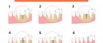
There are 3 methods of performing sinus lift surgery:
- Open is a complex surgical procedure performed for severe bone deficiency greater than 8 mm. The algorithm of actions assumes:
- a hole is created in the gum and bone underneath;
- the sinus floor is raised;
- placing a bone substitute in the vacant space after the sinus has been retracted;
- returning to the place of a previously separated part of the bone.
- Closed - prescribed for bones with a height of 7-8 mm. Operation stages:
- in the place where the implant will be inserted, a hole is prepared through a small puncture;
- an osteotome is used to move the sinus;
- the vacated space is filled with granulated osteoplastic substance.
- Balloon - indicated when the bone height is 3-4 mm. Immediately after completion of this procedure, implants can be installed. The balloon method is performed similarly to the closed method. Its difference is in the installation of a catheter with a balloon under the mucous membrane. The balloon is gradually filled with radiopaque liquid, which ensures smooth, gentle exfoliation of the mucosa and the formation of free space for the bone substitute.
Old perforations
If the perforation of the maxillary sinus was not promptly identified and eliminated, then after 2-4 weeks the stage of acute manifestations will subside, and a fistula will form in the area of the defect, connecting the sinus cavity with the surface of the gum.
This process is simultaneously accompanied by symptoms of chronic sinusitis:
- constant dull pain in the sinus area radiating to the orbit and temple;
- nasal congestion on the affected side;
- purulent discharge from the nasal cavity, as well as from the fistula;
- Sometimes patients have swelling of the cheek on the side of the damaged sinus.
Most patients also complain of a sensation of air moving through the fistula when talking or sneezing, difficulty pronouncing certain sounds, and liquid food entering the nasal cavity from the mouth.
Treatment of such chronic perforations with fistulas presents some difficulties, since the presence of a chronic focus of inflammation in the maxillary sinus significantly reduces the effectiveness of therapy and quite often leads to relapse and re-formation of the fistula canal.
Such patients are indicated for surgical intervention, which includes opening the maxillary sinus with removal of all non-viable tissues and foreign bodies from its cavity, excision of the fistula and plastic closure of the defect. Antibiotics after removal of the fistula are prescribed for a course lasting 10-14 days with the simultaneous use of anti-inflammatory and antihistamine drugs, and the use of physiotherapeutic methods of treatment.
Consequences of perforation
Perforation of the maxillary sinus is a fairly serious pathology that often has to be treated in a hospital. Attempts to independently treat it with folk remedies at home without medical assistance can lead to the development of serious and dangerous consequences:
- The development of a pronounced inflammatory reaction in the sinus cavity with the spread of infection to the surrounding bone tissue and the formation of foci of osteomyelitis of the upper jaw.
- Spread of inflammation to other sinuses of the skull (frontal, sphenoid and ethmoid).
- Loss of healthy teeth located in the area of untreated perforation.
- Formation of purulent foci (abscesses, phlegmons).
Due to the close location of the maxillary sinus and the brain, after perforation, infection may spread to the meninges with the development of meningitis or meningoencephalitis, which threatens the patient’s life.
Possible complications
To minimize the risk of re-development of the pathology, anastomosis plastic surgery is performed exclusively in a hospital setting.
Despite the fact that doctors at the clinic constantly monitor the stages of healing, postoperative complications are possible:
- Inflammatory lesions of the airways. Gradually, the lesion infects the surrounding bone structures, and signs of osteomyelitis of the upper jaw appear. The affected area is disinfected with antiseptic compounds, and drug therapy is prescribed.
- Extension of the localization of inflammation to the paranasal sinus of the frontal bone . The problem cannot be corrected without repeated surgery.
- Loss of healthy units of the jaw row located near the perforation.
- Abscess (local accumulation of pus). Occurs due to insufficient sterility or due to the penetration of pyogenic particles into the thickness of the tissue. A complex clinic requires an integrated approach to treatment.
Preventive actions
Prevention of perforations of the floor of the maxillary sinus consists of:
- in a full examination of the patient before complex dental procedures;
- in the correct assessment of the anatomical and topographical characteristics of each person;
- in strict adherence to the technology of therapeutic manipulations.
Timely detection of signs of perforation and its adequate treatment is the key to a favorable outcome for the patient. Incorrect therapeutic tactics or self-medication can aggravate the course of such a complication and cause the development of severe negative consequences.