Teeth and numbering methods in dentistry
All units located in the dentition are divided into the following varieties:
- To solve various dental problems, tooth numbering was developed to facilitate diagnosis
fangs;
- molars;
- premolars;
- incisors.
They all perform a specific function:
- grind food;
- hold;
- take a bite;
- tear off pieces of food;
- make a grab.
There are a total of 32 teeth in a row. The molars-eights are the last to erupt in adulthood.
They do not take part in the process of chewing food, but are susceptible to caries no less than others. It happens that an already erupted molar shows signs of caries.
To solve various dental problems, tooth numbering was developed to facilitate diagnosis, enter treatment data into the patient’s chart, and transmit information by phone or e-mail.
There are several counting methods, almost all of which are based on dividing the jaw into segments.
There are four of them in total; the conventional boundaries are determined by the upper and lower jaws and the central gap on the front incisors. The segments are counted clockwise, starting from the right side of the upper jaw.
Dental clinics use generally accepted numbering systems:
- Universal;
- Haderupa;
- Two-digit (Viola);
- Zsigmondy – Palmer (another name is a square-digital circuit);
- Alphanumeric.
Why do you need to number your teeth?
Any numbering of teeth is developed taking into account the specific structure of human jaws. It should also be taken into account that each tooth has its own shape depending on the function it performs.
How dentists number teeth
Doctors came up with the idea of numbering teeth to make it more convenient to diagnose dental diseases, as well as record the results of examinations in medical documents.
Dental indices:
| Index of level of oral cleanliness, periodontal condition or whether the patient needs treatment - CPITN | Index of the number of caries in a patient |
«0»no treatment needed | checkpoint(Number of fillings and cavities) |
«1»improve oral hygiene | KPV(Caries with temporary bite) |
«2»preventing plaque from appearing on teeth | Krp(Amount of destroyed surface) |
«3»gum treatment, inflammation reduction | CPU(Caries Filled Extracted Teeth) |
«4»Re-treatment of gums is required | “Note! The KPU includes the level of pain on a scale of one to ten. From six to ten is considered severe pain with increased caries damage.” |
One part of the teeth is used exclusively for biting food, and the other is used for grinding. When entering information about them into the patient’s chart, it is important not to make a mistake and clearly indicate which tooth needs treatment. This is where numbering helps.
Functions of teeth
How to count teeth in an adult?
Assigning a number to the teeth on the jaw begins by counting from the middle of the row in the directions to the right and left:
- You should start counting correctly from the right side of the upper jaw towards the molars
the front two incisors are responsible for biting off food (marked with the number 1);
- the other two, located on either side of the central ones, perform the function of grasping and biting (marked with the number 2);
- fangs, which have a cone shape, ensure biting and tearing pieces from solid foods (marked with the number 3);
- premolars participate in the chewing process, which is ensured by two tubercles on the outer side of the surface, they are also called small molars (marked with numbers 4 and 5);
- molars are large molars, having four tubercles on the outer surface for crushing food; they perform the main chewing function (marked with numbers 6, 7, 8).
The dentition of a child differs from the jaw of an adult in anatomy, so the numbering is developed according to a different principle.
The diagram indicates a total of 20 milk units, of which:
- incisors – 8 pcs.;
- fangs – 4 pcs.;
- molars – 8 pcs.
The peculiarity of a child's jaw is the absence of premolars and extreme molars, better known as wisdom teeth.
During the period when baby teeth erupt, the rudiments of molars are formed. On an x-ray before the age of 6, both can be clearly seen.
Dairy plants also have a root system, but after they fall out during the replacement period, the roots resolve on their own, which eliminates root removal surgery.
Schematically, the numbering of the children's jaw by sector is indicated by the following digital values:
- 1 segment – fifty;
- Segment 2 – sixties;
- 3 segment – seventies;
- Segment 4 – eighties.
You should start counting correctly from the right side of the upper jaw towards the molars. The numbers on the bottom row are assigned in a similar way.
Constant units are designated by numbers from 11 to 48.
What are the types of teeth?
Depending on the purpose and configuration, teeth are divided into 4 types.
- Incisors . There are 4 teeth of this type on the lower and upper jaws. They are located in the front middle of the jaw. There are lateral and central incisors. They are flat in shape, with sharp edges and one root. Designed for biting food.
- Fangs . Placed immediately behind the incisors. They are stronger and thicker than incisors, but are also designed for biting food.
- Premolars . Follow the fangs. They perform the function of chopping food.
- Molars . The last three teeth, with the help of which the final grinding of food occurs before swallowing.
Types of teeth
Some more interesting facts
Order of teeth in the oral cavity
Video - Anatomy of teeth
Adult teeth numbering scheme
The table shows the numbering of teeth in an adult:
| Numbering of teeth in the mouth of adults | ||||
| Name | Top right row | Top left row | Bottom left row | Bottom right row |
| Incisors | 11, 12 (center) | 21, 22 (center) | 31, 32 (center) | 41,42 (center) |
| Fangs | 13 (behind cutter 12) | 23 (behind cutter 22) | 33 (behind cutter 32) | 43 (behind cutter 42) |
| Premolars | 14, 15 (behind canine 13) | 24, 25 (behind fang 23) | 34, 35 (behind fang 33) | 44, 45 (behind fang 43) |
| Molars | 16, 17 (behind premolar 15) | 26, 27 (behind premolar 25) | 36, 37 (behind premolar 35) | 46, 47 (behind premolar 45) |
| Wisdom teeth | 18 (side) | 28 (side) | 38 (side) | 48 (side) |
Numbering of adult teeth
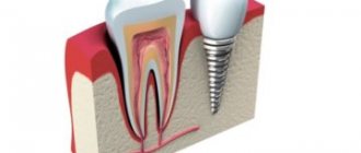
Root canal treatment
Filling cavities inside the root is one of the main conditions for the successful treatment of pulpitis and periodontitis. The stages of a doctor’s work are as follows:
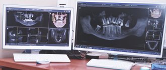
- Determination of length. The doctor removes the pulp and, using special, thin instruments, measures the length. In good clinics, the process is carried out under the control of an apex locator - a device whose display reflects the moment the instrument reaches the root apex.
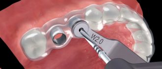
- Treatment for expansion, preparation for filling. The procedure is done manually or using an endodontic handpiece.
- Medical treatment using disinfectants administered through a thin needle.
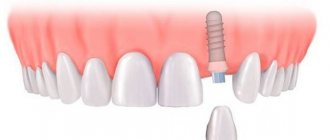
- Filling with gutta-percha material. The pin is selected according to the size of the expanded space, it is filled with paste, the pin is installed and secured.
- X-ray quality control.
- Removal of excess, installation of temporary filling.
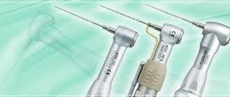
Tools for processing channels.
Standards for the provision of dental care do not allow simultaneous filling of canals and dental cavities. The crown should be restored at the next visit.
Treatment is not an easy task. It often leads to complications:
- Trauma in the area of the apex of the tooth root: damage to the walls with instruments, inaccurate removal of the pulp, penetration of antiseptics into the tissues surrounding the apex.
- Poor fillings: Fillings do not reach the end of cavities, allowing bacteria to continue to grow in those areas. This is evidenced by pain and swelling of the gums.
- The filling material penetrates beyond the apex.
- Root perforation, which occurs due to a doctor’s error or with curved canals that are difficult to treat.
Most often, the method of correcting errors is re-sealing, which involves re-opening the cavities. To avoid this, you need to carefully choose the clinic and doctor who will treat pulpitis. The best option is to prevent the development of the disease by following the rules of hygiene and visiting a doctor for preventive purposes.
- Cohen S., Burns R. Endodontics. E-book, 8th edition, 2007.
- Borovsky E.V. Therapeutic dentistry. Moscow, 2003.
Numbering of a child's teeth
The table shows the numbering of a child’s teeth:
| Numbering of teeth in children - how to count them correctly | ||||
| Name | Top right row | Top left row | Bottom left row | Bottom right row |
| Incisors | 51, 52 (front) | 61, 62 (front) | 71, 72 (front) | 81,82 (front) |
| Fangs | 53 | 63 | 73 | 83 |
| Molars | 54, 55 | 64, 65 | 74, 75 | 84, 85 |
Numbering of baby teeth
Process and stages of root canal treatment
The root canal treatment process has a clear sequence of stages:
- Initially, it is necessary to correctly diagnose the disease. In difficult cases, the dentist sends the patient for an x-ray.
- After this, the doctor carries out the necessary preparatory procedures in the oral cavity for treatment.
- Canal treatment is a rather painful procedure, so the tooth must be numbed by injecting anesthesia (usually into the gum near the diseased tooth).
- After this, complete asepsis of all instruments is carried out and the diseased tooth is separated from the rest using a special rubber film (caffedram).
- Next, the diseased tooth is opened using a drill, providing maximum access to the diseased canals, after which they are initially cleaned, the infected pulp is removed, and in parallel the canals are treated with special medications.
- Afterwards I dry them and seal them with special materials.
If the doctor has any doubts (usually this happens when the tooth is inconveniently positioned and instruments have difficulty accessing it), he puts a temporary filling , after which he sends the patient for an x-ray, using a photo of which he checks whether all the infection has been removed and whether all the canals have been removed. cleaned it. The permanent filling is then placed approximately two weeks later.
This whole procedure, of course, is not very pleasant, but it allows you to save the tooth. Its duration depends on the location of the tooth, the number of canals in it, the complexity of the developed infection and usually takes from thirty minutes to one hour. And success depends on the professionalism of the doctor and the high-quality work he has done, since it is necessary to remove all the affected pulp from the canals without leaving a drop of infection, otherwise it can develop again and tightly seal the tooth so that nothing else can get into the cleaned cavity.
Tooth numbering systems
Modern dental clinics use five generally accepted systems that determine the serial numbers of units.
Haderup system
This method of assigning a number involves conditionally dividing the jaw into 4 equal parts, numbering begins with the front incisors. A sign is added to the serial number: “+” or “-”. A plus signifies the top row, a minus signifies the bottom row.
For adults, the numbers consist of one digit and a sign, while the children's version consists of a two-digit number. It is formed by adding a zero to the serial number (for example, bottom: – 0.1; – 0.2, etc.; top: +0.1; +0.2, etc.)
Square-digital (Zsigmondy-Palmer system)
The system was developed in 1876 and is actively used today for diagnosing dental diseases in adults and children.
Its principle is to designate permanent teeth with Arabic numerals (from 1 to 8), and milk teeth with Roman numerals (from I to V).
Counting starts from the front incisors towards the outer molars. The diagram has a conditional division of the jaw into 4 sections (for adults the numbers are from 1 to 8, for children - from I to V).
Alphanumeric
The universal system provides for the use of letters (Latin) in addition to digital designations.
Designations used by dentists:
- I defines Incisors;
- C – fangs (Canines);
- P – premolars;
- M – molars.
Since several incisors or molars can be located in a segment, each letter is supplemented with an index of a number that indicates the serial number.
This gives the overall picture:
- eight letters I;
- four letters C;
- eight letters P;
- twelve letters M.
The children's diagram uses Latin letters from A to K.
The numerical value entered next to the letter indicates the serial number of the tooth in the segment.
Alphanumeric numbering of teeth
International two-digit Viola system
The number assignment method was founded in 1971, does not involve the use of complex formulas, and therefore remains relevant to this day.
The basic principle is to conditionally divide the jaw into 4 segments:
- 1st – tens (top right);
- 2nd – twenties (top left);
- 3rd – thirty (bottom left);
- 4th – forties (bottom right).
Each conditionally divided part consists of 8 teeth. In the adult version, the following indicators are used: 1, 2, 3, 4. In the children's version, each section includes 5 units; for counting, the following values are used: 5, 6, 7, 8.
An incisor or molar is signed with a number consisting of two digits: the first indicates the section number, the second indicates the serial number of the tooth.
In the adult scheme we get: tens, twenties, thirties and forties. Children's version: fifties, sixties, seventies, eighties.
Universal system
The simplest and most understandable system for a doctor and any patient. You just have to remember which tooth the count begins with and where it is located. The order of number assignment begins with the right eighth molar in the top row.
The calculation on the upper jaw ends with the number 16. The numbering of the lower row begins with the molar located under tooth No. 16. The movement continues clockwise (one, two, three, four, five, six, seven, etc.).
Universal tooth numbering system
What are channels?
Each tooth has a certain number of roots located under the gum.
How many roots do teeth have? The answer to this question depends on several factors - the position of the unit, the person’s age, heredity, even race. It is known that Mongoloids have more roots than Caucasians.
The standard quantity is as follows:
- Incisors, canines – 1.
- Premolars – 1-3.
- Upper molars – 3-4.
- Lower molars – 2.
- Third molars – 3-5.
Read also: Tooth socket curettage, what is it?
Inside the crown there is a pulp - tissue consisting of blood vessels and nerve endings. They pass into the pulp through the apical foramen, located at the apex of the root, and through canals, narrow cavities inside the root. Their number is not always equal to the number of roots.
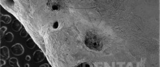
The photo shows the beginning of the root canals.