NeoStom – Dentistry website
The purpose of these studies ? clarification of the diagnosis, substantiation of targeted effective treatment and prevention of the occurrence of a new disease. Clinical blood test ? one of the most common laboratory tests. Determination of the number of erythrocytes, hemoglobin content, color index, number of leukocytes, and leukocyte formula make it possible to assess the severity of the disease and the nature of the body's reactivity.Cytological examination makes it possible to study the dynamics of the state of the mucous membrane under the prosthesis, as well as to carry out the necessary differential diagnosis of inflammatory processes of the oral mucosa and various oncological diseases. The advantage of a cytological study over a histological study is the following: it is possible to observe more subtle structural changes in individual cells, taking material for research is less traumatic for the patient, and the result of the study can be obtained much faster. Histological examination is used to establish or confirm the diagnosis in the presence of infiltration, tumor, difficult-to-heal ulcers, etc. The material for research should be taken from the border area between healthy and pathological tissue. Examination of a patient in the presence of metal inclusions . For orthopedic treatment, stainless steel, cobalt-chromium, silver-palladium, gold, gold-platinum, titanium alloys, and solder are used to connect denture parts. These materials contain over 10 metals. It is necessary to carefully follow the manufacturing technology of bridges and crowns, maintain temperature conditions, prevent spreading and splashing of solder, achieve high-quality casting and high-class polishing. If one of the components is violated, pathological reactions to metal dentures may occur, associated with the influence of denture materials that are different in nature on the tissues of the oral cavity and the human body, on the one hand, and with the reactions of biological environments on dentures? with another. The reasons for the entry of microelements from dentures into saliva are: galvanic corrosion, wear of the metal surface, local corrosion, etc. When two different metals and saliva are present in the oral cavity, a galvanic element is created that produces an electric current, the strength of which depends on the pH of the saliva. Microcurrents contribute to the occurrence of leukoplakia, lichen planus, and paresthesia.
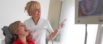
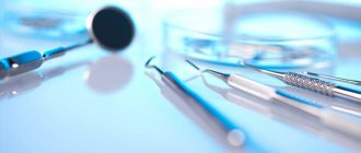
During a clinical examination of persons with the presence of metal inclusions in the oral cavity, the following data must be entered into the medical record of the dental patient: nature, intensity, time of onset of burning, metallic taste or other subjective signs of pathology, time of prosthetics, material of the manufactured prosthesis, appearance of the prosthesis, number of units in the prosthesis and the number of soldered joints, the condition of the mucous membrane. In addition, it is necessary to find out whether the patient was treated for this reason before this treatment, and what the results of this treatment were. It is also necessary to provide data from clinical and laboratory studies. In some cases, it is necessary to conduct a general clinical examination of the patient: blood, urine, gastric juice tests, and also obtain a conclusion from a therapist or neurologist. When questioning, special attention is paid to identifying the subjective symptoms of the disease, the time of its appearance and duration, as well as the period of pronounced subjective sensations. During an objective examination of the patient, the condition of the oral mucosa is assessed? color, moisture, swelling, presence of erosions or other pathological changes. To clarify the diagnosis, the difference in electrochemical potentials is measured, the pH, viscosity and electrical conductivity of saliva, and the taste lability of the tongue receptors are determined. Method for determining electrochemical potentials using a biopotentiometer . The technique is as follows: one electrode (hydrochloride or similar with a known potential) is brought into contact with the sublingual area, and with the help of the second, pre-selected areas of the oral mucosa or existing artificial inclusions (inlays, fillings, dentures, etc.) are alternately touched. The potential difference normally should not exceed 50 mV. When registering large values, a thorough comprehensive examination is carried out to identify and eliminate (remove the prosthesis, remove a filling, etc.) the causative metal inclusion.
The method for determining the threshold of taste lability of the tongue receptors is based on assessing the state of the taste analyzer in terms of the duration of the latent period of sensations and the thresholds of taste sensitivity. To determine the threshold for sweets, use a glucose solution of concentrations of 0.1, 0.5, 10, 20% and higher, for salty? table salt solution 0.1, 0.5, 1% and higher, for sour? solution of concentrated tartaric acid 0.15, 0.5, 1% and higher, for bitter? solution of hydrochloric acid quinine concentrations of 0.0002, 0.002, 0.01% and higher. The taste threshold is taken to be the minimum concentration value correctly characterized by the subject. The study is carried out 1.5-2.0 hours after the patient has eaten. Saliva examination is carried out to clarify the preliminary diagnosis. The quantity, rate of secretion, pH, viscosity, content of organic and inorganic components, and amount of sediment are determined. The daily amount of saliva usually averages one liter. If a patient complains of dry mouth or, conversely, increased salivation, then the rate of salivation is determined. Oral fluid (saliva) is collected in the morning, on an empty stomach, preferably at a certain time. Patients passively spit saliva into a clean, sterile graduated tube for 20 minutes, from which the amount of mixed saliva is determined. The rate of secretion is determined by dividing the amount of oral fluid in the test tube by 20 minutes. The pH of mixed saliva is determined potentiometrically. Instrumental research methods: determination of the angle of inclination of the teeth; electroodontic diagnostics; rheodentography; thermoodontodiagnostics; rheoparodontography; echoosteometry; measuring the endurance of the supporting-retaining apparatus of teeth to vertical and horizontal loads; polarography; gnathodynamometry, etc. are used to establish and confirm the diagnosis.
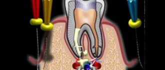
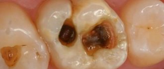
Electroodontodiagnosis is a method for assessing the excitability of the sensitive nerves of a tooth when they are irritated by electric current. In intact teeth, the threshold for irritation of the sensitive nerves of the tooth by electric current lies in the range of 2-6 μA. The threshold of irritation for deep caries is up to 20 μA, for inflammation in the pulp? 20-50 µA, with the death of the coronal part of the pulp? 50-60 µA, with the death of the root pulp? 100 µA and above. It should be remembered that the digital indicators of electroodontodiagnosis depend on the patient’s age, his mental state and the excitability of his nervous system, as well as the anatomical dimensions of the tooth and the correct application of the active electrode. Thermo-odontodiagnostics ? assessment of the state of excitability of the sensitive nerves of the tooth using temperature stimuli. For this purpose, chilled water, air flow, and heated gutta-percha are used. When exposed to temperature stimuli, a quickly passing pain reaction indicates a carious process in the teeth. Long-lasting severe pain suggests the presence of an inflammatory process in the dental pulp. Rheodentography . This method of assessing the condition of the dental pulp is based on graphical recording of the pulsating blood flow through the vascular system of the dental pulp. The method is intended for differential diagnosis of medium and deep caries, deep caries and pulpitis, various forms of pulpitis, as well as to determine the vitality of the dental pulp. Rheodentography is used to monitor the condition of the dental pulp after odontopreparation for an artificial crown or the condition of the tissues under artificial crowns.
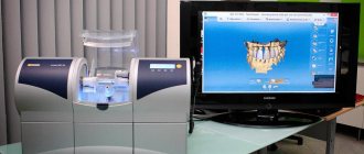
Diagnostic models of the patient’s jaws serve as an integral part of the diagnostic and treatment process in an orthopedic dentistry clinic for most nosological forms. Models are necessary for making a diagnosis and for monitoring treatment (assessment of the initial state, after odontopreparation, after application and fixation of dentures). A number of measuring techniques are more convenient to carry out on models, and some are possible exclusively on them. Models make it easier to make a diagnosis and develop a treatment plan, and allow you to judge its results. Using diagnostic models, it is possible to clarify the surface relief of the alveolar process and alveolar part, the degree of atrophy and its nature, and deformation after injury.
Related materials:
- Assessment of the condition of the mucous membrane of the prosthetic bed
- Assessment of occlusal contacts
- Methods for studying periodontal condition
- X-ray examination of teeth
- Determination of tooth mobility
Next >>
Types of laboratory tests
Cytological
They help determine the nature of the cellular material, detect caries pathogens or specific cells that cause a pathological process in the oral cavity. Exfoliative material is obtained from the surfaces of ulcers, areas of leukoplakia, and from surgical discharge. Aspiration – from tumors, lymph nodes, foci of inflammation. In addition, swabs are used for research. The material is fixed, stained and examined under a light microscope for its constituent components.
Mycological and microbiological
Biomaterial is taken from areas of damage to the tooth or mucous membrane. The objective of the study is to obtain the most detailed information about the microecology of the oral cavity, salivary gland duct, periodontal pocket or other biological niche. These methods help to identify the causative agents of the pathological process and determine the sensitivity of microorganisms to antibacterial agents.
Virological
This group of methods is aimed at identifying traditional pathogens that cause the most common diseases of the oral mucosa. The presence of specific IgG and IgM antibodies in certain reactions will indicate that the disease is viral in nature. During acute infection and during relapse, they manifest themselves differently, which makes it possible to differentiate the stage of the pathological process.
Immunochemical
This includes various research methods, including the indirect enzyme immunoassay method. It is used to determine the titers of specific antibodies to fungi, viruses and bacteria in saliva and blood serum.
DNA polymerase
The material being studied is tested using high-precision methods for the presence of DNA pathogens.
Immunological
The methods are focused on checking the level of general (systemic) and local immunity of the patient and allow further adjustment, for example, of the treatment of pulpitis of primary teeth depending on the immune response. Instead of skin tests, today laboratories often resort to enzyme immunoassay methods.
General clinical laboratory tests are also carried out to diagnose dental diseases. They study the physicochemical and microscopic properties of blood, urine, feces and other fluids and separated substrates from the body.
Areas of dentistry where physiotherapy is used
An integrated approach and adherence to physiotherapeutic protocols - these are the principles that guide the specialists of the dental department of the Doctor Nearby clinics. They use physical therapy to address the following:
- minimizing or eliminating pain symptoms;
- stimulation of regeneration after implantation and sinus lift surgery;
- eliminating emergency conditions for facial pain of unknown nature;
- treatment of teeth and gums;
- treatment of TMJ diseases;
- restoration of the mucosa after surgery;
- stimulation of muscle tone and nerve conduction.
Types of physiotherapy in dentistry
| Name | What is? |
| Laser therapy | Provides exposure to red and infrared rays on the gingival area, oral mucosa and facial skin. Allows you to eliminate swelling, reduce inflammation, slow down the growth of pathogenic bacteria, eliminate pain symptoms, etc. |
| Electrophoresis and galvanization | Involves the use of continuous direct current of low strength and low voltage for the purpose of administering pharmacological drugs through the skin and mucous membranes. Allows you to stimulate blood flow, start bone tissue restoration processes, and improve the flow of oxygen to the tissues around the affected tooth. |
| Magnetotherapy | It is based on the use of a low-frequency magnetic field for the treatment of inflammatory processes, edema, elimination of hematomas and the consequences of traumatic injuries to the maxillofacial area. After injuries, it can be used already on the second day and provides a good effect. Wounds and postoperative sutures are not a contraindication for use. |
| Ultrasound therapy | Based on the therapeutic effects of mechanical vibrations of ultra-high frequencies. Allows you to normalize metabolic functions and stimulate the supply of oxygen. The antispasmodic effect provided by ultrasound makes it possible to reduce pain symptoms. |
Goals and objectives of physiotherapeutic treatment in dentistry
Physiotherapeutic techniques are used in dentistry independently or as part of complex treatment.
First of all, they are aimed at stimulating blood supply processes and metabolic functions of the body at the level of cell tissue. This approach provides optimal conditions for the restoration of organs and systems affected by the disease. Physiotherapy gives good results after tooth extraction and dental surgery, for pathological conditions of the oral cavity and for eliminating pain symptoms of various natures. It is prescribed even for such serious diseases as functional pathology of the temporomandibular joint.
The correct selection of physiotherapeutic techniques plays an important role and makes it possible to restore the functioning of not only the organs of the maxillofacial area, but also the entire body.