3213
Among dental diseases there are a number of those that can be characterized as tumors. Epulis belongs to them. Despite the fact that the appearance of epulis is possible regardless of age, in both men and women, in the latter this formation is diagnosed several times more often .
What is epulis?
Epulis is a formation on the gum in the form of a tumor. It has synonymous names: supragingival, epulid, giant cell granuloma. Most often, epulis occurs on the alveolar jaw process, in the area of small molars; There are cases of occurrence on the body of the jaw (usually the lower one). Occurs both in adults (several times more often in women) and in children
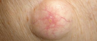
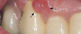
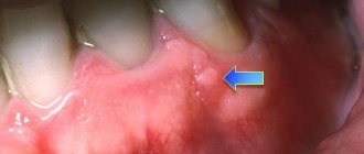
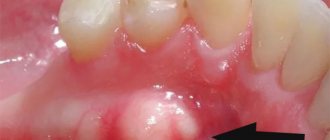
Photo: what epulis looks like on the gum
Childhood and pregnancy
Childhood and pregnancy are two periods during which the risk of developing epulis reaches its maximum.
In childhood, children under 5 years of age and adolescents are most often affected by the disease. The child constantly puts everything into his mouth, often injuring the mucous membrane.
Protective functions at this age are not fully formed, and a tumor forms at the site of injury . In addition, epulis is observed during the eruption of milk crowns, but as it is completed it disappears on its own.
In older children, the appearance of epulis is most often associated with hormonal changes in the body. The main feature of the course of this disease in childhood is that one type of pathology can quickly flow into another. Also, relapses of inflammation most often occur in children.
Hormonal imbalances are the main cause of the appearance of tumors during pregnancy. During pregnancy, the disease can progress several times faster than under normal conditions.
Causes: why does a tumor form?
Epulis occurs under the influence of the following factors:
- Injury to the gums through: an overhanging filling;
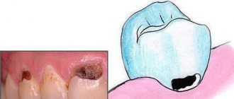
- edges of a decayed tooth;
- tartar;
- burn;
- bruise;
- poor quality prosthesis.
Read also:
Everything you need to know WOLMAR FOR DOGS
Epulis in dogs is a growth of connective tissue. The localization of this formation is considered to be the gums or the thickness of the jaw. In another way, epulis is called supragingival. This pathology can have different sizes. As a rule, epulis occurs as a result of systematic mechanical irritation of the gum mucosa. We are talking about gnawing sticks, injury from the edges of a damaged tooth, etc. The development of this pathology is predisposed by an incorrect bite and a violation of the location of the tooth.
[custom_ads_shortcode3]
Symptoms and signs: what does epulis look like?
In general, epulids in diameter can reach from several mm to 3 cm or more. The color of the supragingival can be reddish-brown, brown, bluish , and also match the color of the gums . Often, ulcers may appear on the surface of a giant cell granuloma, which is most often caused by permanent trauma (sharp edge of a tooth, denture).
The shape of epulid is mushroom-shaped. The formation is located on a stalk, sometimes on a broad base. When pressed, it may move slightly. Epulis can be both soft and hard to the touch. Sometimes the teeth in the area of formation are mobile and slightly displaced.
There are two clinical forms of epulis: benign and malignant. Each of them has its own symptoms. Signs of a benign epulis tumor are:
- Slow growth;
- Diameter up to 2 cm;
- Usually asymptomatic course of the disease.
Malignant supragingival tissue is characterized by:
- Swelling of the gums;
- Rapid growth;
- Soreness;
- Destruction of dental root canals in tissues close to inflammation.
Clinical forms
In addition to the listed types, there are 2 more types of pathology, differing in the nature of the inflammatory process.
Benign
It is characterized by the formation of a node up to 3 cm in diameter and slow growth. Most often, symptoms are smoothed out or completely absent.
Malignant
It is characterized by severe symptoms and active growth . With this type of pathology, the node is painful on palpation and chewing. In this case, the pain radiates to the temple area . The growth of tissue is accompanied by damage to the dental tissue of the roots of adjacent teeth.
White pinpoint lesions are visible on the tumor. When exposed to the area of growth, profuse bleeding appears, the formation of cracks and ulcers.
Diagnostics
The diagnosis is made based on examination by a specialist and histological examination. A differential diagnostic method is used to exclude the presence of hypertrophic gingivitis in the patient. It has similar symptoms to epulis. An x-ray is also used for diagnosis, where, with epulide, rarefaction and destruction of bone tissue due to inflammation are observed.
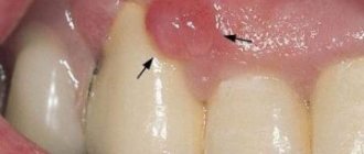
Epulis can often be confused with gingivitis
Symptoms of epulis in dogs
The disease does not have specific signs, often they are only indirect: the dog develops bad breath, excessive salivation, nasal discharge, the owner notices the appearance of blood in a bowl of water, on toys. The dog may be bothered by something in the mouth area, soreness appears - the animal is reluctant to eat. When the tumor reaches a large size, it begins to “bulge” the cheek or bulge outward. As a result, asymmetry of the muzzle occurs. Also, this could be a completely random find.
Classification
There are several types of epulis. Each of them has its own number in ICD-10 (International Classification of Diseases, 10th revision).
Fibrous (K06.82 - benign neoplasms):
Location area: vestibular side of the gum;
- Round or oval shape;
- Slow growth;
- Surface smooth/lumpy surface;
- Doesn't bleed;
- Wide base;
- Thick consistency;
- Light pink (to match the gum color).
Angiomatous:
- Location area: neck of the tooth;
Giant cell (K06.81 - other specified changes in the gums):
- Location area: alveolar part;
Classification by shape
The disease can be either benign or malignant (the latter is much less common).
Benign
In most cases, the formations are benign. They are characterized by the following characteristics:
- slow growth;
- absence of random bleeding;
- intact mucous membrane.
However, even if there is no evidence of degeneration, it is necessary to eliminate the disease in a timely manner. It is dangerous not only due to its growth and malignancy, but also due to the possibility of bacterial infection due to mechanical damage.
Malignant
Malignancy of the tumor can be assumed by the following signs:
- rapid increase in size;
- the appearance of swelling of the gums;
- sensitivity and pain when touched;
- ulceration, random bleeding;
- the appearance of whitish foci in the tumor area;
- enlargement of regional lymph nodes.
Epulis is dangerous due to the possibility of a bacterial infection being attached to mechanical damage.
In some cases, the lymph nodes may become enlarged and painful not because of metastases, but because of the addition of a bacterial infection, so it is necessary to consult a doctor in time for a reliable diagnosis.
Treatment methods, how the tumor is removed
Surgical removal
Treatment with epulid is carried out by removing the tumor. An incision is made at a short distance from the epulis, after which it is removed along with the periosteum. If surgery is performed on a giant cell granuloma, an area of bone tissue is also removed. The procedure is carried out using a bur or cutter. If the formation is more than 1 cm, then removal is often carried out under anesthesia with pharyngeal tamponade. The operated area is covered with iodoform gauze. Teeth are removed only when there is great mobility and root exposure. The dentist is the doctor you should contact if you have symptoms of epulis.
After stopping the bleeding, cauterization with alcohol, piocide, thermocautery, or an electrical regulator may be suggested. Treatment of angiomatous epulis can be carried out through sclerotherapy (urethane-quinidine mixture).
Laser treatment
One treatment option is laser removal of the granuloma. The procedure is performed under infiltration anesthesia. Excision of the body of the epulis is performed using a calibrated laser; removal of the body is done with tweezers.
Folk remedies
It is impossible to cure the supragingival tissue with folk remedies (without surgery). Folk remedies will only help in tissue healing. The composition of rinses for quick recovery should be agreed upon with the dentist. Effective recipes:
- Chamomile / sage / eucalyptus / calendula. Two tablespoons of dry plant + a glass of boiling water. Wait until the solution cools slightly. Rinse several times daily.
- A teaspoon of soda + a glass of warm water (prevents suppuration);
- A teaspoon of salt + a glass of hot water. Wait until it cools down (anti-swelling).
Treatment
The doctor chooses the optimal treatment method in a particular situation after a thorough diagnosis. It includes visual inspection, palpation, x-ray and microscopic examination.
Medication
It is impossible to completely eliminate the tumor with medication. The main goal of drug treatment is to stop its growth. It is also prescribed after surgical interventions to accelerate tissue regeneration.
The most commonly used antiseptic drugs are:
- Dimexide;
- Polycresulene;
- Chlorhexiine;
- Resorcinol.
All of the listed remedies are external, prescribed in the form of ointments, rinses, irrigations or applications.
It is impossible to completely eliminate a tumor with medications
Are traditional methods effective?
Folk remedies are effective only for preventing the development of infection, as well as for accelerating tissue healing after surgery. Among them, for example:
- Infusion of calendula, sage, chamomile, eucalyptus. 15 grams of the mixture is poured into a glass of boiling water and used for rinsing 4-5 times a day.
- A tablespoon of soda is dissolved in 0.5 liters of warm water. Rinse up to eight times a day.
- A tablespoon of iodized salt is dissolved in 0.5 liters of water until the crystals disappear. Use for rinsing three times a day.
Surgical treatment of epulis
The optimal method of treatment is removal of the tumor, which is performed using a surgical or laser scalpel. The doctor makes an incision and completely removes the tumor, often including the periosteum. The edges of the wound are closed, the area is treated and covered with a sterile napkin.
Sometimes adjacent teeth may also need to be removed. This situation occurs due to their increased mobility. During pregnancy, operations are usually not performed unless there are critical and urgent indications.
The optimal treatment method is removal of the epulis
Using laser
Recently, a laser scalpel is often used for dental operations, including the removal of epulis. This solution can be considered optimal for several reasons:
- the edges of the wound are cauterized directly during surgery;
- disinfection occurs at the same time;
- The time of the operation itself and the rehabilitation period are reduced.
How does it occur in children?
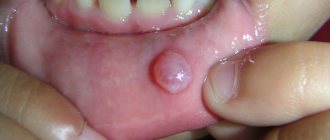
In babies, epulis may appear during teething. Most often this is an angiomatous type of disease, but others can occur less frequently. It is advisable to contact a specialist as soon as possible to prevent the danger of tumor growth. Epulis must be removed through surgery. Sometimes the angiomatous appearance is treated with sclerotherapy
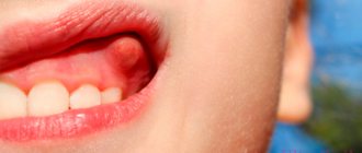
Epulis during teething in a newborn
Types of pathology
In veterinary practice, specialists usually encounter such types of tumor-like growths on the gums as fibrous, fibro-osseous, ossifying and giant cell epulis.
Fibrous epulis in dogs
Fibrous epulis in dogs on the gums is the most common type of pathology and is characterized by the fact that dense connective tissue is involved in the process of metaplasia. Bone and osteo-like structures account for less than half the volume in this type of tumor.
Fibro-osseous
A type of fibrous formation can be considered fibro-osseous, in which the proportion of osteoid structures in the formation of a tumor formation increases. This type has a dense consistency and usually spreads over 2-3 teeth.
Ossifying
In the case when the proportion of bone cells forming a tumor on the gum increases to 80-90%, experts talk about the development of epulis ossificans in the dog. This type of pathology does not pose a serious danger to the health and life of the pet.
Angiomatous
With the growth of thin vessels penetrating the gums, the dog may develop angiomatous epulis. The formation can be smooth or lumpy, bright red in color. Bleeding and the presence of erosions and ulcers on the surface are often detected. As a rule, this type of tumor is localized in the neck of the tooth.
Due to the fact that the tumor leads to the destruction of connective tissue and can damage bone structures, this type of disease is an aggressive tumor and requires immediate surgical removal.
giant cell
During histological examination of the tumor, veterinary specialists often find giant multinucleated cells, as well as osteoblasts. In this case, we are talking about the formation of giant cell epulis.
Epulis during pregnancy
Giant cell epulide is most common among pregnant women. Other types of disease are also common in expectant mothers. The body of epulis in pregnant women is subject to accelerated growth due to hormonal changes in the body (occurs for the same reason). Epulis should not be confused with a fistula (a white pimple-like formation with an opening from which white pus flows). In this case, folk remedies will only help speed up healing after excision. There is evidence that after pregnancy, epulis decreases significantly.
Course of the disease
Angiomatous epulis is common in children. But other types may also arise. The tumor is more common in girls than in boys. The disease usually affects young patients during teething. Why is this happening? Teething is accompanied by severe trauma to the gums and mucous membrane. The tumor grows greatly in the oral cavity and begins to seriously bother. The child’s parents need to seek help from a specialist as soon as possible. The faster help is provided, the easier the consequences will be.
In adolescents, epulis can also occur as a result of trauma. Another reason is hormonal changes in the body. Most often, tumors occur during puberty. It can also occur as a result of taking inappropriate medications.
It’s interesting, but in children, one form of epulis often turns into another. For example, when granulomatous epulis occurs, this type eventually turns into angiomatous and then fibrous. This transition is observed in 14% of cases of the disease.
Due to hormonal imbalance or changes in the body, epulis occurs in pregnant women. If we add trauma to this, the tumor grows quite quickly.
FAQ
Epulis and bleeding: normal or not?
Yes, this is the norm . Angiomatous epulis most often bleeds heavily, while giant cell epulis bleeds moderately. If the supragingival body is injured, bleeding may occur.
Can the tumor go away on its own?
No, reverse development is almost impossible. It is necessary to consult a doctor to remove epulid. Most often, experts recommend excision of the tumor.
Can epulis reappear after cauterization?
Yes, relapses are not excluded . If a formation occurs, you should contact your dentist again.
How to distinguish epulis from hypertrophied gingivitis?
With hypertrophied gingivitis, the gums swell and enlarge, acquiring a scarlet color. There is also bleeding of the gums, heavy plaque on the teeth, an unpleasant odor, and discomfort when chewing solid food. With epulis, such changes in the gums do not occur.
Is it possible to place implants if you once had epulis?
Yes, you can.
How is epulis different from gum cancer?
Symptoms and consequences. Signs of gum cancer are:
- Large swelling;
- Whitish lesions;
- Painful sensations when touched;
- Pigmentation of the seal;
- Ulcers and cracks around the formation;
- Heavy bleeding.
Fatalities are extremely rare, but they do happen. Sometimes the signs of gum cancer are blurred, so if swelling occurs, you should consult a doctor. In the early stages, the disease is treated much faster and easier, but an advanced form can lead to many complications.
Will my gums heal after surgery?
Yes, it is overgrown. It is necessary to follow the wearing of the bandage and all the doctor’s recommendations for good healing.
Epulis on the gum under the denture
The reasons for the appearance of lumps under the prosthesis can be:
- Expired prosthesis;
- Improper dental care (lack of hygiene);
- Gum injury;
- Taking strong medications.
Reasons for appearance
The occurrence of epulis can be provoked by the following factors:
- permanent injury to gum tissue : chipped crown, poor-quality filling, prosthesis, etc.;
- injury;
- burn;
- abnormal bite ; pathological development of the dentition;
- hormonal imbalance;
- diabetes.
In the comments there are reviews about curettage of periodontal pockets. And also the opinion of experts.
What antibiotics should I take for tooth flux? Here's the list.
The link https://zubovv.ru/detskaya-stomatologia/desnyi-ds/gnoynik-u-rebenka-etiologiya-i-lechenie.html tells what to do if a child has an abscess on his upper gum.
Epulis on the gum under the denture
The formation of a tumor under a prosthesis is not uncommon. Most often the reasons for this are:
- poorly made prosthesis;
- poor installation of the structure;
- poor oral hygiene;
- rubbing of the mucous membrane , which often happens during the period of addiction;
- the use of low-quality means for fixing prostheses.
Symptoms
Symptoms of epulis
Clinical manifestations depend on the type of supragingival, which is localized mainly in the area of premolars (small molars). The average size of the formation is 2–3 cm.
The cap can be seen when you smile, but the stem attached to the tooth can only be seen by a specialist. With a significant size of the formation, patients may complain of problems with swallowing and impaired chewing.
The teeth in the area of the tumor are mobile and loose. The surface of the neoplasm may ulcerate, especially in the giant cell form and malignant course. Ulcers are often caused by a poorly fitting denture, a chipped edge of a tooth, or a protruding filling.
The consistency is from soft to dense, the color is from pale pink to purple-bluish, the mucous membrane is covered with a whitish coating. Epulis on the gums does not affect the full opening of the mouth and does not lead to enlargement or changes in regional lymph nodes.
In the congenital form, the formation appears on the alveolar ridge and may decrease over time. In a child, gum tissue can grow during teething, which is accompanied by injury to the mucous membrane. The tumor bothers the baby when sucking. In adolescents, in 14% of cases one form of the disease passes into another. For example, the granulomatous form becomes angiomatous, then fibrous.
In the absence of treatment, the supragingival can be complicated by bleeding (weak or heavy), relapses, suppuration of adjacent tissues, severe swelling, the formation of pockets, cracks, and ulcers.
Giant cell formation
Photos of epulis on the gum would additionally familiarize you with the appearance of the formation, but this is a rather unpleasant sight. Here is a medical description of the pathology:
- Most often it is localized in the alveolar part of the gum.
- Adults aged 40-60 years suffer from this type of tumor. The vast majority of them are women.
- Color - brownish-brown or bluish-brown.
- It is a round or oval formation in shape.
- Bleeding occurs occasionally. Quite moderate, not strong.
- The surface is lumpy.
- The patient feels pain both when pressing on the tumor and when it is otherwise irritated.
- The consistency of the formation is elastic and dense.
What it is?
Epulide is a tumor of the periodontal ligament . This is a structure that “attaches” the tooth to the jaw. The disease is specific to dogs. These tumors are extremely rare in cats. Neoplasms of this type are (usually) benign, but they still cause a lot of harm to the dog. Animals of all ages and breeds are susceptible, but statistics show that they are most often diagnosed in dogs aged six years and older. No gender predisposition was identified. Veterinarians have also found that brachycephalic breeds are much more likely to develop the disease.
This is especially true for boxers, in whom this disease can be considered specific. Unfortunately, to this day, no objective reason for the development of tumors of this type has been identified. There are three types of epulides recognized, and they are grouped by tissue origin.
- Fibromatous . Only the ligamentous apparatus develops. The most common and simplest variety.
- "Ossifying" . Despite the tendency of these types of tumors to harden, they also do not pose a serious danger to the health of the pet.
- Acanthomatous epulis . Despite their benign origin, such neoplasms manifest themselves as quite aggressive tumors that contribute to the destruction of ligamentous and bone tissue.
Possible complications
Most often, tumor removal does not bring additional problems to the patient. However, the following complications cannot be ruled out:
- Re-formation of epulis. Some forms of tumor are prone to recurrence. They also appear with unstable hormonal levels and continued chronic injury to the mucous membrane.
- Postoperative swelling. As a rule, this is a temporary phenomenon that goes away either on its own or after using folk remedies.
- Bleeding. Blood and ichor may be released from the wound for some time, which brings the patient some discomfort in the postoperative period. But, again, these are temporary inconveniences.
- Suppuration on the soft tissues in the wound. It is caused by the contact of pathogenic organisms with damaged vulnerable matter. To prevent this from happening, you need to pay close attention to oral hygiene during the recovery period.
To avoid such troubles, you should scrupulously follow the dentist’s recommendations and take the medications prescribed by him. If a complication is diagnosed, contact a specialist as soon as possible!
Diagnosis of epulis in dogs
First of all, this is an examination of the dog’s oral cavity. Special diagnostic methods include morphological (cytological and histological) and radiological. The simplest is x-ray. It is carried out to determine the boundaries of the formation and is important for choosing treatment tactics and the size of tissue for excision. A cytological examination is a simple injection into the tumor. After collecting the material, on the same day you will receive a preliminary diagnosis and will be able to consult with a doctor about further prospects and treatment.
With the help of histological examination, the doctor-morphologist accurately diagnoses epulis. But, such a study is carried out within 2-3 weeks and is possible only after surgical removal of the formation.
Diagnosis and treatment of oral lesions in newborns
Infants often develop lesions in the oral cavity, which cause discomfort for themselves and cause anxiety for their parents. The most common disorders and diseases include congenital and neonatal teeth, various oral mucous cysts in newborns, ankyloglossia and congenital epulis of the newborn. In this article we will look at the features of diagnosis and treatment of this type of disorder and try to give readers an idea of the correct methods of treating and counseling young patients and their parents.
During their practice, doctors encounter various cases of oral lesions in newborns: from physiological characteristics associated with the development of the child to cancerous tumors. Awareness of such disorders plays an important role in correct diagnosis, counseling and treatment planning. The purpose of this article is to inform healthcare professionals about the diagnosis and treatment of the most common oral disorders in newborns.
Congenital and neonatal teeth
The eruption of the first baby tooth occurs approximately six months after the baby is born. But some babies reach this age already having congenital (the baby is born with them) or neonatal (erupted during the first month of life) teeth in their mouths.
Almost all congenital teeth (about 90%) erupt in the incisor area of the lower jaw. As a rule, they have the correct shape, but may be characterized by discoloration and an uneven surface. Their typical distinguishing feature already during the development period is increased mobility due to the absence or short length of roots. Most of the congenital teeth are subsequently included in the row of twenty primary teeth, but about 10% of them turn out to be supernumerary. Congenital teeth are rare: one case in two to three thousand births of healthy children, and, as a rule, this deviation is random. But in some cases, the appearance of congenital teeth can be a symptom of certain syndromes, malformations and gingival tumors.
If the congenital tooth turns out to be supernumerary and is not included in the row of baby teeth (this can be determined using an x-ray) or interferes with breastfeeding, it is recommended to remove it. Excessively mobile teeth should also be removed to prevent possible aspiration. In addition, congenital teeth can cause traumatic ulceration of the ventral surface of the tongue (Rigi-Fede syndrome), but this disorder is not an indication for tooth extraction and is cured by smoothing the rough cutting edge of the congenital tooth.
Newborn cysts
To refer to oral mucous cysts in newborns, many terms are used that replace each other, causing some confusion. But, based on the different histogenesis of the lesions, all of them can be divided into two categories: palatal and gingival.
Palatal cyst of a newborn
The palatal plates are bilateral rudimentary processes that join along the midline of the oral cavity in the eighth week of fetal development to form the hard palate. They also fuse with the nasal septum, resulting in complete separation of the oral and nasal cavities. In this case, the connective epithelial lining between the plates is destroyed under the action of enzymes, providing the possibility of fusion of the connective tissue. Neonatal palatal cysts, or Epstein's pearls, form from epithelial inclusions along the fusion line of the palatine plates. This disorder is characterized by high prevalence and is observed in 65%-85% of newborns. Cysts are small (1-3 mm) yellow-white bumps along the palatal suture, especially often located at the junction of the hard and soft palate. Histological examination reveals that these cysts are filled with keratin. No special treatment is required, since the cysts atrophy and disappear soon after their contents are removed.
Gingival cysts of newborns
Gingival cysts develop from the dental lamina (ectodermal ligament), which serves as the basis for the formation of primary and permanent teeth. Its remains can proliferate to form small cysts and subsequently cause the development of various odontogenic tumors and cysts. Depending on the location of formation, cysts that appear on the gums of newborns are called Bohn's nodes (present on the buccal and lingual surfaces of the alveolar ridges) or gingival cysts (formed on the process of the alveolar ridge).
Neonatal gingival cysts have a high prevalence: for example, Taiwanese infants screened within three days of birth had a 79 percent prevalence of the disorder.
Typically cysts look like small whitish lesions of constant size. Those that form on the anterior ridge of the lower jaw can be mistaken for congenital teeth. No separate treatment is required as cysts often rupture due to secondary trauma or friction.
Ankyloglossia
The term “ankyloglossia” (tongue-tied) describes the clinical situations of fusion of the tongue with the floor of the oral cavity or insufficient length of the frenulum of the tongue, limiting its mobility. Ankyloglossia can occur in representatives of various age groups, but is most often observed in newborns. According to research, the frequency of this disorder in newborns ranges from 1.7% to 10.7%, in adults – from 0.1% to 2.1%. Based on this, it can be assumed that some milder forms of ankyloglossia resolve with age.
Ankyloglossia of an infant can cause difficulty in breastfeeding and even cause pain in the nipple area for its mother or wet nurse. The preferred treatment for this disorder in newborns is simple frenectomy, where the frenulum is cut off at its thinnest point with small scissors. The procedure can be performed under superficial anesthesia, which ensures minimal discomfort and reduces the likelihood of bleeding. But bleeding is not necessary. Thus, according to the results of a study involving 215 newborns who underwent frenectomy without anesthesia, 38% of children had no bleeding, and 52% had only a few drops of blood. In 80% of cases, nutrition improved within 24 hours after the start of the procedure.
Congenital epulis of the newborn
This disease is a rare tumor of unknown histogenesis. As a rule, the lesion forms on the alveolar ridge of newborns. The course of the disease is as follows: the tumor does not increase in size from the moment of birth, sometimes it can decrease over time, which indicates a reactive rather than a neoplastic etiology. Most often, this tumor is found in the frontal part of the alveolar ridge of the upper jaw and has the appearance of a round attached formation, usually less than 2 cm in diameter (but sometimes larger ones are found), with a smooth lobulated surface. These types of tumors are more common in girls, which may indicate the influence of hormones, although estrogen and progesterone receptors have not been identified. In 10% of cases, multiple lesions may occur, confirming the need for a thorough oral examination.
As a result of histological studies of congenital epulis, large granular cells with small nuclei were identified. Unlike granular cell tumors, staining with the S100 protein antigen in congenital epulis gives a negative result. Other markers of neurogenic origin also showed negative results, confirming a nonspecific mesenchymal origin of the tumor. Surgical removal is recommended for the treatment of congenital epulis, especially if there is difficulty breathing or feeding problems, or if there is a need for histological confirmation of the diagnosis. For smaller tumors, a wait-and-see approach is acceptable, since cases of spontaneous regression of the tumor are known. There were no cases of relapse, even with incomplete removal of the tumor, or malignant degeneration.
Authors:
Van Heerden, Van Zyl