What causes ulcerative necrotizing stomatitis of Vincent
Vincent's ulcerative-necrotizing stomatitis is caused by a symbiosis of the spindle-shaped rod and Vincent's spirochete.
Under normal conditions, these microorganisms are representatives of the resident microflora of the oral cavity and are detected in small quantities in all people with teeth. They are found mainly in the gingival groove, periodontal pockets, carious cavities, and crypts of the palatine tonsils. The development of Vincent's ulcerative-necrotic stomatitis is associated with a sharp decrease in the body's resistance to infection due to viral diseases (acute respiratory infections, herpetic stomatitis, pneumonia, etc.), vitamin deficiencies, stress, overwork, and poor nutrition. Necrotizing ulcerative gingivitis often complicates the course of severe general diseases (leukemia, agranulocytosis, pneumonia, infectious mononucleosis).
It can occur as a complication of exudative erythema multiforme and erosive allergic stomatitis. When specific and nonspecific defense mechanisms in the body are disrupted, the virulence of fusobacteria and spirochetes increases. Their numbers increase to such an extent that they become dominant compared to other microflora.
A decrease in the overall resistance of the body negatively affects the resistance of the oral mucosa. It cannot act as a reliable barrier to the introduction of infection, and the violation of its integrity in an unsanitized oral cavity due to the presence of local traumatic factors (sharp edges of teeth, dentures, tartar deposits, etc.) creates conditions for the introduction of fusobacteria and spirochetes. Therefore, Vincent's ulcerative necrotizing stomatitis often develops in people with an unsanitized oral cavity.
This disease is considered non-contagious, although there are known cases of group incidence of Vincent's stomatitis (in military units, schools, kindergartens). Such cases are explained by similar unfavorable living conditions (malnutrition, lack of vitamins, lack of hygienic measures for oral care, etc.).
Reasons for the development of stomatitis
What causes stomatitis? The causative agents of Vincent's ulcerative-necrotic stomatitis are saprophytic microorganisms, which are considered opportunistic. They live in the thickness of the tooth canals, in the interdental spaces and on the oral mucosa, even in healthy adults and children. But under the influence of certain external and internal factors, a decrease in immunity occurs.
This causes active proliferation of saprophytic bacteria and the appearance of an infectious lesion in the oral cavity.
To understand what causes stomatitis, you need to check the following factors:
- damage to the lining of the oral cavity;
- the presence of massive dental plaque, carious cavities;
- use of dental structures for bite correction and dental prosthetics;
- endocrine pathologies with severe course;
- problematic wisdom teeth, the eruption of which is accompanied by pain and inflammation of the hood.
Why does Vincent's ulcerative necrotizing stomatitis appear?
Anaerobic microorganisms are sensitive to changes in the functioning of the immune system. As soon as the protective function of the mucous membrane decreases, they begin to actively multiply, moving deeper into the tissues. In this case, the production of specific antibodies occurs, triggering a hyperreaction of the immune system, part of which is damage to the vascular walls and the formation of necrotic areas.
What is Vincent's ulcerative necrotizing stomatitis?
Vincent's ulcerative necrotic stomatitis (stomatitis ulceronecroticans Vincenti) is an inflammation of the oral mucosa caused by spindle-shaped bacilli Bacillus fusiformis and Borellia vincentii.
It is described under various names: ulcerative gingivitis, ulcerative stomatitis, ulcerative membranous stomatitis, fusospirochetous stomatitis, Plaut-Vincent stomatitis, “trench mouth”, ulcerative membranous stomatitis, etc. According to the modern classification, the disease is called “Vincent ulcerative necrotic stomatitis” or “Vincent stomatitis”.
Symptoms of stomatitis in the mouth
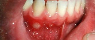
Vincent's ulcerative-necrotizing stomatitis can be acute, subacute and chronic. The course of the disease occurs in several stages. Stomatitis begins with a slight deterioration in general health. Signs of gum inflammation appear in the oral cavity. They may not only turn red, but also bleed.
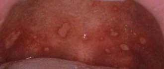
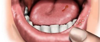
The severity of clinical manifestations increases gradually. Patients complain of loss of appetite and physical weakness. Later, painful ulcers appear, which have a yellow coating on top. During the first 2-3 days, it changes its shade to gray or greenish. Areas of necrotic tissue form, which most often appear in the lower jaw or on the cheeks, where the line of closure of the teeth passes. Ulcers may also appear on the side of the tongue.
As the disease progresses, the depth of tissue damage increases. A severe form of necrotizing ulcerative stomatitis affects the muscular layer of the oral cavity. If no measures are taken and first aid is not provided, the infectious process spreads to the bone and can lead to osteomyelitis.
Some people develop muscle lockjaw due to the disease and cannot even open their mouth. The infection can spread to the oropharynx and tonsils, causing the development of Simanovsky-Plaut-Vincent angina.
Diagnosis of ulcerative-chronic stomatitis
The diagnosis is made on the basis of the clinical picture and the identification of fusospirillary symbiosis.
Biopsy analysis reveals two zones:
- superficial – necrotic;
- deep – inflammatory.
In the superficial layer of necrosis, the flora is rich and diverse (cocci, rods, fusobacteria, spirochetes, etc.), in the deeper layer, which is adjacent to living tissues, fusospirochetosis sharply predominates. These tissues are in the phase of acute inflammation. Inside living tissue there are only spirochetes.
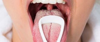
The cytological picture of a scraping from an ulcer with Vincent's stomatitis is similar to a non-specific process of inflammation.
How to diagnose necrotizing ulcerative stomatitis
The diagnosis is made by the dentist based on the results of the examination and laboratory tests. The specialist detects characteristic ulcerative-necrotic changes in the mucous membrane. The edges of the ulcers are torn, their bottom is covered with a gray-green coating. An additional sign is enlargement and hardening of the lymph nodes.
Diagnostic procedures include the following studies:
- laboratory blood test (ESR and leukocytes are increased, the leukocyte formula is shifted to the left);
- bacterioscopic examination (increased number of Borrelia Vincent and Fusobacteria);
- cytological diagnosis (signs of a nonspecific inflammatory process, increased number of neutrophils were found).
https://www.youtube.com/watch?v=cckZDjeiLG4
Differential diagnosis is carried out with drug-induced stomatitis, malignant tumors, secondary symptoms of blood diseases, immunodeficiency conditions and diseases of the gastrointestinal tract. If concomitant diseases are detected, consultation with specialized specialists may be required: infectious disease specialist, gastroenterologist, immunologist.
Diagnostics
Diagnosis of ulcerative stomatitis begins with a visual examination. The doctor assesses the condition of the oral epithelium and the ulcers that have formed. After this, the dentist prescribes the following examinations:
- biochemical and general blood test;
- tissue sampling for cytology;
- microbiological analysis:
- PCR – examination of a swab from the oral cavity.
Since the causes of Vincent's stomatitis are numerous, the patient is referred to specialists in several fields: a gastroenterologist, otolaryngologist, allergist, endocrinologist.
Treatment of ulcerative necrotic stomatitis of Vincent
Local treatment consists of removing traumatic factors, necrotic tissue, influencing the microflora and stimulating regeneration processes of the oral mucosa.
Treatment of the oral cavity should begin with application or injection anesthesia. For this purpose, anesthesin, pyromecaine, trimecaine, lidocaine are used. Then all mechanical irritants are eliminated: sharp edges of teeth and dentures are ground, tartar and plaque are removed. Carious cavities are treated with antiseptic solutions.
Removal of decayed teeth should be postponed until the ulcers are epithelialized, since this intervention in the infected oral cavity of a patient with necrotizing ulcerative stomatitis is fraught with serious complications (alveolitis, periostitis, abscess, phlegmon). Ulcerative surfaces are cleaned of necrotic tissue using proteolytic enzymes: trypsin, chymotrypsin, lysoamidase, deoxyribonuclease.
The entire oral cavity is treated with warm antiseptic solutions (0.5% hydrogen peroxide solution, 0.25% chloramine solution, 0.5% etonium solution), as well as antimicrobial drugs: 0.02-0.06% chlorhexidine solution and its combined forms (lizoplak, parodium), 0.5% metronidazole solution (Flagyl, Metro Gil, Klion)
Gum pockets, the surface of the ulcer, interdental spaces and sub-hood spaces are best rinsed with a high-pressure jet. On the first visit, the entire oral cavity should be treated. Subsequently, daily processing is carried out. The patient is prescribed oral baths with antimicrobial agents and Gildent metro applications to the affected areas of the mucous membrane at home.
For mild ulcerative-necrotizing stomatitis of Vincent, local treatment is sufficient. In more severe cases, general treatment is necessary.
As antibacterial therapy, metronidazole (Trichopol, Flagyl, Klion) is prescribed orally, 0.25 g 2 times a day for 7-10 days. Broad-spectrum antibiotics are also used: chloramphenicol 0.5 g 3-4 times a day, course of treatment is 7-10 days; sumamed according to the scheme for 5 days; Rulid 150 mg 2 times a day for 7-10 days.
Antihistamines are prescribed (tavegil, suprastin, fenkarol, diazolin), as well as multivitamins.
They recommend drinking plenty of fluids and high-calorie, non-irritating foods.
With proper treatment, patients' condition improves within 24-48 hours: pain decreases or disappears, patients can eat and sleep. Swelling and hyperemia of the oral mucosa decrease, epithelization of ulcers begins, which, with a mild degree of the disease and satisfactory condition of the oral cavity, is completed by the 3-6th day.
In an unsanitized oral cavity, epithelization of ulcerative surfaces proceeds more slowly. After the general condition of the patient has improved and acute inflammatory phenomena have disappeared, it is necessary to carry out a thorough sanitation of the oral cavity with the removal of tartar, tooth roots, treatment of carious teeth and periodontal diseases.
Relapses of ulcerative-necrotic stomatitis can occur if foci of chronic infection remain in the oral cavity (periodontal pockets, hoods over incompletely erupted third large molars) or traumatic factors (overhanging fillings, carious cavities, roots of decayed teeth, tartar, poor-quality dentures, etc. .). The cause of relapse may be unsatisfactory hygienic condition of the oral cavity.
Treatment of symptomatic necrotizing ulcerative stomatitis in blood diseases, allergic conditions, mercury intoxication consists mainly of general treatment of the underlying disease causing these changes.
With timely and correct treatment, the prognosis is favorable. Epithelization of ulcerative surfaces in an acute process occurs after 3-6 days, in a chronic process - somewhat later. In an unsanitized oral cavity, in the presence of many traumatic factors and untimely or improper treatment, receding (retraction) or deformation of the gums, resorption of bone tissue of the alveolar process can occur. These changes contribute to the further progression of periodontitis.
Patients who have had Vincent's stomatitis are subject to active monitoring for a year. The first examination is carried out after 1-2 months, subsequent examinations after 6 months.
Main symptoms
Initially, there is a slight malaise with a headache and a slight increase in temperature. In this case, gingivitis occurs in the form of redness, the mucous membrane becomes dry, and the gums bleed. This condition, depending on the subsequent form, can last several days or hours. Gradually, weakness begins to increase, lethargy and apathy appear, performance and appetite deteriorate, and the patient sleeps poorly.
Ulcers, thickly covered with a yellow coating, form on the oral mucosa. After 3-4 days they become greenish-gray, which indicates necrotic processes occurring in the ulcer. This film is firmly attached to the underlying tissues, and its removal exposes the bleeding surface. This increases salivation.
In mild forms of the disease, the affected area is limited and is only catarrhal in nature. The pain in the oral cavity when touched is insignificant, the state of health is not affected, the temperature may remain normal or rise slightly. Bleeding gums are especially noticeable when eating food. The gums become swollen, a lot of saliva forms in the mouth, but necrosis affects only certain parts of the tissue between the teeth. The patient remains quite active. Basically, more serious problems are observed in the patient with necrotizing ulcerative stomatitis, which occurs in more advanced stages.
With moderate severity of the disease, catarrhal manifestations are replaced by the formation of an ulcerative surface, the temperature rises sharply, which is accompanied by chills. At the same time, a person’s performance decreases, the ulcerative lesion grows, and is also covered with a necrotic film. The size of the wounds can reach 5-6 cm, they have uneven and soft edges.
After this, necrosis gradually develops, the edges of the gums become greatly altered, this disorder persists even after treatment. The gums bleed a lot, and a yellowish coating forms on them, which is quite easily removed. There is a foul odor from the mouth, pus is released, and a headache. There is also soreness in the mouth, and the face takes on a grayish-pale tint. Lymph nodes enlarge, become denser, and severe weakness appears.
The very last stage of the disease is characterized by the fact that the ulcers reach the deep muscle layer, the temperature rises and severe weakness is observed. Nausea and abdominal pain appear. If treatment is not carried out at this stage, the ulcer may reach the bone and osteomyelitis of the jaw may develop. In addition, there is a limitation in opening the mouth, this occurs as a result of damage to the masticatory muscles, so it becomes almost impossible to eat. Necrosis may spread to the tonsils.
Traditional methods
Rinsing with a solution of propolis tincture disinfects the mucous membrane. At an early stage of the disease, it helps to remove existing sores and prevent new ones from appearing.
It is recommended to smear the tongue, cheeks, and palate with fresh aloe or Kalanchoe juice. This helps relieve inflammation. You can simply chew the leaves.
Gargling with carrot or cabbage juice half and half with water also helps fight inflammation.
As for medicinal rights, use decoctions of St. John's wort, calendula, and chamomile. Oak bark, sage, and calamus have proven themselves well. For rinsing, you can use individual herbs or a collection. Dosage is at your discretion.
For more severe forms, traditional medicine recommends treatment with a strong decoction of onion peels. Almost any wound or sore will heal faster with the use of rosehip or sea buckthorn oil.
Fresh honey is considered an effective remedy. Honey ointment based on egg whites helps with inflammation of the gums, relieves general swelling, and helps regenerate the mucous membrane. Honey rinses will soothe the mucous membranes and relieve inflammation.
However, despite centuries of experience in curing diseases, traditional medicine is not able to offer an effective substitute for antibiotics, which are used to cure Vincent’s stomatitis. Therefore, folk remedies are good as an addition to main therapy.
Causes
The most common causes that can cause ulcerative stomatitis include infectious diseases of various types, namely influenza, diphtheria, measles, adenovirus, herpes, and so on.
What is ulcerative stomatitis
But other factors can also influence the development of the disease:
- weak immune system;
- lack of vitamins in the body, in particular vitamin B and C, which are responsible for oral health;
- diseases of the gastrointestinal tract (gastrointestinal tract);
- allergic reaction to external irritants;
- hereditary factor;
- the presence of pathologies of the oral cavity (tartar, pulpitis, caries, and so on);
- injuries to the oral cavity that led to damage to the mucous membrane (chewing hard foods, brushing teeth with a hard brush, biting, and so on);
- burn of the oral mucosa.
Stomatitis in an adult
On a note! If children most often suffer from aphthous stomatitis, then more mature patients, whose age ranges from 20 to 35 years, are usually exposed to a chronic form of this dental disease.
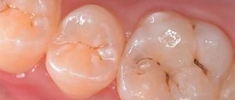
Photo of ulcerative stomatitis
Prognosis and prevention
The prognosis for the treatment of ulcerative stomatitis is generally positive, since timely therapy can eliminate undesirable manifestations of the disease. If the pathology is not treated, then the following complications may develop:
- Development of pleurisy;
- Inflammation of the hearing aid;
- Gastroenteritis;
- Endocarditis;
- Progressive rhinitis.
Prevention of inflammation is based on compliance with the recommendations below:
- Maintaining a healthy lifestyle;
- Maintaining oral hygiene;
- Quitting bad habits (drinking alcohol, smoking);
- Regular intake of vitamin complexes containing all necessary substances and microelements;
- Timely treatment of chronic diseases;
- Preventive examinations to identify various pathologies;
- Elimination of possible allergens;
- Carrying out mouth rinses for dental diseases and disorders of the digestive system.
What is ulcerative stomatitis
Ulcerative stomatitis remains an incompletely studied disease. Doctors believe that the pathology is infectious in nature, and its causative agent is the fusiform bacillus. The bacterium is found on the mucous membranes of healthy people, but it becomes the cause of the problem when it predominates over other types of microbes. The fusiform rod is in a dormant state.
On this topic
- Stomatitis
Find out how long it takes for stomatitis to go away
- Maria Konstantinovna Tevs
- July 26, 2020
It is activated by sudden changes in the body:
- with hormonal imbalances;
- during pregnancy;
- during puberty ;
- during menopause;
- with a sharp decrease in immunity.
It has been scientifically proven that hypovitaminosis C provokes the occurrence of ulcerative stomatitis. Outbreaks of morbidity are observed in the spring (April-May). At this time, a person lacks vitamin C, which he does not get from food.
Ulcerative stomatitis belongs to the category of independent diseases and is divided into the following forms:
- chronic;
- spicy;
- I'll sharpen it up.
Pathology is classified depending on its course. According to severity, ulcerative stomatitis is divided into:
- easy;
- heavy;
- medium-heavy.