Breast granuloma is a small, benign lump that appears against the background of inflammation and is localized in the mammary glands. It is a hollow nodule. The pathology is diagnosed quite rarely and is characterized by accumulations of infiltrates in the mammary glands.
Disease code according to ICD-10 No. 60.80.
The neoplasm appears as a result of the enlargement and division of cells that carry out phagocytosis. They are the ones who transform into granuloma. The nodule is not a tumor, but tissue hyperplasia.
Granulomas are diagnosed in any tissue of the body. They are found on the skin, lips, and lung tissue. There are also granulomas of the teeth and navel of newborns.
Causes of breast granuloma formation
The reasons for the development of formation in the mammary gland can be divided into two types: infectious and non-infectious. The first type includes inflammatory diseases caused by pathogenic microorganisms, the second includes mechanical damage, foreign bodies or substances found in the tissues of the mammary gland.
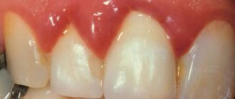
Often, a lump forms after an injury or bruise to the chest. As a result of ruptures of small blood vessels, blood circulation in the damaged area is disrupted. Necrosis begins in this area of tissue, and inflammation grows around it. Often it goes away on its own. Sometimes a capsule of fluid forms in the affected tissue. If the contents of this capsule become infected, suppuration may occur and a compaction may form as a result. A photo of a diseased mammary gland shows reddish or bluish areas of skin at the site of formation of the node.
The development of the disease is promoted by:
- Long-term use of silicone implants. Studies have shown that after 8-10 years of use, silicone is capable of releasing substances that are toxic to tissues, provoking inflammation and triggering the formation of granuloma nodes.
- Surgical interventions. In this case, the neoplasm is localized in the area of the postoperative suture. The node can be either on the surface or inside the scar. The formation does not cause pain, fever or other alarming symptoms.
- Actinomycosis is a fungal infection of tissues. As the disease progresses, purulent granuloma nodules may form.
- Sarcoidosis of the breast. The disease affects the connective tissue of the mammary gland and in rare cases can provoke the appearance and growth of a neoplasm. Sarcoidosis often does not have severe symptoms and goes away without treatment within 2-3 years, without causing discomfort to the patient.
- Rapid decrease in body weight.
- Hormonal imbalances.
- Use of radiation therapy methods.
Women with large breasts and the elderly are at risk. After a bruise, granuloma develops rapidly. In other cases, several months may pass from the beginning of the growth of the node to the pronounced manifestation of symptoms.
Formation of postoperative scar granuloma
Tissue damage occurs due to the penetration of substances into the body that can cause infection with subsequent inflammation. The focus of the inflammatory process is called granuloma.
After surgery, some patients notice a new growth in the area of the sutures.
Granuloma of a postoperative scar in most cases is formed due to the use of non-absorbable threads by surgeons for suturing.
Causes
- A small lump may appear due to recent surgery. The neoplasm is localized both outside and inside the scar. The size of such a compaction rarely reaches large sizes; often the granuloma on the scar feels like a pea or a bump.
- The reason for the appearance of granulomatous compaction lies in the use of non-absorbable materials when suturing the wound. There are no other symptoms, such as pain and fever. In this case, there is no reason to worry. The lump is benign and will disappear on its own within a short period of time.
- Another cause of granuloma can be the entry of foreign particles into the wound, such as talc and starch, which are used for rubber gloves. If sanitary rules are not followed during surgery, microbes can enter the body and can trigger the onset of an inflammatory process.
- Infection of tissues by microorganisms will certainly have its consequences. There is an increase in body temperature, general malaise, and pain in the operated area. In this case, an urgent consultation with a doctor will be required to select the necessary treatment. You should not resort to home treatment using traditional methods. There is a risk of developing serious complications.
Redness of the skin, high temperature, acute pain - all these signs may indicate the appearance of pus. It is recommended to undergo an examination and additional examinations to differentiate a granuloma from a seroma or fistula.
Lipogranuloma of the mammary gland, the so-called steatogranuloma, can develop as a consequence of surgical intervention, be it breast surgery or sectoral resection.
It is fat necrosis of lipocytes. Lipogranuloma of the mammary gland can also appear due to injury.
Injury leads to impaired blood circulation inside the gland, which causes the development of the inflammatory process.
Granulomatous formations may appear asymptomatically. For this reason, it is necessary to regularly palpate the suture area to be able to diagnose the seal in the early stages.
Symptoms and possible complications
The symptoms of granuloma are quite obvious, but in most cases patients do not pay attention to them. That is why it is recommended to visit a doctor in the first months after surgery of any kind to examine the sutures.
Main symptoms:
- redness of the skin of various types from light pink to brown
- the appearance of a pink nodule inside or outside the scar
- roughness of the skin in the suture area
- painful sensations on palpation
- increase in body temperature
- general malaise: increased fatigue, weakness
A complication of granuloma can be its destruction. The capsule bursts, as a result of which fluid may leak out at the site of inflammation under the influence of suppuration. A burst granuloma can cause the development of fistulas. As the capsule disintegrates, it releases pus, which provokes the appearance of ulcers, cysts and other pathological formations.
Another possible complication is granulomatous mastitis, which mostly occurs in the postpartum period. However, it can also appear after breast surgery. Characterized by the formation of granulomatous capsules inside the lobules of the mammary gland. In some cases, granulomatous features indicate breast carcinoma.
A granuloma on a postoperative scar cannot have serious complications if diagnosis and treatment were carried out in a timely manner.
Ignoring the problem often leads to the accumulation of pus in the capsule of the neoplasm, which is dangerous.
However, in most cases, the lump resolves without outside help. The only thing that is necessary is to monitor the condition of the granuloma.
Diagnostics
To diagnose granuloma of a postoperative scar, it is enough to carry out three main procedures:
- anamnesis
- visual inspection
- palpation
However, in the presence of serious symptoms, such as high fever and acute pain, differential diagnosis will be required to exclude various pathologies.
The main methods used for differentiation are:
- Aspiration is a virtually painless procedure and is performed without anesthesia. A needle is inserted into the seal and biomaterial is collected - liquid or parts of cells. The procedure allows you to study the structure and consistency of the neoplasm and differentiate a cyst from a granuloma. The extracted material is sent to the laboratory to study the nature of the tumor.
- Ultrasound examination - allows you to distinguish a granuloma from a malignant tumor.
- Biopsy is a procedure aimed at extracting biomaterial from a lump using a needle for further examination for the presence of cancer cells.
- Mammography is a procedure performed to study the nature of the lump in the mammary gland. Mammography allows you to exclude or confirm the presence of breast cancer, and also provides information about the nature, structure and stage of development of the pathology.
Treatment and prevention
As mentioned earlier, postoperative scar granulomas are not dangerous to health and go away without any treatment. Only in cases where any discomfort appears and the symptoms are pronounced, should you consult a specialist and begin treatment.
Granuloma of a postoperative scar can negatively affect the condition and functioning of nearby organs and tissues. Localization of a large number of granulomas in one place often leads to circulatory dysfunction.
In this case, it will be necessary to excise the connective tissue of the granulomatous neoplasm and scar.
If the nodule is formed due to the suture material, then during excision the remaining threads are additionally removed to avoid relapses.
Before surgery, the patient must undergo an MRI and ultrasound of the inflamed area, as well as a blood test to determine the presence of infection in the body.
The operation is performed under local or general anesthesia. Recovery after surgery is quite fast. Many patients go home the very next day.
If any complications are suspected, the doctor may leave the patient under observation for a few more days.
The doctor prescribes antibacterial and anti-inflammatory medications after surgery. Usually these are ointments that need to be used to treat seams. To treat the suture area, you will need hand disinfection and sterile gloves.
To prevent granulomas of a postoperative scar, doctors strongly recommend that in the first months after surgery, regular tests are performed to determine the presence of microorganisms and inflammation in the body.
To avoid the formation of granulomas after surgery, it is advisable to use absorbable material. Consult your doctor in advance and select hypoallergenic threads for stitches. Sometimes nodules on the scar cause an allergic reaction.
Doctors advise seriously strengthening your immune system. High immunity will protect against neoplasms following surgery. And most importantly, forget about self-medication and traditional methods when it comes to serious pathologies.
Source: https://bolivgrudi.ru/patologicheskaya-mastodiniya/granulema-posleoperacionnogo-rubca.html
Classification
Granulomas are diagnosed in many pathologies. The most significant granuloma breast diseases are:
- chronic granulomatous mastitis
- granulomas caused by foreign bodies
- rare mycoses
- tuberculous mastitis
- autoimmune disorders (polyarthritis nodosa, Wegener's granulomatosis)
- parasitic diseases
Depending on the etiology and characteristics of the course of the disease, there are several types of mammary granulomas:
- Injection. Occurs due to the entry of oils, fats, silicone or other substances into the tissue.
- Post-traumatic. Develops as a result of injuries, bruises, blows to the mammary gland. The occurrence of traumatic granuloma is possible after improper massage.
- Parainflammatory. In this case, the pathological process is localized around the source of inflammation.
Granuloma of the postoperative scar of the mammary gland is also isolated. It can develop after surgery when suturing using non-absorbable suture material. A foreign object in the form of remnants of threads will be rejected by the body. Ultimately, the development of an inflammatory process in the scar tissue is possible.
Granulomas of postoperative scars can appear due to the ingress of talc from surgical gloves or harmful microorganisms into the surgical wound if asepsis requirements in the operating room are not observed. If the cause of the neoplasm is an infection, the pathology will be characterized by malaise, weakness and fever.
If there is suppuration, pain and redness in the suture area, additional examination is required to exclude more serious postoperative complications - fistula or seroma.
Causes
The etiology of each type of granuloma (especially infectious) follows from the name itself. Let's take a closer look at the reasons for the formation of granuloma in a postoperative scar:
- Use of non-absorbable suture materials during intervention. In our medical institutions, these occur quite often.
- Untimely removal of sutures - patients sometimes consult a doctor late, or removal is delayed for other reasons.
- Suppuration of the scar or suture material - in this case, the granuloma is a protective reaction that delimits the focus of granulomatous inflammation.
- Getting talc from operating gloves into a wound is extremely rare today, since before the intervention the gloves are treated with an antiseptic.
- Dehiscence of sutures after surgery - failure to comply with the protective regime may cause the suture to dehisce. This additional damage to the skin can become the basis for the formation of granuloma.
Knowing the main causes of pathology, it is possible to formulate principles for its prevention.
Symptoms of the disease
In the initial stages of the formation of a node, women practically do not feel any signs. Often attention is paid to pathology when the shape of the breast changes in a certain area. When palpating the mammary gland, a small, painful, round lump is felt. As the formation increases, new symptoms appear:
- severe pain and discomfort, increasing with tumor growth
- feeling of heaviness in the mammary glands
- retraction or reshaping of the nipple
- enlargement of local lymph nodes
- in case of tissue destruction, pits form over areas of necrosis
- Post-traumatic granuloma is characterized by a red or bluish discoloration of the skin at the site of injury and possible loss of sensation over areas of necrosis
- discharge from the mammary glands
- general weakness, chronic fatigue
- with granuloma of a postoperative scar, a lump forms in a certain area of the suture, blood circulation in the adjacent tissues is disrupted, this can lead to cell death and the formation of an area of necrosis
Causes of subcutaneous granuloma of the face
Subcutaneous granuloma of the face is an inflammatory process in the subcutaneous tissue.
It is characterized by sluggish flow and focal localization. In medical terminology, there is such a definition of this pathology as migratory granuloma, but it has been proven that migration is not characteristic of subcutaneous granuloma. Subcutaneous granuloma of the face is a disease that is very difficult to ignore. According to medical statistics, every fourth person seeks medical help in the first months of its development; another third visit the doctor by the second or third month of the disease. The causes of subcutaneous granuloma are the basis for the following classification:
- odontogenic - a secondary disease that complicates, for example, chronic periodontitis; pathogenic microflora spreads from the focus of chronic infection into the subcutaneous tissue, causing granulation tissue and individual granulomas to grow;
- non-odontogenic - an inflammatory process that originates from foreign bodies present in the subcutaneous tissue, which is noteworthy, of organic origin.
The localization of odontogenic granuloma is associated with the location of the causative tooth. Usually in half of the patients the inflammation originates from the molars of the lower jaw. Slightly less common is the origin of granuloma from the large molars of the upper jaw; even less often, the pathology spreads from the small molars of the upper jaw.
Odontogenic subcutaneous granuloma of the face is characterized by several stages of progression:
- infiltrative - in the skin and subcutaneous tissue there is a dense, mobile, low-painful infiltrate;
- abscess stage - an abscess is formed from the infiltrate concentrated in the tissue and is located on the surface of the skin.
With the formation of an open abscess, the patient begins to complain of pain, itching and burning at the site of formation of the subcutaneous granuloma; however, the general condition is not disturbed.
Another classification of subcutaneous granulomas is based on the course of the disease. There are:
- single granuloma,
- multiple granulomas.
Granulomas differ in size, but they are always united by the following criteria:
- clear boundaries
- mild pain on palpation,
- The skin covering the granuloma is thinned, shiny, sometimes changes in color, and is characterized by unevenness and folding.
The causes of subcutaneous granuloma of the face, in addition to those described above, include a traumatic or allergic factor, the influence of ultraviolet radiation or medications. The doctor can come to such conclusions, taking into account that there is no infection or foreign body at the heart of the particular case.
Subcutaneous granulomas are characterized by the so-called false fluctuation syndrome. At first glance, it may seem that the neoplasm contains pus, but an autopsy will show its absence in favor of pathological growth of granulation tissue. When a granuloma is opened, bloody or bloody-purulent contents are released from it, and abundant granulations are visible in the wound.
The course of subcutaneous granuloma is defined as chronic and sluggish. It should be noted that medicine does not know cases of self-healing from facial granuloma, therefore, at any stage of the development of the disease, seeking medical help cannot be avoided.
Diagnosis of inflammation
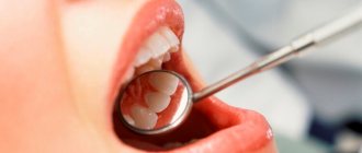
A mammologist examines and treats breast pathologies. The first step in diagnosis is taking an anamnesis. During it, it is determined whether there were bruises or other injuries to the chest. Then the mammary glands are examined and palpated. Laboratory and instrumental research methods are also prescribed:
- Ultrasound diagnostics. The method determines the location, parameters and nature of the tumor. Ultrasound is not informative enough in the early stages of the formation of mammary granuloma, since at first it is similar to an oncological tumor.
- Mammography. In the early stages, the nodule looks like a malignant tumor on mammography images. The main diagnostic sign in this case is the presence of a calcareous shell of the granuloma, which forms in the later stages of the disease.
- Histological examination of breast tissue. In this case, a biopsy is performed if it is impossible to distinguish a benign neoplasm from cancer. The study confirms or excludes oncology.
- Analysis for tumor markers. It involves the determination of substances in the blood, the presence of which indicates a malignant formation.
Diagnostics
The diagnostic search depends on the location and cause of the formation of the skin defect. Here are just a few possible diagnostic methods:
- Examination using a dermatoscope is carried out by a dermatologist.
- In case of formation of oleogranuloma on the mammary gland, mammography is performed to exclude malignant neoplasms.
- If the breast is affected, MRI and ultrasound may also be needed.
- Puncture biopsy or excision of the formation - if the malignant nature of the process is suspected.
- Examination of the dentist and radiography of the jaw during the formation of dental granuloma.
- Blood tests for markers of infections and general clinical tests - if an infectious nature of the disease is suspected.
When examining a postoperative granuloma, additional diagnostic methods may not be needed at all. A characteristic clinical picture and a connection between the formation and a history of surgery are sufficient.
Treatment
Treatment of mammary granuloma begins after examination for the presence of an oncological process. Therapy can be conservative or surgical. Conservative treatment involves prescribing the patient a course of antibacterial agents, corticosteroids, immunostimulants and vitamins. Its goal is to eliminate areas of inflammation and activate the body's defenses. Conservative therapy gives positive dynamics extremely rarely. Therefore, the surgical method is more often prescribed.
Surgical intervention
The operation to remove granuloma is performed under local or general anesthesia. The area of tissue removed depends on the size of the tumor. After excision of a small nodule, the shape of the breast remains virtually unchanged. When the formation is large, sectoral resection is performed. In this case, a significant proportion of the tissue to which necrosis has spread is removed.
After resection, it is possible to perform mammoplasty to restore the shape of the glands and apply cosmetic sutures, thanks to which a postoperative scar is not formed. A certain amount of excised tissue is sent for histological examination.
According to the technique of execution, the operation to remove a granuloma is considered to be a lung operation, and therefore gives complications in rare cases. In the postoperative period, patients are advised to use special supportive underwear and regularly change dressings.
To prevent relapse, a course of vitamin and mineral complexes and hormonal therapy are prescribed. Antibiotics are used to prevent infectious complications. A woman should avoid injuring her breasts.
If fluid is detected in areas of tissue destruction, aspiration is allowed. During the procedure, a needle is inserted into the tumor, with which fluid is pumped out of the granuloma. As a result, the membranes of the node become thinner and the formation disappears. The possibility of aspiration depends on the etiology, characteristics of the course of the disease and the general condition of the woman’s body.
Alternative Methods
In addition to surgical and medicinal treatment methods, there are other methods of treating granuloma:
- Cryotherapy. In this case, the node is removed by deep freezing it with liquid nitrogen.
- Laser therapy. Excision of the granuloma occurs using a laser beam.
- Diathermocoagulation. In this case, the tumor is removed using electric current. The method is relevant only for small tumors.
In folk medicine, nodular formations are treated with the help of sweet clover herb, potato starch, lilac flowers, St. John's wort herb, burdock leaves and cabbage.
Giant cell granuloma in a 4-year-old girl: a clinical case
Giant cell granuloma is a fairly rare benign bone lesion that can develop into a malignant one. This article describes a clinical case of giant cell granuloma in a 4-year-old girl, located in the anterior lower part of the mandible. In children with a mixed dentition, pathological overgrowth of bone tissue can cause delayed tooth eruption, pathological tooth mobility or premature loss of baby teeth. When treating children with pathological tooth mobility and impaired tooth replacement, it is necessary to carry out a differential diagnosis with malignant diseases, in particular with giant cell granuloma.
Introduction
For most practicing doctors, diagnosing such diseases presents certain difficulties, due to the fact that they are quite rare. However, the diagnosis is simplified by the fact that many cystic, metabolic, osteodystrophic, microbial, cancerous and cancer-like diseases of the oral cavity are often accompanied by giant cell lesions.
Giant cell granuloma is a relatively rare benign bone lesion, occurring in less than 7% of benign jaw lesions. This disease was first described in 1953 by Jaffe, who called it giant cell reparative granuloma; the word "reparative" is not currently used. Unlike giant cell cancer of the long bones, giant cell granuloma of the jaw most often affects children, and more often girls (65% of cases) than boys.
Giant cell granuloma is often confused with giant cell carcinoma. However, giant cell carcinoma in most cases develops between 25 and 40 years, affects the long bones, is more aggressive in nature, and is prone to recurrence after curettage. The diagnosis of giant cell granuloma is made on the basis of histological findings. Differential diagnosis of giant cell granuloma is carried out with an extensive list of diseases and pathological conditions, including: radicular dental cyst, adenoameloblastoma, calcifying epithelial odontogenic cyst, desmoplastic ameloblastoma, fibrous dysplasia.
Treatment for giant cell granuloma includes excisional biopsy, safety margin curettage, partial or complete resection of the affected bone, and corticosteroid injections into the affected area.
Symptomatic therapy includes antibiotics, analgesics, a 10-day postoperative course of corticosteroids, and postoperative X-ray examination to monitor disease progression.
Description of a clinical case
Rice. 1. Cone beam computed tomogram.
A 5-year-old girl was taken to the department of pediatric and preventive dentistry at the clinic named after. Bharati Vidyapiti (Pune, India) with complaints of a painless tumor of the anterior part of the lower jaw that appeared 6 months ago. According to the parents, 1 year ago a formation of the same nature was removed under general anesthesia. During the removal of the formation, the central and lateral incisors were extracted on both sides. From the moment of surgery until the time of examination, the formation continued to increase in size. Family history is unremarkable. According to the general examination, the girl was practically healthy; asymmetry of the face in the anterior part of the lower jaw was visually noted. On palpation, the tumor is dense, the skin over it is hyperemic. There were no signs of an infectious disease. When examining the oral cavity, a tumor-like formation measuring 3x2 cm with a smooth bluish surface was determined in the anterior part of the lower jaw.
A general blood test did not reveal any pathology.
Rice. 2. Cone beam computed tomography reveals resorption of the buccal cortical plate with deep-lying tooth germs of the 31st, 32nd, 41st and 42nd teeth.
Rice. 3. Cone beam computed tomogram.
An orthopantomogram did not reveal a clear extension of the cortical plate. An occlusal radiograph of the mandible revealed lingual extension of the cortical plate. After this, a cone-beam computed tomography was performed, in which a zone of radiolucency with clear contours, without division into chambers, was determined in the anterior part of the lower jaw. The zone extended mediolaterally from the first right mandibular molar to the first left mandibular molar. There was also slight lingual displacement of the tooth germs of the permanent central and lateral incisors with minor buccal extension. X-ray diagnosis was made of a solitary bone cyst with the need to carry out a differential diagnosis with a residual cyst and odontogenic keratocyst.
Rice. 4. In the intraoperative photograph, the formation involves the tooth germs of the 41st and 42nd teeth.
Rice. 5. The preoperative photograph shows that the mass extends from the medial part 83 to the medial part 74.
The patient was referred to the Department of Oral and Maxillofacial Surgery for a fine-needle aspiration biopsy. The resulting tissue sample was sent for histological examination; Microscopic examination of the sample revealed cholesterol crystals, after which an inflammatory process of unknown etiology was diagnosed. An excisional biopsy of the lesion was planned.
Subsequently, the patient underwent curettage of the lesion under general anesthesia. An interrupted suture was placed to prevent hematoma formation. The patient's condition after the operation is satisfactory. The postoperative period was uneventful, and the patient was discharged on the 3rd day.
Rice. 6. Remote education.
Rice. 7. Postoperative suture with vicryl.
The tissue sample was sent for histopathological examination. Microscopically, the connective tissue of the fibrovascular stroma was determined, consisting of large fibroblasts with cavernous nuclei. Various multinucleated cells (10-15 nuclei) with clear boundaries were identified, presumably multinucleated giant cells. A large number of macrophages and several mast cells were also detected. Thus, histological examination confirmed the diagnosis of giant cell granuloma.
Discussion
Giant cell granuloma develops predominantly in the bones of the facial skull and jaw bones, although it can also have a different localization. As a rule, it develops asymptomatically and is detected during routine x-ray examination or by the patient himself (or his parents, as in this case). As a rule, tumor growth is quite slow, although cases of rapid tumor growth have also been described in the literature.
As a rule, the disease occurs without pain; the phenomena of paresthesia do not develop. However, according to the literature, from 5% to 11% of cases occur with pain. It can develop in patients of any age group, but is more common in patients under 30 years of age. As a rule, the disease develops in young children, which often causes late diagnosis of the disease. In pediatric dental practice, especially in patients with mixed dentition, anatomical structures often overlap, which often complicates the radiographic diagnosis of the disease, which, in turn, causes late diagnosis of the disease.
The standard treatment for giant cell granuloma is curettage or enucleation. However, the disease often recurs, which, given the extensive nature of the disease, leads to significant deformation of the jaw and facial features. In many cases, if the lesion involves an unerupted tooth (as in this case), the standard of care includes tooth extraction in addition to tumor removal. Martin et al. described a case of curettage with preservation of the rudiments of permanent teeth, which subsequently developed into normal permanent teeth.
In this case, surgical curettage was performed as the most preferred treatment method for small tumors. For aggressive lesions, surgical resection of the affected area is more often performed.
For multiple or large masses in children, weekly intralesional corticosteroid injections, daily subcutaneous injections of calcitonin, and oral interferon-alpha may be suggested as treatment options. This technique in some cases allows one to avoid surgical intervention. However, this method has a significant drawback: it is time-consuming and requires regular visits to the doctor.
Radiation therapy is contraindicated because there is a risk of tumor malignancy.
Authors:
Ashwini Tak (student), Preetam Shah (professor)
Dental College and Hospital, Pune, India
How to treat subcutaneous granuloma of the face?
Treatment of subcutaneous granuloma usually does not involve conservative therapy, but surgery is necessary. It will not be possible to avoid removing a tooth from which the inflammatory process has spread. The tooth socket will need to be carefully scraped out.
Skin and subcutaneous tissue affected by granulomatosis are also subject to excision. The depth of curettage is measured by the depth of the granulation shaft. The latter is presented as dense whitish tissue, from which the pathological formation is easily separated with a surgical spoon. After this manipulation, the granulation shaft is excised.
After the subcutaneous granuloma is finally removed, the patient undergoes local plastic surgery. In case of extensive skin damage, a free skin graft is an alternative.
It is noteworthy that you can eliminate the risk of developing an unpleasant pathology by promptly starting the treatment of caries and, especially, complex dental pathologies.