What to do if the braces cannot be removed?
If the brace has come off and you are unable to remove it, then it is better not to do anything until you visit a doctor. A dangling element will, of course, cause you some discomfort. But once you reach a specialist, this problem will be solved. Remember that parts of the brace system are not attached independently; this must be done by the orthodontist.
If there is little time left before your appointment with the doctor, you can try to fix the loose system with orthodontic wax. This measure will allow you not to worry that the design will interfere with you while sleeping or eating.
But remember that fixing braces with wax is only a temporary way to reduce discomfort. This material does not guarantee the previous fixation of the system element to the teeth, and the effect of braces will be disrupted. Therefore, do not put off visiting your orthodontist under any circumstances! In the final video, we invite you to listen to the story of a patient who was faced with the problem of her braces coming off.
The easiest way for an orthodontist to work on the process of removing metal braces is that sapphire and ceramic braces create a little more difficulty and time in this regard. The reason lies in the fact that the adhesion of these systems is stronger and stronger than that of metal ones; this has a favorable effect on treatment, since accidental unsticking of the bracket is almost completely excluded.
At the same time, removing these systems requires the orthodontist to exert more effort, and therefore the patient has to experience not the most pleasant sensations. They are also associated with the risk of chipping and breaking the structures themselves, because they are more fragile than metal ones. This does not create any serious problems, it will just take a little more time for the technician to remove the system.
There is no need for any special preparation on the part of the patient who will soon lose his braces. In fact, all that is required of him is to stock up on a little time and a little more patience, since he will still have to face some small unpleasant moments. The fact that the structure has been removed does not mean that the patient can leave the dental chair with a clear conscience. Do not forget about cleaning your teeth and polishing the enamel, the need to coat your teeth with remineralizing drugs, as well as installing a retainer.
You will have to spend at least 10 minutes on the removal of the system itself, this is in the simplest cases, then from 5–10 minutes, usually more, up to half an hour, will need to be spent on treating the teeth, plus approximately the same time will be spent on installing retainers . In total, it turns out that the minimum duration of a visit to the dentist’s office to remove braces will be about 40–45 minutes.
For a specialist, the procedure for removing braces is not simple and easy; it requires serious attention from him. The methodology of this process is well established, but has a number of certain nuances that cannot be ignored.
So, what should a patient know about removing braces:
- This process does not cause any painful sensations; in the worst case, some discomfort is possible, except in situations where the patient creates unnecessary problems for himself;
- removal is carried out quickly, but a lot depends on the characteristics of specific models of braces, in particular, metal systems are removed faster than ceramic or sapphire ones;
- the most important point is the restoration of the structure of the tooth enamel, which receives serious loads during the period of wearing the braces system; it is this consideration that causes the need for grinding the enamel and its remineralization;
- The cost of the braces removal service is influenced by many factors: it is important to take into account what restoration procedures were performed, what type of retention device was used, plus it is necessary to remember the type of installed and removed structure.
Many wearers of braces often wonder whether they can remove the braces themselves without resorting to the professional help of an orthodontist. There is, of course, a technical possibility for this, but it’s absolutely not worth doing. The reason is that such actions can seriously damage the surface layer of tooth enamel and the consequences of this violation can be extremely unpleasant and very serious.
Removing braces completes the main stage of treatment, but this is not a signal for its completion, since the patient still has to go through the so-called retention period. Its necessity is due to the danger of repeated curvature of the teeth, which, feeling “free,” can return to their original places.
Typically, the duration of wearing braces depends on the severity of the pathology and the age of the patient. The more complex the clinical case and the older the person, the longer it takes to straighten the teeth. However, the first changes are noticeable within a few months after fixing the system. When it is clear that it is time to remove the braces, the doctor will inform you about the expected end date of treatment.
The process of correcting the bite can take from several months to 2 years. An experienced and highly qualified orthodontist is able to select the most optimal correction scheme that allows you to achieve your goal in the shortest possible time. If you strictly follow all the instructions, the day when you need to remove your braces will not be long in coming.
Most patients know virtually nothing about how braces are removed. The procedure is simple and involves several manipulations. Before the doctor begins work, you can ask him how long it takes to remove braces. As a rule, regular clasps are removed in about twenty minutes, and a lingual system in just under an hour. Any orthodontist can remove braces, regardless of whether the person is his patient or not.
The process of removing braces involves several stages.
- Preparation. A retractor is installed for the patient to protect the oral mucosa from damage.
- Removal of the structure. The ligatures and arches are removed first, then the braces themselves.
- Grinding. Remains of glue are removed from the teeth by polishing, and the enamel is coated with a remineralizing composition.
Does it hurt to remove braces? Definitely not. Dental procedures cause some discomfort, but the discomfort is quite minor and mainly depends on the individual pain threshold.
The query “How to remove braces at home?” found on the Internet quite often. But it should be noted that without special skills and tools it is impossible to correctly remove the system. Removing the structure yourself can cause damage to the mucous membrane, teeth and gums.
DETAILS: Metal braces cost in Moscow
Even if you have theoretical knowledge of how to remove braces yourself, you should not do this, since the procedure is followed by the mandatory installation of a retainer, which you cannot purchase and secure yourself. In addition, at home it is also impossible to remove glue residues and polish the surface of the teeth.
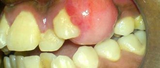
Even one loose bracket cannot be removed without the help of a doctor without damaging the entire structure. It is necessary to make an appointment with an orthodontist as soon as possible to repair the system, since the absence of one element can disrupt its balance, and the effectiveness of treatment will decrease. Before going to a specialist, it is better to fix the fallen lock with medical wax, sold in a pharmacy.
Sometimes braces are removed faster than planned. This is due to the anatomical features of the jaw structure and accelerated biological processes in the human body. However, such cases are extremely rare.
As a rule, the question “Is it possible to remove braces ahead of time?” doctors answer negatively. The patient must put up with certain inconveniences and undergo full treatment. Otherwise, the teeth will return to their previous position, and time and money will be wasted. In addition, removing the brace system ahead of schedule can lead to disruption of the temporomandibular joint.
Previously, braces could only be removed under certain circumstances.
- Correcting the bite does not bring results, and surgical intervention is required.
- There is an urgent need to replace one system with another for some reason - for example, a breakdown or an allergy to the material.
- The doctor is unable to cope with the anomaly and is forced to redirect the client to another orthodontist.
Is it possible to remove braces temporarily if important public events are coming up? Theoretically this is acceptable, but from a practical point of view it is inappropriate. Firstly, after removal the structure becomes unsuitable for secondary use. You will have to purchase a new system and spend money on its installation. Secondly, after repeated fixation, the time required to correct the bite increases.
Future newlyweds are interested in the question of whether it is possible to remove braces for the wedding. You should take care of the problem in advance and opt for ceramic or sapphire devices or transparent silicone aligners. Then the design will not spoil the festive image.
The removal of the system is carried out by the attending physician in the same clinic where it was installed, so the patient does not have to look for a dentist to carry out the procedure.
With turnkey treatment, there is no need to find out in the capital's centers how much it costs to remove braces, since the service is already included in the final price. In addition, the amount includes the costs of the device itself and its installation. If all stages of bite correction are paid separately, then the cost of removing the device is about 10,000 rubles.
After wearing braces, you will need to put in a lot of effort to maintain a beautiful smile. However, those who have patiently gone through the entire path of bite correction from braces to a retainer confirm that the end result justifies any difficulties of the process!
Sequence of procedure
What to do if the plate comes off? If the brace system breaks down, efforts aimed at straightening the row or correcting the bite will be in vain. The design on the teeth will change the pressure and direction of action, which will cause harm to the dentition.
Do not be alarmed if you find a part of the structure that has come off in your mouth; you should immediately consult a doctor. It will help restore the integrity of the system and tell you what to do next so that the situation does not repeat itself.
If you can’t see an orthodontist, you can try to repair the broken bracket yourself. There are 2 options for this repair: glue the plate using special wax or remove the lock.
There are not many options for action that can be taken in a situation with a loose brace. It is important to keep the brace intact and see your doctor as soon as possible so that he can install the lock in place.
If the loose lock was located on one of the outer teeth, you can simply remove it. If the tooth is in the middle of the dentition, then you will not be able to remove the braces yourself.
If the braces are metal, most likely there will be no problems with gluing the old brace - they are quite strong and usually do not break.
The now popular ceramic braces are many times more fragile and can break. Then you will have to buy a new bracket and glue it on. The doctor will inform you about this need, and, as a rule, you can buy a new lock from him.
A problem with the system can occur even when the orthodontist is out of town, on vacation, on a business trip, or somewhere else. What to do in this case? First of all, you need to contact the clinic to find out about the return date of the attending physician. You can walk without braces for about a week, so if time allows, you can wait.
If the braces come off within a couple of days after they are installed, then the problem is in the actions of the orthodontist.
The fact is that the lock is not glued to the “bare” tooth, but to the filling material. To glue it evenly and firmly, the surface of the tooth is dried from saliva. If, despite all the doctor’s efforts, the tooth is not perfectly dry, the braces may begin to come off on the same day. This is an ordinary situation, the lock is glued in place, and payment for gluing is usually not charged.
Treatment with braces requires careful hygiene. For patients, brushing their teeth 3-4 times a day is commonplace, because they need to clean their mouth after every meal. In addition, most people use irrigators during this period. If you put too much pressure on your teeth, your braces can start to come off. To prevent this from happening, you need to clean with gentle movements, and set the optimal water pressure for the irrigator.
The most common reason why braces come off is failure to follow dietary restrictions. The lock is attached with glue, which may not withstand excessive stress caused by hard food.
What you will have to limit yourself to so as not to provoke the braces to come off:
- nuts, including chocolate with them;
- tough meat;
- hard fruits, for example, are definitely not worth biting into an apple; it is better to cut it into pieces;
- hard vegetables such as carrots, cucumbers;
- crackers;
- other foods that are too hard.
Meanwhile, during treatment you can not deny yourself your usual foods. Meat, vegetables, fruits should be cut into pieces and eaten calmly. You can even chop nuts and not worry that the locks will come off.
In this situation, some patients begin to panic. They don’t know what to do if the bracket on the last tooth comes off. This situation is not uncommon, because people chew with their back teeth. The danger here is that the lock can easily fly off the arc and be swallowed.
Fortunately, the wearer of braces usually feels the moment when the lock begins to come off. What to do if the last bracket comes off? The main thing here is to catch it and bring it to the doctor safe and sound as soon as possible.
DETAILS: Teeth straightening in adults - prices for orthodontic treatments
If you couldn’t catch it, you’ll have to buy a new one. Otherwise, the situation in which the last bracket came off is completely normal, no different from others. There is no need to worry about a swallowed brace; it will leave the body after one day. The most important thing is not to choke on it.
No one is immune from troubles on vacation. What to do if the braces come off, and the doctor, and even the clinic itself, is far away? The best option is to contact your orthodontist. As a rule, doctors understand that force majeure is possible and leave their personal contact information to patients.
The orthodontist can recommend a local clinic, since the doctors know each other closely, or offer another solution to the problem.
Orthodontists are unanimous in their recommendations: if a patient’s braces come off, he needs to immediately visit his doctor. The permissible period that can be spent without braces is a week. Teeth move constantly and the changes can be difficult to correct.
In recommendations on how to prevent the structure from coming off, orthodontists also unanimously state: the main thing is to follow a gentle eating regimen, avoiding hard and hard foods.
Treatment with braces, like any other treatment, requires strict adherence to the doctor’s recommendations. If your braces come off, regardless of why, you should contact your orthodontist as soon as possible to have them reattached. Any delay can increase the treatment period, which is already quite impressive.
Removing braces is much faster than installing them and does not cause any pain to the patient. First, the orthodontist removes the curing material, this is done mechanically using dental forceps. Each lock of the system must be opened so that the orthodontic arch can be released, and after this each bracket is removed from the teeth.
The dental glue is removed along with the braces, and its remains are cleaned off by the doctor using polishing machines. At this moment, not very pleasant sensations may also arise, but nothing more. This procedure usually takes no more than five minutes. The longest time you have to deal with is the lingual brace system; it takes about 30 minutes to remove it; in situations with ligature structures, everything is done much faster.
It is not possible to answer this question unequivocally, since much will depend on the individual characteristics of the patient, the severity of his disease and a number of other factors. On average, the period of wearing braces ranges from 4 months to 3 years; it is also important to remember that only a doctor can decide when to begin removing the structure.
Structurally, retainers are divided into two types:
- non-removable - made of metal in the form of a braided arch fixed on the inside of the teeth, this design is invisible to others and after about one to one and a half weeks of wearing it is no longer felt by the patient in the mouth;
- removable - plastic transparent mouth guards, which can be removed and put on again as needed.
Usually, after removing braces, the condition of the teeth leaves much to be desired - glue residues, stains and cracks spoil the impression of the treatment result. White areas on the enamel after removing braces indicate a lack of microelements in the tooth tissues during the period of wearing the orthodontic apparatus.
There is no need to worry about aesthetic defects caused by wearing the system. After the braces are removed, the enamel is restored by applying a remineralizing composition, and irregularities are removed by polishing. In some cases, the installation of veneers is used or the method of artistic restoration is used in order to obtain ideal teeth from an aesthetic point of view.
The treatment process does not end with getting rid of the orthodontic structure. The doctor will perform several procedures to restore your teeth to their natural color and attractive appearance.
With the help of professional hygienic cleaning after removing braces, the remaining glue, plaque and tartar that have accumulated under the clasps while wearing the device are removed from the tooth surface. Remineralizing therapy and fluoridation saturate the enamel weakened by constant stress with the necessary substances.
After removing braces, teeth whitening is recommended no earlier than a month later, when the condition of the dental tissues improves. Premature lightening threatens increased sensitivity and prolonged pain.
Without the pressure of the orthodontic system, the teeth will try to return to their original position if not prevented. To prevent the teeth from becoming crooked after the braces are removed, the patient is fitted with a special retainer that fixes the dental units in the desired position to secure the result. Such structures are divided into removable and non-removable.
- Fixed retainer. A thin wire is glued to the inside of the teeth using a filling material. The arch is absolutely invisible when worn, does not affect diction and does not cause discomfort.
- Removable retainer. We are talking about a silicone mouthguard, which is made according to individual impressions of the patient’s jaws, or retention plates after removing braces, intended for use while sleeping.
The retention period exceeds the duration of treatment by at least 2 times. Some patients wear a retainer throughout their life.
Few people like the way their teeth look after braces are removed. However, aesthetic defects can be easily eliminated. A more serious problem is physical discomfort. After removing braces, teeth hurt from trying to return to their previous position, especially after installing a retainer, which blocks any movement.
In addition, weakened and sensitive enamel reacts sharply to temperature changes, which also causes discomfort. Dizziness or nausea after having braces removed may be related to retainers. If such symptoms occur, be sure to consult a specialist.
Principles of prevention and treatment of dental anomalies
Prevention and early treatment of dental anomalies are the fundamental basis of modern orthodontics (see Dentistry).
There are general and special prevention of dental anomalies. General prevention consists of creating favorable conditions for the growth and development of the child, starting from the embryonic period, as well as eliminating all kinds of bad habits in children (thumb sucking, lip biting, etc.).
Special, or local, prevention includes treatment of carious primary and permanent teeth, grinding of unworn cusps of primary canines or other teeth, removal of retained primary and supernumerary teeth, timely rational prosthetics (see Dentures).
Orthodontic treatment is carried out in the presence of pronounced dentofacial anomalies, causing functional and aesthetic disorders and often traumatic for the patient’s psyche. Severe congenital and acquired anomalies of the jaws are subject to surgical or complex (surgical and hardware) treatment.
Contraindications
Orthodontic treatment includes inflammatory processes in the teeth and periodontal tissues, as well as severe concomitant diseases, for example, epilepsy.
Orthodontic treatment includes: moving incorrectly positioned teeth in various directions (vestibular, oral, or palatal, medial, distal, vertical, rotating the tooth along the axis); expansion of narrowed dental arches; correction of bite in the sagittal, transversal and vertical directions; normalization of the function of chewing and facial muscles, as well as the muscles of the tongue.
Orthodontic treatment methods can be divided into four groups: myotherapy, hardware treatment (use of orthodontic devices), myotherapy in combination with hardware treatment and complex (surgical and hardware) treatment. The choice of treatment method depends on the type of anomaly, the period of development of the dental system (deciduous, mixed, permanent bite), the severity of the anomaly, and the general condition of the patient.
Myotherapy consists of long-term and systematic exercises of insufficiently or improperly functioning groups of masticatory and facial muscles, which stimulates the growth of underdeveloped parts of the jaws and promotes the proportional development of the entire facial skeleton. The main exercises are aimed at activating the orbicularis oris muscle, the muscles that lift and extend the lower jaw, as well as the muscles of the neck and shoulder girdle. To normalize swallowing and speech articulation, special exercises are prescribed for the muscles of the tongue, soft palate, and pharynx.
Myotherapy is most effective in preschool age (during the periods of primary and mixed dentition), starting from 4-5 years, when intensive growth of the bones of the facial skull occurs. At a later date, this treatment method is less effective.
The hardware method is the main one in orthodontics. During periods of primary and mixed dentition, it is often combined with myotherapy, and with permanent dentition - with surgical interventions (removal of individual teeth; osteotomy, decortication or compact osteotomy of the jaws, etc.).
Rice. 1. Microslide of a fragment of a decalcified lower jaw of a dog: the process of resorption of the alveolar bone in the pressure zone, accompanied by the appearance of osteoclasts (1) on the surface of the resorbing bone beams (2); hematoxylin-eosin staining; x 50. Fig. 2. Microslide of a fragment of a decalcified lower jaw of a dog: the process of building alveolar bone in the traction zone, carried out by osteoblasts (1), located along the edge of the bone beams under construction (2); hematoxylin-eosin staining; X 200. Fig. 3. Microscopic specimen of a fragment of a dog’s hard palate with surrounding tissues: the process of building bone tissue during the expansion of the upper jaw - along the edges of the palatal suture (1) newly formed bone beams are visible (2); Hematoxylin-eosin staining x50.
The hardware method is based on the mechanical force acting on the teeth, as well as on the targeted redistribution of functional and mechanical load on the teeth and various parts of the dental system (alveolar processes, jaw bones, temporomandibular joint). As a result of the changed mechanical and functional load, a restructuring of the tissues of the dental system occurs, ch. arr. jaw bone tissue. This is expressed in the resorption of bone in pressure zones, where osteoclasts accumulate, carrying out the resorption of bone beams (Fig. 1), and the construction of bone tissue by osteoblasts in traction zones (Fig. 2). When the upper jaw is expanded with the help of orthodontic devices, the median palatal suture is opened, along the edges of which newly formed bone tissue is built (Fig. 3). Thanks to these processes, the teeth move in the desired direction, the shape of the dentition is corrected and their relationship in the anteroposterior, vertical and transversal directions is normalized.
Hardware treatment consists of two phases - active treatment and retention. In the first phase, the mechanical devices of the device are activated (screw, elastic wire, rubber rod) or its functionally guiding elements are corrected (inclined planes, bite pads, shields). In the retention phase, the device acts passively, consolidating the achieved treatment results. Retention devices are also used for this purpose.
Active treatment can last from several weeks to 2-3 years, depending on the nature and severity of the anomaly. The duration of the retention period depends on the severity of the anomaly, as well as on the speed of active treatment: the faster active treatment is carried out, the longer the retention period and vice versa. This is explained by the fact that with a short period of treatment, the morph and restructuring of periodontal tissues do not have time to occur and therefore longer retention is required. The criterion for the end of retention is the absence of a tendency to relapse after removal of the retention apparatus.
Orthodontic devices are divided into preventive, therapeutic and retention. Preventive devices include spacer devices and disconnecting plates, or mouthguards (see).
Treatment devices can be removable or non-removable, intraoral, extraoral and combined. According to the principle of operation, they are divided into devices of mechanical action, functionally operating, and combined action, including elements of the first two groups.
Mechanical devices exert force (pressure and traction) on the teeth through special devices: an orthodontic screw, an elastic wire arch, a spring, a rubber rod. The strength of such devices is regulated (dosed) by the attending physician. Mechanical devices include Angle, Mershon, Ainsworth devices, and expanding plates.
Rice. 4. Engle’s apparatus: 1 - elastic wire arc, 2 - nut, 3 - screw thread, 4 - screw for adjusting the diameter of the bandage ring, 5 - bandage ring, 6 - horizontal tube. Rice. 5. Schematic representation of the application of Angle devices with an oblique intermaxillary rubber rod: 1 - elastic wire arches, 2 - a hook for strengthening the oblique intermaxillary rubber rod, 3 - bandage rings, 4 - vertical lines showing the sagittal discrepancy of the dentition, 5 - oblique intermaxillary rubber thrust (arrows indicate the direction of the applied force).
The device proposed by Angi (EH Angi, 1886) consists of an elastic wire arch (0.8-1 mm in diameter) made of stainless steel, nuts screwed onto the arch, and bandage rings or stamped crowns fixed to the supporting teeth (Fig. 4). The Angle apparatus is called universal; it is used to expand the dentition and normalize their relationship, as well as to move individual teeth in different directions. Separation (spreading apart) of the supporting and moving teeth is carried out using a wire ligature.
To move the dentition or lower jaw in the anteroposterior direction, Baker (G. Baker, 1892) proposed fixing Engle’s apparatus on both dentitions and connecting them with an oblique intermaxillary rubber rod (Fig. 5).
Rice. 6. Schematic representation of the application of the Ainsworth apparatus: 1 - elastic arch, 2 - ring for fixing the apparatus on the teeth, 3 - tangent beams; arrows indicate the direction of the force developed by the apparatus. Rice. 7. Schematic representation of the action of the Mershon apparatus, used for: a - expansion of the dentition; b - moving the incisors forward; c — medial movement of teeth (left); d - distal movement of teeth (right): d, e - rotation of teeth along the axis (section of the dentition); 1 — rings for fixing the device on the teeth, 2 — elastic processes, 3 — lingual arch. The arrows show the direction of tooth movement during treatment.
In 1904, G. S. Ainsworth developed an apparatus for expanding the dentition without ligature tying of teeth. The device consists of rings placed on the supporting teeth and an elastic wire arch, the edges of which are fixed on the vestibular surface of the rings. Tangent beams are soldered to the rings from the oral surface; their length depends on the length of the area of narrowing of the dentition (Fig. 6).
The device, created by Merschon (JV Merschon, 1909), consists of a supporting lingual arch, elastic processes made of thin wire attached to it, actively acting on the teeth, as well as rings for fixing the device on the supporting teeth (Fig. 7). When activating the processes, the device has a gentle, weak effect on the teeth (with a force of 1-5 g). It is used to expand the dental arch and correct the position of individual teeth.
Rice. 8. Schematic representation of the application of the Begg apparatus on the lower dentition (a section of the dentition is shown): 1 - elastic vertical loop; 2 — lock fastening; 3 — ring for fixing the device on the teeth. Rice. 9. Schematic representation of an expanding plate with a Coffin spring: 1 - vestibular retraction arch, 2 - expanding Coffin spring, 3 - sawing of the palatal plate, 4 - palatal plate, 5 - clasps. Rice. 10. Schematic representation of the Kalvelis apparatus with arm-shaped springs for distal movement of teeth: 1 - palatal plate, 2 - arm-shaped springs, 3 - clasp. The arrows indicate the direction of the acting force. Rice. 11. Kalamkarov mouth guard for distal movement of teeth: 1 - metal crown on the tooth being moved; 2 - sectors of the mouthguard for sequential movement of teeth; 3 — plastic mouthguard; 4 - pieces of Angle wire arcs with screw threads and nuts for activating the device.
The device proposed by Begg (P. Begg, 1965) is an elastic arc made of thin steel wire. A special feature of the design of this arch is the many vertical loops and other bends that make it possible to exert a constant and very gentle effect on the teeth (Fig. 8). The Begg appliance can be used to correct various misaligned teeth, dental deformations and malocclusions.
Mechanical devices also include expansion plates with orthodontic screws or a Coffin spring (Fig. 9), which serve to expand the dentition and move teeth. For distal movement of teeth, a Kalvelis apparatus with arm-shaped springs is used (Fig. 10), as well as a Kalamkarov mouth guard, consisting of a metal crown fixed on the last (moved) tooth, and a plastic mouthguard covering the dentition. In order to move the next tooth, a segment is cut from the mouth guard, which is activated using segments of Angle's arches (Fig. 11).
There are two types of functional devices. Devices of the first type redistribute the force of chewing pressure. Their design includes inclined planes, or bite pads (surfaces), with which individual teeth or groups of teeth come into contact during contraction of the masticatory muscles. In this case, the teeth perceive increased load and move in the direction determined by the inclined plane. Teeth excluded from occlusion (closing) remain underloaded. As a result of the redistribution of the functional load, a corresponding tissue restructuring occurs in the dental system, and the deformation is corrected.
Rice. 12. Plaster model of the upper jaw with a Katz bite block for the treatment of progenic bite: 1 - plastic plate, 2 - flip clasps, 3 - plaster model of the upper jaw, 4 - inclined plate. Rice. 13. Schematic representation of the Katz guide crown, put on a moving incisor, when closing the dentition: 1 - metal crown, 2 - inclined wire plane.
The devices of this group include the Katz bite plate (Fig. 12), the Katz guide crown, consisting of a metal crown fixed on the tooth being moved and a wire inclined plane soldered to it (Fig. 13), Bynin's mouth guard, applied to the lower dentition and having an inclined plane only at the teeth that are to be moved, etc. These devices are used to correct the oral inclination of the upper incisors and treat progenic bite.
Rice. 14. General view (a) and schematic image in sagittal section (6) of the vestibular plate of Corbitz: 1 - handle, 2 - vestibular plate itself, 3 - teeth.
Devices of the second type help restore the disturbed myodynamic balance between the muscles of the tongue, on the one hand, and the muscles of the lips and cheeks, on the other, normalize nasal breathing, and eliminate bad habits (finger sucking, lip biting). The second type of apparatus includes vestibular plates (Fig. 14), placed in the vestibule of the mouth (between the lips and teeth).
Combined-action devices contain elements of mechanical and functional devices. Such devices include the Andresen-Heupl activator, the Bruckle apparatus, the Schwartz double plate, functional Frenkel regulators of 3 types, etc.
Rice. 15. Andresen-Heupl activator for correcting the disturbed relationship of the dentition (general view): 1 - tooth bed, 2 - plastic monoblock, 3 - vestibular wire arch with U-shaped bends.
The activator proposed by V. Andresen and K. Haupl consists of a plastic monoblock and a vestibular wire arch (Fig. 15). According to indications, the device can be supplemented with an orthodontic screw or a Coffin spring. With the help of the activator, the disturbed relationship of the dentition is corrected: prognathic and progenic bites, as well as deep and open bites.
The device proposed by H. Bruckl is a plate for the lower jaw with an inclined plane in the area of the anterior teeth and a vestibular retraction arch. The device is used to treat progenic bite and correct the position of the upper and lower front teeth.
Rice. 16. Functional Frenkel regulator for correcting the disturbed relationship of the dentition: 1 - vestibular arch; 2 — cheek shields; 3 - loops on the upper canines; 4 - lingual arch; 5 - palatal clasp. Rice. 17. Various types of retention devices: 1 - device for fixing the tooth after rotation along the axis; 2 - apparatus for fixing the tooth after eliminating the palatal position; 3 - apparatus for fixing the tooth after eliminating the vestibular position; 4 - apparatus for fixing the tooth after traction; 5-8 - devices for maintaining the gap between teeth.
The double plate, developed by A. M. Schwarz, consists of two active plastic plates for the upper and lower jaws and is used to treat both prognathic and progenic bites. Functional regulators designed by Frenkel (R. Frankel) consist of cheek shields, lip pads and various mechanical devices: screws, springs (Fig. 16). These regulators are used to treat prognathic and progeic bites and other anomalies.
Retention devices are designed to consolidate the results of active hardware treatment. They can be removable or non-removable (Fig. 17). Removable plates with clasps and a vestibular arch can be used as retention ones. If during the process of active movement of teeth or changes in bite, natural (physiological) retention is established, then retention devices are not needed.
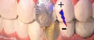
How is tooth enamel polished?
You should not be afraid of grinding your tooth enamel after removing the braces; this is a normal procedure that is not associated with any difficulties or complications. The orthodontist, using polishing attachments, removes the remaining adhesive from the teeth in a circular motion and polishes the enamel. When the structure is removed, pockets of demineralization often form around the braces, which can be easily eliminated with a paste containing calcium and fluoride; if the need arises, teeth whitening can also be performed.
Does it hurt?
Most patients fear that the procedure for removing orthodontic structures will be painful. But this is absolutely not true.
In the process of removing the elements of the apparatus, the orthodontist does not touch the enamel and soft tissues of the oral cavity, so the occurrence of painful sensations is completely excluded. They can only occur if the patient is restless, spinning in the chair while working with forceps and thereby increasing the risk of periodontal injury.
Both vestibular and lingual models can be removed equally painlessly, with or without ligatures. When the clasps are separated from the surface of the tooth, a slight discomfort may occur due to the tight connection of the glue with the enamel.
DETAILS: Braces do not cover the entire dentition
There is no pain even during polishing of the enamel. It can only occur if the gums are accidentally touched by a grinding disc, but only if the patient does not sit still at the time of treatment.
It is not difficult to understand the fear of pain in patients who are about to part with their braces, but it should be said that during this procedure the doctor does not touch either the soft tissues of the oral cavity or the tooth enamel, except for grinding it after removing the structure. Accordingly, pain is almost completely eliminated, but the likelihood of this unpleasant phenomenon still exists and here’s why:
- During the process of removing braces, the patient becomes overly nervous, spins around in the chair and thereby increases the risk of injury;
- when separating the clasps from the tooth surface, discomfort may occur if the connection of the tooth with the glue is too tight;
- During grinding of enamel, pain may occur as a result of the grinding disc touching the gums, however, this is a consequence of the patient’s restlessness in the chair.
It actually hurts quite a bit. My orthodontist said that after the installation my teeth would hurt for about a week. I was sick for a month and a half. No, not 24 hours a day, but always during meals. However, it is different for everyone. A friend of mine had toothache for only three days after getting braces.
I wrote about everything related to painful sensations in a separate short article: Do braces hurt? No, those are not my teeth in the photo;) Mine, if you’re interested, are here.
Procedures during dental treatment
Interproximal teeth separation
Video demonstrating this procedure:
In what cases is separation necessary?
1. The width of the upper incisors does not correspond to the width of the lower incisors. Orthodontists have certain methods for calculating the correspondence of the width of the incisors of the upper and lower jaws. And when the doctor identifies this discrepancy, he suggests that the patient grind the incisors of the upper or lower jaw so that the front and lateral teeth will receive a more physiological closure in the future.
2. When we try to treat without removal (and the risk of removal is high). In this case, the patient is offered a choice - either grinding of all teeth, the jaw where removal is possible, or removal of one or two teeth.
3. When the teeth of the left or right half of the jaw are asymmetrical. This is more common in adult patients who have already passed through numerous hands of dental therapists. When the dentist installed a filling, he did not try to replicate the anatomical shape of the tooth, either had no idea that this should be done, or did not have such an opportunity due to the prevailing conditions in the patient’s mouth.
4. When the teeth are triangular in shape and after correction, black triangles are visible. This is more often used in adult patients. When a patient begins treatment with significant crowding of incisors and canines. At the same time, over many years the gums have acquired the low, forced position in which the teeth stand today, and the patient sees the normal position of the gums at the beginning of treatment. After installing braces, triangular-shaped teeth begin to straighten and at the same time, the gums do not change their position following the teeth. As a result, the teeth are straight, and the gums remain almost in the same place where they were before the treatment began. Black triangles visually appear between the incisors. The patient perceives this as a receding gum. Although it was not the gums that changed their position, but the teeth.
Is it safe?
The volume of separation (grinding) is not large. A small amount of enamel is ground off from the sides of the tooth (0.1-0.5 mm). Each tooth in these places has a fairly thick layer of enamel. There is no need for anesthesia for separation. It's a little unpleasant, but not painful. And this procedure cannot lead to caries. It happens that after the procedure there is a slight sensitivity of the teeth, but after a short period of time this goes away.
How is this done?
After a short straightening of the teeth (for 4-8 months) with a very thin diamond disc, the doctor very carefully grinds off the required amount of enamel from the planned teeth. After this, the ground surfaces are polished with polishing discs. Immediately after separation, the patient sees gaps between his teeth. The doctor puts an elastic chain on all teeth and within 1-4 months all gaps are closed.
Microimplantation
Microimplants are used in orthodontics to increase bone support. They are small screws that are installed into the bone tissue during orthodontic treatment. They differ from conventional implants in size, duration of use and purpose. A microimplant is a temporary element that is installed and removed during orthodontic treatment. The length of the microimplant ranges from 6 to 12 mm (most often we use 6-8 mm).
Installation of a microimplant takes no more than 10 minutes, is performed under local infiltration anesthesia (this is the lightest type of anesthesia, in which only the mucous membrane is anesthetized) and does not require a rehabilitation period. After the procedure, minor pain in the mucous membrane at the installation site is possible (only from the injection), which disappears the very next day.
After achieving the desired result, the microimplant is removed. This can be done by an orthodontist, since anesthesia is not required (there are no nerve endings in the bone tissue). In the area where the microimplant was unscrewed, not even a trace remains after 2-3 days. We recommend rinsing the area with 0.05% chlorhexidine for 3 days and maintaining good hygiene in the area.
The presence of a microimplant requires more careful oral hygiene, otherwise there is a high risk of its rejection. Also, in smoking patients, the frequency of rejection increases significantly, since the tars contained in tobacco smoke settle on the surface of the microimplant, irritating the mucous membrane, inflammation of the mucous membrane spreads to the bone tissue and the microimplant becomes mobile.
So why are microimplants needed?
1. To create space in the case of a tilted tooth towards the tooth removed in front of it and subsequent implantation and prosthetics. 2. In the case of a lowered upper tooth, down into the place of a previously long-removed lower tooth. 3. For immovable support when moving a group of teeth. 4. To align the occlusal line. A line drawn along the edges of the upper incisors and in comparison with a line drawn along the level of the pupils. When the teeth of the left or right half are at different heights.
Example No. 1
The 25th tooth is missing, the free space must be filled by moving the front teeth (24, 23, etc.) back, however, if we give elastic traction between teeth 24 and 26, they will move towards each other (according to Newton’s third law), but we need so that the 26th tooth remains in place. Therefore, we install a microimplant behind the 27th tooth - this allows us to provide support for the 26th tooth, the microimplant does not allow it to move forward.
Example No. 2
In this case, the patient had the 15th tooth removed for orthodontic reasons; the 27th tooth was already missing. We decided to move the 26th tooth back to avoid the removal of another tooth (the 25th tooth) and implantation in the area of the 27th tooth. Thus, the patient will receive perfectly straight teeth and a correct bite in the absence of implants, although it would be much easier for us to carry out a symmetrical removal of 5 teeth and implantation in the area of the missing 27 tooth. Such treatment is only possible with the use of microimplants.
Photographing
Modern orthodontics is on a par with similar specialties such as cosmetology and plastic surgery, which significantly change the appearance of the patient. In this regard, photographic documentation of the treatment history is important for the doctor and the patient. Only photographs of the face and dentition can give an objective assessment of the extent of changes that have occurred during the treatment process. Today, photographic equipment at an orthodontic appointment is as integral a part as disposable shoe covers at the entrance to a medical facility.
So, why does a doctor need to photograph a patient, and what kind of photographs does the doctor take?
The doctor conducts a comparative analysis of the changes occurring in your teeth throughout the treatment. Do you agree that it is quite difficult to remember the position of each tooth and compare them with the original ones after 4-6 months of treatment? What if a doctor has 20-30 patients a day and 25 working days a month? Using simple calculations, we find that the doctor must remember 750 cases of the location of teeth that change their position every month. Photo documentation of each case helps us with this. Taking diagnostic photographs before, during and after treatment is of great clinical importance because... Changes during orthodontic correction occur gradually. In addition, by looking at the photographs taken, the doctor can track the dynamics of treatment and pay attention to the nuances that he missed when assessing the situation in the oral cavity.
Photography takes place during your second visit to the clinic, dedicated to concluding a contract and taking impressions. Thus, based on the analysis of x-rays, photographs and diagnostic plaster models, the doctor draws up a comprehensive treatment plan.
.
During the process of orthodontic treatment, photography of the dentition can be repeated every 2-6 months to obtain a comparative assessment, the effectiveness of the selected mechanics and analysis of possible errors in diagnosis or implementation of the treatment plan. By analyzing the dynamics of changes from photographs, the doctor understands what measures need to be taken to speed up the treatment process or direct it in the necessary direction.
Of course, at the end of treatment, the doctor also photographs the face and teeth to document the result obtained and compare photographs before and after orthodontic treatment.
.
By comparing the photos before and after treatment, we clearly see the result of the work done. In the “Examples of Work” on our website you can see just such photographs of various clinical cases.
.