What leads to impaired pneumatization of the maxillary sinuses?
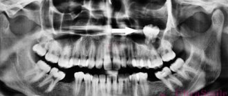
Impaired pneumatization of the maxillary sinuses is associated with the development of inflammation in the maxillary sinus due to the thin structure of its lower wall. The reasons for the violations may be problems with the patient’s teeth. During treatment and filling, filling material sometimes gets into the air cavities, which causes problems. Reduced pneumatization of the maxillary sinus can be seen in photo 1, which shows an x-ray.
All these diseases are different in their form, nature and location.
Thus, there are 4 groups of inflammation of the mucous membrane of the adnexal cavity: sinusitis, sphenoiditis, frontal sinusitis and ethmoiditis. All of them are united under a single name - sinusitis.
These diseases can be either viral or bacterial in nature. Patients suffer from sinusitis as complications after an acute runny nose or infectious diseases, as well as after an injury.
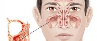
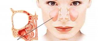
The paranasal cavities through which air passes are located in the bone tissue of the brain and facial skull and connect to the nose to supply air and serve as voice resonators. The following groups are distinguished:
- the paired maxillary cavity is located in the maxillary region;
- the frontal cavity is located in the frontal bone of the skull;
- the sphenoid is located in the body of the main bone;
- The ethmoid labyrinth is formed by the cells of the ethmoid bone.
Photo 1. Pneumatization of the maxillary sinus is reduced.
The accessory nasal cavities begin to develop from the prenatal period until human puberty. Inside them there is an epithelium, the cells of which secrete mucus. The largest sinus is the maxillary sinus. It opens into the middle nasal passage and has 4 recesses (coves). Depending on the pneumatization of the air cavity, additional depressions may become deformed and change it.
The sinus, which is located in the frontal bone, is divided into 2 parts by a septum. The frontal cavity connects with the middle nasal passage, while its sections are pneumatized to varying degrees. The same situation is observed in the sphenoid sinus, which is divided by a septum into 2 parts with varying degrees of pneumatization of the sections.
But what leads to the section of the air cavity is located closer to the upper part of the anterior wall of the sinus and exits into the nasal cavity. The ethmoid labyrinth consists of cells that exit into the upper and middle nasal passages.
To assess the condition of the paranasal sinuses, the doctor will conduct an external examination, during which he will be able to examine the nasal and oral cavity, the palate for swelling, hyperemia, and the presence of a fistula. Palpation will help identify the location of pain. For a deeper examination of the inner surface and branches of the nose, rhinoscopy is used.
One of the most difficult methods for studying the patency of sinus and nasal anastomosis is probing. It is carried out through the frontal cavity, less often through the sphenoid cavity.
Before the procedure, the nose is treated with a local anesthetic and a drug that constricts blood vessels, usually a solution of dicaine and ephedrine. Then a Lansberg cannula probe is inserted through the middle turbinate.
To get rid of fluid, mucus and pus, the sinuses are washed and a special radiopaque substance is injected into them. Sometimes a puncture of the paranasal sinuses is performed using a special Dufault or Kulikovsky needle through the lower nasal passage. The needle is immersed no more than 1 cm into the lumen of the sinus. This is a very complex procedure, where a doctor’s mistake is fraught with serious consequences. The liquid obtained as a result of the puncture undergoes bacteriological testing. The puncture site is washed with an antiseptic.
Exceptions to the rules - pathologies
There are cases of improper development and formation of additional partitions or the absence of some cavities. Excessive pneumatization and pathologies of bone walls are diagnosed. Congenital pathologies do not reveal themselves, but cause complications.
Nasal problems occur after injury directly to the nasal sinus or after a closed intracranial one. The sign is always eye pain, and sometimes the person suffers traumatic shock or fainting. Bone fragments can become dislodged, causing bleeding, and if they touch the labyrinth, liquorrhea occurs. Upon examination, an uneven fracture line and displacement of the shadows of the sinus wall lines are observed.
Inflammation of the nasal air sinuses is accompanied by increased temperature, pain at the site of inflammation, and impaired sense of smell. The patient experiences heaviness in the forehead and eyes, especially when tilting the head forward or down. The patient complains of discomfort with sudden movements of the head, thick nasal discharge. The x-ray shows darkening of the sinus. With the growth of polyps and the presence of more exudate in the mucous membrane, swelling occurs, and this causes a decrease in pneumatization of the sinus.
If a patient suffers from allergies, he is diagnosed with rhinosinusitis, an allergic rhinosinusopathy. The signs of the disease are the same as those of rhinitis, and the photographs show significant swelling of the mucous membrane, which is reflected by darkening of the sinuses. The outlet of the accessory sinus is closed for a long period due to injury, cystic distension (mucocele, pyocele, empyema). Such problems reveal their presence by protrusion or thinning of the lower wall of the mucous membrane, which crunches when touched.
The inflammatory process in the maxillary sinuses is one of the most common diseases, which is becoming more frequent due to possible infection from the teeth.
This is a seasonal disease because the peak of inflammation occurs during outbreaks of influenza and other acute colds. Most often, the cause of sinusitis is considered to be infection through the blood, nose or teeth, or injury to the sinus walls of the nose. There are acute and chronic forms with formations of a catarrhal or purulent nature.
Reasons for the decline
There may be several reasons why pneumatization of the maxillary sinus is impaired:
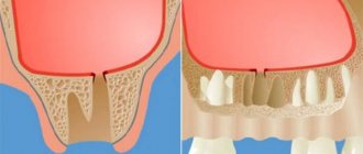
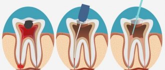
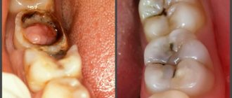
- Dental problems. The main reason why most often there is a decrease in pneumatization of the maxillary sinuses is its thin lower wall, which can be easily damaged, for example, during various dental procedures: dental canal treatment, screwing in maxillary pins, etc.
- Inflammatory process (sinusitis). In total, the maxillary sinus has four small depressions (bays), and its entire internal surface is lined with epithelial cells that constantly produce mucus. As the inflammatory process develops, swelling of the mucous membrane may appear, and it fills part of the affected cavity, displacing air from it. With purulent sinusitis, pneumatization decreases even more due to the accumulation of pus.
- Fractures and injuries of the skull bones. A more rare reason for which the pneumatization of the maxillary sinus is impaired. The worst thing is when bone fragments get into the cavities of the nasal sinuses and remain there, provoking chronic inflammatory processes. In this case, only surgical intervention can radically solve the problem. Sometimes the configuration of the cavity changes due to improper fusion of bones, including after unsuccessful plastic surgery.
- Polyps and other neoplasms. They tend to grow and can gradually fill most of the sinuses with their body. And if the formation is malignant, it can not only grow on its own, but also metastasize even to distant organs.
- Congenital anatomical features. This includes both the curvature of the nasal septum and the structural features of the upper jaw and other bones of the skull. In some cases, the formation of insufficiently deep paranasal sinuses occurs due to disproportionate growth of the facial bones.
In principle, all these causes can be eliminated with the help of properly selected drug treatment or surgical intervention. In some cases, when pneumatization is slightly reduced and there are no active inflammatory processes, no treatment is required at all.
Read more Criminal cases database by last name for free
Pneumatization of the nasal sinuses
Often, after visiting a doctor, a patient sees an incomprehensible term in the medical history - pneumatization of the sinuses. Few people know what this means, so many questions arise. This term cannot be considered a diagnosis, it is just a description of an x-ray of the paranasal cavities. This indicator can be used to determine the inflammatory process in the nasal appendages, the accumulation of purulent contents, swelling and the formation of cysts . Patients diagnosed with sinusitis are referred for an X-ray examination.
Only if the paranasal sinuses are pneumatized normally can the sinuses perform their functions fully. If the air circulation in the paranasal sinuses is impaired, this means that the person has some kind of disease.
What affects pneumatization?
Medical research has proven that the structure and volume of the nasal sinuses are strictly individual.
The volume of air that reaches a person depends on this; this aspect also depends on the structure of the bone structure of the face. Pneumatization of the sinuses is not characterized by the volume of air that passes through the sinuses. Doctors examine the quality of the mucous membrane of the sinus surfaces, as well as the presence of third-party neoplasms that may interfere with stable operation or provoke changes in the bone structure.
https://www.youtube.com/watch?v=0kfkDFm4nZo
Changes and disruptions in pneumatization occur under the influence of infections, bacteria or injuries. Inflammation of the mucous membranes leads to the formation of edema, which interferes with the normal passage of air. Mucus and pus form in the sinuses, which require cleansing. A person inevitably experiences difficulties with nasal breathing, and a feeling of fullness appears around the nose.
Swelling may be the result of an allergic reaction to external irritants. In this case, a lot of mucus is released, pus does not accumulate during an allergy, if there is no bacterial or infectious nature of the lesion.
The degree of pneumatization of the sinuses directly depends on the age of the person. In children, only by the age of 10-12 years, the structure of the nasal sinuses develops to the state of an adult; before this, the process of air circulation is different, we can say that it does not work fully.
What does pneumatization of the sinuses indicate?
Pneumatization of the sinuses is a characteristic of the results of an x-ray examination. This indicator can reveal the presence of an inflammatory process in the paranasal cavities, swelling of the mucous membrane, accumulation of exudate or pus, the occurrence of a cyst and other pathological conditions.
Pneumatization of the sinuses is a characteristic of the results of an x-ray examination. This indicator can reveal the presence of an inflammatory process in the paranasal cavities, swelling of the mucous membrane, accumulation of exudate or pus, the occurrence of a cyst and other pathological conditions.
Paranasal sinuses: anatomy, possible pathologies and diseases
One of the results that helps reduce pneumatization of the paranasal sinuses may be the development of a disease such as sinusitis. This process occurs due to the appearance of various bacteria or viruses. In addition, it can appear after a runny nose that has been suffered but not completely cured.
If the paranasal sinuses are in a healthy state, then a term such as “pneumatized sinuses” is used. They are constantly filled with air that moves through the nasal passages. There are cases when these sinuses become filled with pus or other inflammatory fluid. This may indicate the presence of the following inflammatory processes:
- sinusitis (inflammation of the upper paranasal sinuses);
- frontal sinusitis (occurs as a result of malfunction of the frontal sinuses);
- ethmoiditis (inflammation of the ethmoid sinus, which is the causative agent of this disease);
- sphenoiditis (inflammatory processes affect the main sinus);
- pansinusitis (a disease that indicates the presence of inflammatory processes in all sinuses).
Paranasal sinuses are appendages of the nasal cavity, represented by bony recesses in the skull, covered from the inside with mucous membrane. They connect to the oral cavity and serve to humidify, clean and warm the inhaled air. The paranasal sinuses are also directly involved in the formation of sounds.
In this material I would like to describe the anatomy of such appendages. Let's find out what the paranasal sinuses are for. Their meaning, variations and anomalies will be discussed further. We will also highlight the diseases to which these appendages are susceptible.
The following sinuses adjoin the sides of the nasal cavity:
- maxillary;
- frontal;
- wedge-shaped;
- lattice labyrinth.
Let's look one by one at how the paranasal sinuses work.
The maxillary sinus is located in the thickest bone of the upper jaw. This is the most massive paranasal sinus. In a mature sexually mature person, its average volume is from 10 to 12 cm3.
The shape of the maxillary sinus resembles a tetrahedral pyramid. Its apex is located near the zygomatic process. The lower wall forms the so-called alveolar process, which separates the upper jaw from the oral cavity.
Frontal sinus
Let's continue to find out how the nose and paranasal sinuses are formed. Next, let's look at the anatomy of the frontal sinus. The latter is located between the lamellar bone bodies of the frontal zone. It is divided into equal halves by a special partition.
The size of the frontal sinus varies greatly among individuals. Its average volume can be from 3 to 5 cm3. The development of the presented appendage of the nasal cavity begins in the first years of life and ends by the age of 25.
The sphenoid sinus, also called the main sinus, is located in the thickness of the sphenoid bone of the upper jaw, immediately above the roof of the nasopharynx. It is divided by a bony septum into two unequal parts, each of which has an outlet into the upper nasal passage.
The sphenoid sinus begins to develop immediately after birth. Its formation ends around the age of 20.
Lattice Maze
Describing the paranasal sinuses (the photos presented in the article clearly demonstrate their location), it is worth considering the anatomy of the so-called ethmoid labyrinth.
This sinus is formed by a network of air chambers of different shapes and sizes. They are located in the area between the nasal cavity and the eye sockets.
In the upper part, the ethmoid labyrinth borders the orbital retina and the anterior cranial fossa.
Next, we will find out what pathologies and diseases of the paranasal sinuses exist.
Rhinitis
The most common disease that affects the paranasal sinuses. The disease is acutely infectious and of viral origin. It is characterized by the appearance of copious mucous discharge from the nasal cavity and difficulty breathing.
For rhinitis, drug therapy is used. In the most complex, advanced cases, doctors resort to surgery. The need for such treatment arises in the presence of deformation of the nasal septum, as well as pneumatization of the middle and hypertrophy of the superior turbinates.
Sinusitis
Under this definition, tissue inflammation is known, which causes pain in the paranasal sinuses. The causative agent of the disease is allergies and infections. Main symptoms: persistent increase in body temperature, constant nasal congestion, headaches, loss of smell, feeling of pressure on the eye sockets. In the most severe cases, acute toothache occurs, as well as swelling of the face.
When treating sinusitis, the use of immunomodulatory drugs, vasodilating drops, antibiotics, and drainage of the paranasal sinuses is indicated. Without timely treatment, sinusitis can develop into more acute forms, known as frontal sinusitis, sinusitis, and ethmoiditis. These complications lead to inflammation of the bone walls and mucous membranes of the respiratory tract.
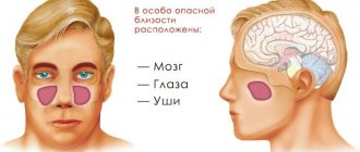
In severe sinusitis, an abundance of purulent masses are released from the affected sinuses. Without proper treatment, the infection can spread to the tissues adjacent to the nasal sinuses, in particular reaching the brain, which can have the most serious consequences.
Polyps are new tissue formations that gradually grow on the mucous membranes of the nose. They arise against the background of a variety of ailments. Most often, chronic tissue inflammation leads to their formation.
Among the main symptoms of polyposis it is worth noting:
- obstruction to free breathing;
- periodic attacks of complete nasal congestion;
- tissue inflammation;
- changing the timbre of the voice to a more nasal one;
- impairment of smell and then hearing.
The growth of polyps is stopped by surgery. If pathology is detected in the early stages of formation, it is possible to use conservative methods of therapy, in particular taking antihistamines and anti-inflammatory drugs, vitamin complexes.
The root of the pathology is the deviation of the nasal septum from its midline in both or one direction. The disease can be acquired or hereditary in nature. If the nasal septum is deformed, a person may experience breathing problems and regularly suffer from sinusitis and rhinitis.
Treatment is carried out through surgery. To restore health, an operation is performed to correct the nasal septum - the so-called septoplasty. The main goal of the procedure is to improve nasal breathing.
Finally
As you can see, the paranasal sinuses actually form a single network of air-conducting cavities. It is not surprising that all pathologies covering the presented area are similar in etymology.
It is extremely difficult to independently determine which disease has affected the paranasal sinuses. Methods that produce results for some pathologies may be completely ineffective for others.
If you have symptoms of the above ailments, it is better to immediately seek diagnosis from a qualified doctor, which will avoid complications.
When assessing the level of air filling of the sinuses, it is necessary to take into account the person’s age. Thus, in early childhood, the paranasal sinuses are not fully formed. By the age of two, only the maxillary sinuses can be detected in a child, while the other 3 sinuses are formed only by the age of 12. According to doctors, in most cases the sinuses are not sufficiently pneumatized due to the presence of a certain inflammatory disease.
The paranasal sinuses may be poorly pneumatized after the onset of sinusitis, which is caused by pathogenic microorganisms. In addition to sinusitis, incompletely cured rhinitis (runny nose) can affect the level of air filling of the sinuses.
In the absence of any pathologies, the sinus is completely healthy, that is, it effectively performs the tasks assigned to it. In this case, the doctor says that the sinuses are pneumatized. In other words, the air circulates normally within them.
However, the sinuses can often be filled with clear secretion (mucus) and even pus. This means that the cavity is affected by an inflammatory process. Most often, the following diseases lead to pathological changes:
- inflammatory process in the upper paranasal sinuses (sinusitis);
- inflammation of the ethmoid sinus (ethmoiditis);
- disruption of the normal functioning of the frontal sinuses (frontitis);
- damage to all existing sinuses (pansinusitis);
- inflammation in the main sinus (sphenoiditis).
Mycotic sinusitis
Recently, there has been an increase in the number of fungal sinusitis. Chronic forms occur under the guise of polypous recurrent sinusitis, the MRI picture is nonspecific, and laboratory diagnosis is difficult. There may be a change in the bony walls of the sinuses due to hyperostosis or destruction of the sinus wall as a result of prolonged pressure from the fungal body.
characterized by parietal thickenings caused by hyperplasia of the mucosa and partial fibrous changes in it. The thickness of the mucous membrane ranges from 4-5 mm.
Pneumatization of the nasal sinuses is preserved: what is it and what does it mean
If the paranasal sinuses are pneumatized, the patient experiences difficulty breathing, headaches, and discomfort near the bridge of the nose. Body temperature may increase due to inflammatory diseases.
In what cases can the level of pneumatization change? What reasons influence this important indicator and evaluation criterion?
For a doctor, such an image serves as a means of detection and indicates the location of inflammatory processes, swelling of the mucous and purulent fluid in a patient diagnosed with sinusitis.
What is it, pneumatization of the paranasal sinuses? This is the filling of the sinuses with air, which is possible due to direct communication with the nose. This process is involved in respiration.
The cavities are located on the front part of the skull, depending on their location they are divided into the following types:
- maxillary sinuses,
- frontal sinuses,
- sphenoid sinus,
- ethmoid cavity.
The nasal cavities play an important role in the functioning of the human body. They are involved in breathing, effectively moisturize, warm and clean the inhaled air, and protect against large particles from entering the mucous membrane using the protective mechanism of sneezing. They provide a good sense of smell and allow you to distinguish odors. Participate in the formation of the voice and its timbre.
Based on the results of an x-ray examination, pneumatization of the sinuses can be determined as increased or decreased. If it is saved, this means that there are no violations.
The degree of air filling of the sinuses is assessed in comparison with the healthy side (maxillary sinuses) or the transparency of the orbit. It is the last indicator that is best taken as a basis when assessing the survey results, since it is unchanged. If the description of the image indicates that pneumatization, or the filling of air in the sinuses, is not impaired, there is no need to worry.
In addition, the age of the patient must be taken into account. After all, the degree of air filling of the sinuses in adults and children can differ significantly, which is associated with anatomical features.
What is it - pneumatization of the paranasal sinuses? This is the filling of the sinuses with air, which is possible due to direct communication with the nose. This process is involved in respiration.
- maxillary sinuses;
- frontal sinuses;
- sphenoid sinus;
- ethmoid cavity.
The paranasal or paranasal sinuses are one of the most important structures of the external respiratory tract. Their clinical significance is extremely great. By participating in the breathing process, they actively moisturize, warm and purify the passing air.
We recommend that you immediately consult a doctor if you identify characteristic symptoms of inflammation of the paranasal sinuses. If you ignore the problem for a long time and let the disease take its course, you cannot avoid dangerous complications and a significant deterioration in the clinical picture.
The swollen epithelial tissue begins to thicken, narrowing the lumens of the paranasal sinuses. The viscous exudate, having no outlet, accumulates and further blocks the nasal passages. A large amount of secretion displaces air from the cavities, thereby leading to a decrease in pneumatization. At this stage, a secondary infection may occur. An acute inflammatory process develops, and nasal discharge becomes purulent.
Depending on which cavity is involved in inflammation, the following types of sinusitis are diagnosed:
- sinusitis - inflammation of the maxillary or maxillary sinuses;
- frontal sinusitis - frontal sinuses;
- ethmoiditis – labyrinth of the ethmoid bone;
- sphenoiditis – main sinus (sphenoid);
- pansinusitis - the mucous membrane of all nasal sinuses.
The phrase “paranasal sinuses are pneumatized” can be seen as a result after an x-ray of the facial bones. If pneumatization is preserved, then there are no complications, but if pneumatization is increased or decreased, we can talk about pathology.
Diagnosis and treatment methods
X-ray is an effective diagnosis of the condition of the paranasal sinuses.
Diagnosis and treatment are prescribed by an otolaryngologist. The doctor examines the nose, palpates the sinuses, identifies painful areas, and also collects anamnesis. The main diagnostic method is x-ray of the paranasal facial sinuses.
Inflamed sinuses with impaired pneumatization appear as dark spots on x-rays. If the sinuses are filled with pus, then they speak of a total darkening.
Treatment methods depend on the causes of pneumatization disorders:
- Antibiotics. Antibacterial therapy is necessary when pus appears in the sinuses, as well as to prevent complications and bacterial infection. For sinusitis, Amoxicillin, Ciprofloxacin, Amoxiclav, Sumamed are most often prescribed. The dosage and drug are prescribed by the doctor depending on the age and severity of the patient’s condition.
- Local treatment. Local treatment consists of using anti-inflammatory hormonal sprays, creams and drops. They are effective for sinusitis, but for other types of sinusitis and cysts they may be powerless. Sprays and drops are also often used to flush out mucus and pus from the paranasal sinuses.
- Vasoconstrictor drugs. With a decrease in pneumatization, severe nasal congestion is observed. To relieve it, you have to use vasoconstrictor drugs based on xylometazoline, oxymetazoline, and phenylephrine. They help quickly relieve swelling, but are addictive.
- Surgical intervention. Puncture of the paranasal sinuses is sometimes the only method of normalizing pneumatization. The puncture allows you to remove fluid from the sinuses and immediately introduce disinfectants. The effect comes quickly.
- Folk remedies. Traditional recipes can help in the initial stages of inflammatory sinus diseases. For tumors and abnormal structure of the facial bones, folk remedies are powerless. Typically, warming compresses and homemade drops from herbs, juices, and honey are used. They help with sinusitis, but drug treatment cannot be ruled out.
Mycotic sinusitis
Recently, there has been an increase in the number of fungal sinusitis. Chronic forms occur under the guise of polypous recurrent sinusitis, the MRI picture is nonspecific, and laboratory diagnosis is difficult. There may be a change in the bony walls of the sinuses due to hyperostosis or destruction of the sinus wall as a result of prolonged pressure from the fungal body.
Swelling of the mucous membrane of the right maxillary sinus with fluid level
Main causes of violations
The shape and structure of the paranasal sinuses are unique to each person. The causes of impaired pneumatization can be congenital, but most often they are a consequence of the penetration of viruses and microorganisms into the nasal cavity, which results in the appearance of a runny nose and nasal congestion. The appearance of vasomotor rhinitis leads to swelling of the nasal passage and swelling due to impaired vascular tone.
When PPN is violated, swelling in the nose and eyelids is often observed. The person begins to experience headaches and a feeling of pressure. Symptoms may worsen when tilting the head. The patient's lacrimation increases and the temperature rises. With a bacterial infection, green discharge with particles of pus appears.
Pneumatization of the sinuses is a characteristic of the results of an x-ray examination.
This indicator can reveal the presence of an inflammatory process in the paranasal cavities, swelling of the mucous membrane, accumulation of exudate or pus, the occurrence of a cyst and other pathological conditions. When PPN is violated, swelling in the nose and eyelids is often observed. The person begins to experience headaches and a feeling of pressure. Symptoms may worsen when tilting the head. The patient's lacrimation increases and the temperature rises. With a bacterial infection, green discharge with particles of pus appears.
When does decreased or increased pneumatization occur?
This condition is observed with the development of an inflammatory process in the sinus, accumulation of fluid (exudate, pus), the appearance of a cyst or their congenital underdevelopment.
Depending on the cavity in which the inflammatory process is localized, the following can be diagnosed:
Pansinusitis can also be diagnosed, a condition in which all paranasal sinuses are involved in the inflammatory process. As a rule, it is in this case that there is the greatest risk of complications.
The most common inflammation of the frontal cavities or maxillary cavities. This is due to the peculiarities of their placement. An X-ray examination is required to make a diagnosis. A decrease in pneumatization of the maxillary sinuses makes it possible to finally confirm this diagnosis. The main cause of the disease is the penetration of various viruses or bacteria into the cavity, and hypothermia.
If the inflammatory process is accompanied by an accumulation of exudate or pus, an x-ray can show its level relative to the nasal cavity. In this case, there will be no air in the lower part of the sinus. This can only be noticed when the patient is in an upright position during the examination. Both paranasal cavities or one may be involved in the inflammatory process.
There are other causes for this condition. A disorder such as decreased pneumatization of the maxillary sinuses can develop as a result of dental diseases. This is due to the thin partition between them. During dental treatment, particles of medications and filling material can penetrate into the cavity. These foreign substances provoke the occurrence of an inflammatory process.
The frontal nasal sinus and other paranasal sinuses are also characterized by a decrease in pneumatization when an inflammatory process occurs in them. Therefore, the X-ray method of examination is one of the main ways to establish a diagnosis. In this case, not only its results should be taken into account, but also the patient’s complaints about deteriorating health.
If the maxillary sinuses are pneumatized beyond normal, this may indicate that the patient has serious diseases of the endocrine system. Therefore, further treatment should be carried out not by an ENT doctor, but by a specialized specialist. Most often, this condition is observed when a person has pathologies such as gigantism and acromegaly.
For some patients, after an examination, the ENT makes a final diagnosis, the formulation of which includes “impaired pneumatization of the maxillary sinuses.” It comes as a real shock to many. In fact, there is nothing terrible - most often this is only one of the symptoms of sinusitis, and with proper treatment it can be eliminated.
Read more Registration of property complex Rosreestr
The content of the article
Symptoms that indicate the need to see a doctor
If you experience any symptoms that indicate possible inflammatory processes, you should immediately consult a doctor for diagnosis and examination.
Delayed or untimely treatment can lead to various complications and deterioration of health. The close location of the nose to other vital organs, and therefore inflammatory processes in it, can have a negative impact on the normal functioning of such organs and systems.
Symptoms that appear if the paranasal sinuses are pneumatized and require contact with a specialist:
- a feeling of painful discomfort in the forehead, eyes or nose (as a rule, the pain intensifies when the head is tilted forward);
- a bursting sensation in the area of the nose and eyes in the absence of discharge;
- Nasal congestion is often observed, which persists for a long period of time;
- discharge of pus from the nose;
- excessive tearing, eyes react painfully to light;
- increased body temperature;
- rapid fatigue, which is accompanied by loss of appetite and a feeling of apathy;
- swelling of the eyelid or cheek, redness of the eye from the inflamed cavity may be observed;
- attacks of dry cough, which are especially pronounced at night.