Periodontal abscess or radicular cyst of a tooth is a common complication of untreated pulpitis and periodontitis. This is a cavity under problem roots filled with pus. At first it does not make itself felt, but over time pain arises. Swelling is possible where pus comes out.
Without treatment, a tooth with a cyst cannot be saved. Moreover, infection beyond the abscess can lead to serious complications. How to avoid an unfavorable outcome - read our article.
What is a dental cyst
Translated from Greek, “cyst” means “bubble”. This pathological neoplasm is a cavity with a fibrous wall and contents. Roughly speaking, it’s a sac filled with pus. Its dimensions vary from a few millimeters to several centimeters.
The cyst that appears above the tooth is attached to the apex of the root and is located in the periodontal space - between the root and the bone tissue. Over time, it increases in size and affects the bone. Moreover, this happens faster in the upper jaw, since the bone tissue there is more porous.
Such neoplasms appear as a response of the body to infection in the canals. The cavity holds dead bacteria and prevents them from spreading further. But without treatment, rupture of the “bubble” is inevitable.
How does a tooth root granuloma form?
Granuloma has the following development mechanism:
- The first stage: the appearance of a dental disease and bringing it to an advanced state. The presence of a long inflammatory process leads to the formation of a large number of microorganisms in the pulp. The inflammatory process occurring in the pulp gradually leads to its death.
- Second stage: microbes go into further action. Gradually, the infection enters the bone tissue area. As a result, a new formation appears, which gradually turns into the granuloma in question.
- The third stage: the bone begins to retreat from the source of infection, a capsule of connective tissue is formed at this site, quite dense in structure. A serious inflammatory process continues inside the capsule, as a result of which bacteria rapidly multiply. The stage is characterized by rapid tissue growth. Over time, the bacteria turns into pus. If you consult a doctor at the last stage, he will diagnose “acute granuloma”.
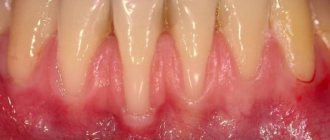
The disease greatly aggravates weakened immunity. Against this background, the tooth becomes abnormally mobile. Eventually its roots will be exposed.
Infections reach the tooth root in another way: through periodontal pockets. These pockets appear when hard tartar forms. The stone contains a huge variety of bacteria. They provoke the appearance of a gap between the socket and the gum. It is through this that the infection goes to the root. At the very base of the root, tissue grows that is completely filled with pus. This granuloma.
Causes
As has already been written before, a dental cyst is a direct confirmation that there is an infection in the canals. Through the root apex it enters the periodontal tissue. Why is this happening? There are several reasons:
- untreated caries and pulpitis - turns into periodontitis;
- chronic periodontitis;
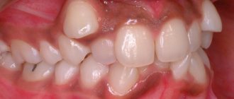
- heavy teething of the figure eight;
- a previous infection, the causative agent of which reached the periodontal tissues through the bloodstream;
- chronic inflammation under an artificial crown;
- tooth injury due to impact, fall;
- complication of chronic sinusitis;
- poor quality endodontic treatment.
The last reason should be discussed in more detail. According to statistics, in 60-70% of cases, the canals are filled poorly. A cyst under a tooth may appear because the canals were not filled to the very top of the roots (due to the dentist’s carelessness or due to impassable canals) or due to periodontal infection with non-sterile endodontic instruments.
Tooth cyst on x-ray
Causes of granuloma
Granuloma occurs after the following factors:
- Untreated caries.
- Untreated pulpitis, which appeared against the background of an inflamed pulp.
- Inflammatory process in the periodontium (the tissue that surrounds the tooth).
- A tooth fracture that results in infection in the internal area.
- Poor tooth antiseptics, which over time led to infection.
- Poor quality antiseptic treatment after pulp removal.
- Poor quality antiseptic treatment after root canal treatment.
Secondary causes of granuloma:
- Stress.
- Severe physical stress.
- Sudden climate change.
- Severe hypothermia.
- Serious cold infection.
The inflammatory process of periodontium can begin as a result of improper treatment. For example, even due to unprofessional dental filling.
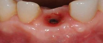
Often the disease occurs after tooth extraction. Why? The disease appears against the background of inflammatory processes and a complete lack of necessary preventive measures. Over time, the site of the extracted tooth begins to be covered with tissue. Microbes quickly get inside it, which leads to further periodontal inflammation. If prevention is not done, the granuloma will quickly grow and fill with pus. If treatment is neglected, the granuloma will begin to move along the entire length of the gum. This course of the disease leads to infective endocarditis, a rather dangerous disease that often ends in death.
Granuloma often occurs after the removal of children's baby teeth. Childhood does not mean that the patient cannot have granuloma.
Classification
For reasons
- radicular cysts (at the root of the tooth - at its apex or on the lateral surface) - a complication of periodontitis;
- retromolar or paradental - a consequence of difficult eruption of the “eight” due to chronic inflammation of its rudiment;
- residual - can form after incomplete removal, after which parts of the root remain;
- eruption cyst – normally occurs in all children aged 7-10 years and resolves itself during the process of formation and eruption of the bud;
- primary or keratocyst - appears when tooth development is disrupted, consists of residual cells of dental tissue;
- tooth-containing or follicular - a consequence of an infection that affects unerupted rudiments or supernumerary teeth.
By location
The most common cysts are wisdom teeth, frontal teeth (purulent cavities at the roots of “fours” often grow into the maxillary sinuses), and roots under artificial crowns.
To size
- granuloma or cystogranuloma – up to 0.5 mm in diameter;
- cyst – more than 0.5 mm in diameter.
Tumors of the jaws
Benign odontogenic tumors of the jaws
Ameloblastoma
is the most common odontogenic tumor of the jaws, prone to invasive, locally destructive growth. It primarily affects the lower jaw in the area of its body, angle or branch. It develops intraosseously and can grow into the soft tissues of the floor of the mouth and gums. It often appears between the ages of 20 and 40.
In the initial period, ameloblastoma is asymptomatic, but as the size of the tumor increases, jaw deformation and facial asymmetry occur. Teeth in the affected area often become mobile and shift, and toothache may occur. A tumor of the upper jaw can grow into the nasal cavity, maxillary sinus, or orbit; deform the hard palate and alveolar process. There are frequent cases of suppuration, recurrence and malignancy of ameloblastoma. The clinical course of jaw tumors such as ameloblastic fibroma and odontoameloblastoma resembles ameloblastoma.
Odontoma
most often occurs in children under 15 years of age. Typically, tumors are small in size and asymptomatic, but can cause delayed eruption of permanent teeth, diastema and trema. Large tumors can lead to jaw deformation and fistula formation.
Odontogenic fibroma
develops from the connective tissue of the tooth germ; occurs more often in childhood. Tumor growth is slow; localization - on the upper or lower jaw. Odontogenic fibroma is usually asymptomatic; in some cases, aching pain, tooth retention, and inflammation in the tumor area may be observed.
Cementoma
– a benign tumor of the jaw, almost always fused to the root of the tooth. Most often develops in the area of premolars or molars of the lower jaw. It occurs asymptomatically or with mild pain on palpation. Occasionally, multiple giant cementoma occurs, which may be a hereditary disease.
Benign non-odontogenic tumors of the jaws
Osteoma
may have intraosseous or superficial (exophytic) growth. The tumor can spread to the maxillary sinus, nasal cavity, or orbit; interfere with the fitting of dentures. Osteomas of the mandibular localization cause pain, asymmetry of the lower part of the face, impaired jaw mobility; maxillary localization - nasal breathing disorders, exophthalmos, diplopia and other disorders.
Osteoid osteoma
accompanied by intense pain, worsening at night and during meals; facial asymmetry. When examining the oral cavity, bone protrusion is determined (usually in the area of premolars and molars of the lower jaw), hyperemia of the mucous membrane.
Osteoblastoclastoma
(giant cell tumor of the jaw) mainly occurs at a young age (up to 20 years). The development of the clinical picture is characterized by an increase in pain in the jaw, facial asymmetry and tooth mobility. The tissue over the tumor becomes ulcerated; fistulas form; there is an increase in body temperature. Thinning of the cortical layer leads to pathological fractures of the lower jaw.
Hemangioma
jaw is relatively rarely isolated and in most cases is combined with hemangioma of the soft tissues of the face and oral cavity. Vascular tumors of the jaws are manifested by increased bleeding of the gums, bleeding from the root canals during the treatment of pulpitis or periodontitis, from the socket during tooth extraction, etc. Upon examination, fluctuation, looseness of the teeth, and cyanosis of the mucous membrane may be detected.
Malignant tumors of the jaws
Malignant tumors of the jaws are 3-4 times less common than benign ones. With jaw cancer, radiating pain occurs early, tooth mobility and loss occur, and pathological fractures of the jaw are possible. Malignant tumors of the jaws destroy bone tissue; parotid and submandibular glands and masticatory muscles sprout; metastasizes to the cervical and submandibular lymph nodes.
Carcinoma of the maxilla can invade the orbit, nasal cavity, or ethmoid labyrinth. In this case, recurrent nosebleeds, unilateral purulent rhinitis, difficulty in nasal breathing, headaches, lacrimation, exophthalmos, diplopia, and chemosis are noted. When the branches of the trigeminal nerve are involved, otalgia is a concern.
Malignant tumors of the lower jaw early infiltrate the soft tissues of the floor of the mouth and cheeks, ulcerate, and bleed. Due to contractures of the pterygoid and masticatory muscles, closing and opening of teeth becomes difficult. Osteogenic sarcomas are characterized by rapid growth, rapidly progressing infiltration of soft tissues, facial asymmetry, unbearable pain, and early metastasis to the lungs and other organs.
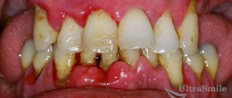
Symptoms
The insidiousness of a dental cyst lies in the fact that its appearance at first is almost asymptomatic. It makes itself felt when the purulent sac has already reached a fairly large size. Active growth of an abscess occurs when the immune system is weakened. People often learn about it during acute respiratory diseases or viral infections, when the body weakens due to the fight against the disease.
In the early stages, pathology can be detected by the following signs:
- slight pain in response to tactile stimuli - biting or pressing on the crown;
- soreness of the gums near the problem area.
When the cavity has increased significantly, acute toothache, swelling and swelling of the gums and cheeks appear. Since this is an infectious disease, there is an increase in body temperature, weakness, and general malaise. A tumor may appear on the gum, which will gradually increase and sooner or later a fistulous tract will appear in it, through which pus from the cyst will enter the oral cavity.
A dental cyst is diagnosed based on the results of radiography. On an x-ray it appears as a dark spot at the apex of the root. Sometimes pathology is detected completely by accident, examining a neighboring tooth.
Maxillary sinus cyst
Why is a cyst under a tooth dangerous and its consequences?
Possible complications:
- root destruction with subsequent tooth loss;
- periostitis - inflammation of the periosteum;
- osteomyelitis of the jaw bone - a growing cyst provokes purulent-necrotic processes that lead to destruction of bone tissue;
- inflammation of the lymph nodes located near the affected area;
- chronic sinusitis (may appear when a purulent sac grows into the maxillary sinus);
- spontaneous fracture of the jaw - occurs when the dental cyst enlarges excessively;
- the appearance of phlegmon - diffuse acute purulent inflammation.
In the worst cases, death is possible. When the contents of the cavity enter the bloodstream, sepsis develops - blood poisoning. In addition, a cyst from a benign tumor can degenerate into a cancerous one.
Formation of a cyst on the root of a tooth
Complications
Untreated granuloma leads to:
- To complete loss of a tooth. This happens due to complete destruction of the root. As a result, soft tissues are drawn into the inflammation process, in which pus accumulates.
- Osteomyelitis of the jaw.
- Formation of a dental cyst.
- Cancerous tumors.
- Infection of other organs and the development of sinusitis, pyelonephritis and infectious myocarditis.
- If pus gets into the skull, meningitis, encephalitis and inflammation of the peripheral nerves may begin.
- The appearance of migratory granuloma. Manifests itself in the form of protrusion of the facial skin. The disease also appears in the form of abscesses and fistulas in different places.
What to do?
Be sure to contact the dentist. Not only the methods of treatment are important, but also the time when it begins. The larger the cyst, the longer and more difficult the therapy will be.
At the present stage, such pathologies are treated in two ways - conservative and operative (surgical).
Surgical treatment
If there is a pin or artificial crown in the root canal, the canals are not filled at the root apex, or the cavity size exceeds 0.8 mm, surgical treatment is indicated. There are several options:
- resection – removal of the root apex and excision of the cyst;
- hemisection - removal of the cyst along with the diseased root, after such an operation prosthetics are required;
- cystectomy – complete removal of the abscess and infected area of the root, the most optimal option in terms of effectiveness, but difficult in terms of technique;
- cystotomy - removal of the anterior wall of the cavity with opening access for its sterilization - a simple method, but is used only for large abscesses and requires long-term postoperative treatment.
After all these procedures, doctors prescribe a course of antibiotics and antiseptic rinses.
If the cyst completely surrounds the roots or the crown is completely destroyed, the tooth must be removed.
Removal of a tooth cyst
Conservative treatment
A dental cyst with a diameter of up to 0.8 mm detected at an early stage can be eliminated without surgery. This is possible if the canals were not previously sealed or if they were poorly sealed along their entire length.
Stages of therapy:
- Preparation of the causative tooth and opening of the canals. If the pulp has not been removed previously, it is removed.
- Medicinal treatment of canals with antiseptics.
- Disinfection of the cyst cavity. The medicine is taken beyond the root directly into the cavity.
- At this stage, you can resort to depophoresis - the destruction of pathogenic flora with a medicine based on copper-calcium hydroxide. Under the influence of a light current, it spreads through the tissues and kills bacteria and fungi.
- Filling canals with antiseptic paste.
- Placement of a temporary filling.
The third and fourth stages are repeated several times. As a rule, treatment takes at least three months. After the course, radiography is performed.
If the cyst has shrunk, the dentist can proceed with permanent filling of the canals and installation of a permanent filling.
If the cyst was too large, an artificial bone preparation is injected into the remaining cavity before filling, which prevents the resorption of bone tissue.