Filling material in the maxillary sinus is accompanied by unpleasant symptoms, if they occur, you should immediately seek medical help. Lack of treatment can lead to serious complications due to inflammatory processes, although the composition of the filling does not pose a health hazard. The symptoms are similar to those of a runny nose, which cannot be cured using conventional therapeutic methods; after diagnosis, surgical treatment is performed.
We recommend reading this article to anyone who has been suffering from a runny nose for a long time and cannot cure it with medications; an examination by a doctor will help identify the cause of the pathology.
Actions to take if filling material gets into the maxillary sinus
Diseases of the ENT organs are considered one of the most common; they are often accompanied by a runny nose, sore throat and are infectious in nature. One of the complications of rhinitis is also considered to be sinusitis, which can develop due to the penetration of foreign objects into the maxillary sinuses, most often this is associated with dental procedures. During dental treatment, filling material can penetrate through the thin bone septum into the maxillary sinus, which is accompanied by unpleasant symptoms.
Inflammatory processes lead to the development of sinusitis, which, if untreated, transforms into a chronic form, threatening the development of serious complications. When the first signs appear, you should undergo a diagnosis; based on the data obtained, the doctor prescribes surgery.
Diagnostic methods
If filling material gets into the maxillary sinus, professional diagnostics are carried out; it plays a leading role in this case.
Diagnostic procedure:
- Questioning the patient - an experienced doctor will be able to identify the inflammatory process based on complaints of pain.
- Collecting anamnesis, in parallel, the fact of visiting the dentist is recorded.
- Inspection of the paranasal sinuses using special instruments for the presence of foreign elements, endoscopy and rhinoscopy are used.
- CT scan – provides the most accurate diagnosis, allows you to identify the presence of a foreign body and other inflammatory processes, the extent of damage to the nasal mucosa and the amount of pus.
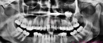
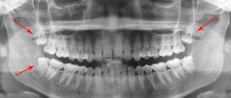
- X-ray of the maxillary sinuses - the images clearly show all the seals, purulent formations and other defects. In this case, you will also need diagnostics and consultation with a surgeon; a comprehensive examination will help identify the problem and prescribe treatment.
Foreign body: symptoms, how does it get into the sinus and what could it be?
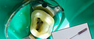
The most common reason why a foreign body can be found in the maxillary sinus should be looked for in the dentist's office. We are talking about filling the upper jaw. The roots of the teeth are located close to the mucous membrane of the sinuses. When pressing, the dentist may not calculate the force and push a foreign object into the cavity. This can happen when installing a filling and when building up the wall of the upper jaw for subsequent implantation.
A foreign body is also commonly understood as cystic formations in the form of a thin bubble. The reason lies in private runny noses, when the mucous membranes become irritated.
Less common are cases of foreign bodies entering the sinuses when injured by a firearm. There are examples in the medical literature when part of a match or toothpick gets into the sinus when picking teeth. Children may have in their sinuses: construction parts, berry seeds, beads and other small objects.
When performing operations in the upper jaw and nasal passages, unscrupulous medical personnel may leave parts of tubes, threads and other materials in the maxillary sinuses.
After some time, the foreign object becomes covered with fungus and causes characteristic symptoms. An inflammatory process occurs. Before this, a person may not notice that there is a problem.
Symptoms of a foreign body entering the sinuses are similar to symptoms of sinusitis, sinusitis and other colds. The person feels the following:
- headache and heaviness, especially when tilting the head forward;
- runny nose;
- thermometer readings 38 and above;
- after sleep the patient feels a “heavy head”, and for many this symptom interferes with sleep;
- discharge of pus; has a putrid odor, and for this reason nausea is observed;
- sometimes toothache when moving the jaw;
- when blowing his nose, the patient feels relief;
- pain in the cheeks and under the eyes;
- decreased appetite and general weakness.
When a person comes to the clinic with such symptoms, the specialist is in a hurry to give him a diagnosis that is not related to the entry of a foreign body.
Only on an X-ray or on a computer screen during a tomography can a problem be detected.
Before this, doctors can treat sinusitis or other diseases for quite a long time and to no avail. Antibiotics and other antiviral drugs will not cope with the fungus, which covers the foreign body on all sides in the maxillary sinus.
Diagnostic measures
Comprehensive diagnostics includes the following activities:
- assessment of patient complaints, analysis of such indicators as the duration of symptoms, the presence of a visit to the dentist and other manipulations in the nasal and jaw area;
- examination using a nasal speculum;
- use of an endoscope to examine the nasal cavity and middle nasal passage;
- examination in the dentist's office of the upper dentition;
- X-ray image, which shows not only the presence of the body, but also pneumatization (presence of air);
- the use of computed tomography;
- consultation with a surgeon.
It is believed that surgery is the only way to remove a foreign body located in the maxillary sinus. It is advisable not to delay the operation and schedule it immediately after an accurate diagnosis. Lack of timely intervention can cause the development of meningitis, sore throat, sinusitis and osteoperiostitis (inflammation of the bones of the upper jaw and orbit).
Pneumatization of the sinuses is not a disease, but a term. With its help, doctors characterize phenomena on an X-ray image or on a tomography screen.
Pneumatization is the level of filling of the sinuses with air, which, under normal conditions, moves without obstruction through the nasal passages.
In the picture you can see that pneumatization:
- saved;
- reduced;
- increased.
In the first case, there are no deviations from the norm and most likely the cause of the disease is not a foreign object.
If a foreign body is present in the maxillary sinus, this will be indicated by a reduced level of pneumatization. At the same time, air circulates with difficulty, which leads to a decrease in oxygen supply to the body.
Symptoms and signs
The symptoms of a foreign body entering the maxillary sinus may vary, much depends on the size, degree of contamination, and characteristics of the body. In some cases, the object is surrounded by a mucous membrane and the pathology does not manifest itself in any way. But, most often in such cases, the body rejects the foreign body, as a result of which the inflammatory process of the mucous membrane starts.
The presence of a foreign object can be recognized by the following signs:
- Heaviness in the nose.
- Pain when chewing, which can radiate to the upper jaw.
- Loss of appetite, pain when swallowing.
- Fever.
- Persistent runny nose that cannot be treated.
- Discharge from the nasal cavity often has an unpleasant odor.
- General intoxication of the body.
The symptoms are very similar to viral/bacterial sinusitis and have common symptoms with rhinitis and frontal sinusitis. The main difference is the infectious nature of these diseases, which can be cured with conventional drugs, but surgical intervention may be required to remove the foreign body.
How can a foreign object end up in the maxillary sinus?
In 40% of cases, the cause of the disease is improper dental treatment, most often due to the close proximity of the upper teeth and the nasal cavity.
The upper wall of the oral cavity is very thin, the filling material easily penetrates into the sinuses. When a hole appears, surgery is most often performed.
A foreign body may end up in the maxillary sinus due to large tooth roots; it can be not only a filling, but also any other small object that the dentist can use during an external examination. In medical practice, there have been cases of matches and toothpicks being found in the paranasal sinuses, but the patient was not always aware of this. In any case, surgical intervention is required; this is the only way to prevent a process that can lead to various complications.
Foreign body in the nasal sinus
How did the elements of the filling, which should close the hole in the tooth, get from the sinus? Naturally, after visiting the dentist’s office and dental treatment, or rather after:
- Prosthetics.
- Installation of seals.
In both cases, most often the walls of the sinuses are thin for some reason. And as a result of the procedure for installing a prosthesis or treating a tooth, they are injured.
It turns out that filling a tooth is not as easy as many people think due to incompetence in this matter. Sometimes the roots of the tooth are very close to the wall of the sinus, and when they are filled, the material ends up in the sinus itself.
Not only during the filling process can the dentist “introduce” material into the maxillary sinus. If the thickness of the bone tissue does not allow for normal installation of the implant, a specialist can “build up” it using artificial material.
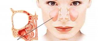
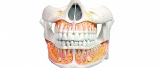
The maxillary sinuses are anatomically located in the upper jaw, just above the teeth. Functionally, this is a very important formation. Thanks to the maxillary sinuses, during normal nasal breathing, the air is warmed to its optimum as it passes through the respiratory system. And already warmed it enters the bronchi and lungs. The lining of the nasal sinuses is represented by the mucous membrane, the role of which is to clean the air from microorganisms and dust.
The presence of various objects in the maxillary sinus is fraught with unpleasant and sometimes dangerous consequences. Therefore, if the question is whether to remove a foreign body or refrain from intervention, doctors give a clear answer - be sure to carefully remove it!
What will happen if left untreated?
The main method of treatment is surgical intervention, and there is always a part of patients who carry a foreign body in their sinus for years, avoid surgery and require drug therapy. Despite the inertness of the filling material, physiotherapeutic procedures are useless in this case; the only way out is surgery.
The consequences of finding a foreign object in the sinus can be quite mild:
- sinusitis of fungal or bacterial origin,
- encapsulation of material, accompanied by the formation of a cyst, the contents of which often become purulent in nature,
- formation of purulent granuloma.
In medical practice, many cases have been recorded where the pathology was accompanied by sinusitis; in the absence of treatment, the disease transformed into a chronic form.
Complications of chronic sinusitis:
- inflammatory diseases of other ENT organs,
- meningitis,
- hypertrophy of the mucous membranes of this area, difficulty in nasal breathing,
- heart disease and mild brain hypoxia due to impaired normal breathing,
- myocarditis – occurs as a result of damage to the heart by harmful microorganisms from the source of inflammation,
- pathologies of the lower respiratory tract with an increased risk of an asthmatic component,
- osteoperiostitis of the head bones,
- sepsis.
All these complications do not always develop; the risk of their occurrence increases against the background of exacerbations, acute infections, overwork, hypothermia, and weakened immunity. At the first signs of damage to the walls of the maxillary sinus, you should consult a doctor.
Foreign body extraction methods
If a foreign body in the maxillary sinus provokes inflammation and causes chronic sinusitis, it must be removed. Otherwise, it can cause serious complications: long-term intoxication of the body, inflammation of the facial bones (with their subsequent destruction), periodontal disease and loss of healthy teeth.
There is no need to be afraid of such a procedure. Moreover, today small foreign bodies are pulled out very quickly through a small hole in the maxillary sinus made with an endoscope. The procedure is performed under local anesthesia and looks something like this:
- Using a control x-ray, the exact position of the foreign body is determined.
- The sinuses are washed and treated with an antiseptic solution.
- The patient sits in a chair and his head is securely fixed.
- The selected form of anesthesia is used: local or intravenous.
- An endoscope is inserted through the nasal passage, at the end of which a mini-camera is installed, transmitting the image to the monitor.
- A small hole is made in the wall of the maxillary sinus.
- The foreign body is captured by the loop and removed out.
- If a large amount of pus has accumulated in the maxillary sinus, rinsing is performed.
- The wound is treated with an antiseptic, and the nasal passage is closed with a sterile swab.
That's it, the operation is over. The doctor gives the patient recommendations for the postoperative period and after 1-2 hours, in the absence of complications and nosebleeds, the patient goes home. A follow-up examination and a repeat x-ray are taken a few days later.
If the foreign body is large, has sharp edges, came in as a result of trauma, etc., a full-fledged surgical intervention is often necessary, which is performed in a hospital, in the department of maxillofacial surgery. The course of the operation and the form of anesthesia are discussed with the surgeon in each specific case.
Sometimes you have to postpone the operation. Contraindications to its implementation are: pregnancy, acute inflammatory processes, weakened immunity, blood clotting disorders, intolerance to anesthesia. In this case, chronic inflammation is relieved with drug therapy. But this course will have to be repeated several times a year until the problem is radically solved.
Methods of treatment, removal of a foreign object
The main method of treatment is the removal of filling material from the maxillary sinus; this should be done immediately after diagnosing the pathology. In this case, there is no need for long trips to hospitals; the intervention of doctors does not take much time. The operation is performed in public and private clinics, the cost depends on the pricing policy and region of residence. It is very important to follow your doctor’s recommendations, monitor your health, and avoid infectious diseases.
There are two main ways to remove a filling:
- The endoscopic method is scraping of the mucous membrane through the nasal cavity.
- Laparoscoria - foreign material is removed through an incision in the gum.
Both methods have been used with great success for several decades and the procedure does not take much time. Both are performed under local anesthesia. After surgery, being under the supervision of a doctor in an inpatient setting is indicated.
Before discharge, conservative therapy is prescribed:
- taking antibiotics, the action of which is aimed at destroying microorganisms that provoke sinusitis,
- filling and sanitation of dental canals,
- flushing the sinuses with medications to remove purulent fluid,
- introduction of vasodilators into the nasal cavity,
- taking painkillers,
- taking immunomodulatory drugs, multivitamins.
Surgery is considered the most effective treatment method, and in order to prevent the spread of infection, complex therapy is prescribed, which includes taking medications.
Conservative treatment
It is applicable both during the preparation of surgical intervention and immediately after its implementation and is designed to minimize the activity of the local inflammatory process.
If it is impossible to immediately remove the filling material from the maxillary sinus, therapeutic treatment is a method of containing microbial aggression until a time favorable for surgery is reached.
Reasons to postpone surgery include:
- the onset and course of pregnancy;
- development of an acute inflammatory (including infectious) process;
- immunity deficiency;
- disorders of blood clotting mechanisms;
- intolerance to certain drugs.
The set of measures includes the use of medications with the following effects:
- antibacterial – suppressing the development of pathogenic microbial flora;
- anti-inflammatory – reducing the activity of tissue destruction, adapting the body to a given level of metabolism;
- antihistamine – reducing swelling and preventing overproduction of mucous gland secretions, as well as the occurrence of allergic reactions to antibiotics used;
- local antiseptic - helping to remove excess mucus from the sinus when washing it.
Recommendations after treatment
After surgery, you should be attentive to your own health, avoid hypothermia, which will prevent relapses of sinusitis.
The patient should adhere to the following recommendations:
- Observation by a dentist will help reduce the risk of caries and other diseases that develop against the background of dental diseases.
- Regular dental and oral care is required.
- Any injuries to the head, throat and nose area are contraindicated.
- Taking multivitamins will help strengthen the body's immune status.
- In order to prevent complications of sinusitis, it is recommended to avoid hypothermia, and timely treatment of the runny nose and other inflammatory diseases of the ENT organs is mandatory.
Treatment methods
As mentioned earlier, a foreign body found in the maxillary sinus is removed through the intervention of a surgeon. Removal in modern medicine is carried out in two ways:
- Endoscopic. An endoscope is used, which is inserted through the nasal passages; in this case, the removal of the foreign body occurs together with the growth of the fungus; it is important to remove it all to prevent its proliferation; The sinuses are also pierced to cleanse them of pus.
- Laparoscopic. The surgeon makes an incision in the upper jaw.
The procedures are performed using local anesthetics and last about 15-25 minutes. Patients may require an additional 2-4 days of hospital stay depending on the severity of the surgery and the severity of symptoms.
It is believed that the first method of removing a foreign body from the maxillary sinus is the most convenient and less traumatic for the patient. But this method cannot always be used for a number of reasons. One of which is an abnormal structure of the nasal septum (congenital or acquired). In this case, it is impossible to get into the maxillary sinus using an endoscope.
After any type of surgery, antibiotic therapy is required.
Based on the results of laboratory tests, the type of fungus that led to inflammation is determined. Only after this is a specific drug prescribed to stop the proliferation of microorganisms.
Both operations are performed in specialized clinics. It is better to choose a modern healthcare facility that has all the necessary equipment to perform the operation with less trauma and greater precision in performing the manipulations.
A cyst is also considered a foreign body, although it does not enter the cavity from the outside, but is formed there. The method of its removal is surgery: classical or endoscopic.
The first method means an incision under the lip. Afterwards, the anterior wall of the sinus is opened. The final stage is when the surgeon removes the formation through a small incision.
This traumatic manipulation is performed under local anesthesia. Disadvantage: disruption of the physiological composition of the mucous membrane. Which can subsequently lead to a number of diseases.
Endoscopic removal is less dangerous. Complications after such manipulation are observed in extremely rare cases.
Preventative measures after surgery
To avoid cases of foreign bodies getting into the sinuses after surgery, the following measures should be taken:
- promptly treat caries and have your teeth checked by a dentist to reduce the likelihood of getting fillings;
- pay attention to runny nose and sinusitis, treat these diseases in a timely manner to prevent the appearance of cysts;
- Avoid head injuries in every possible way, especially in the nose.
Rehabilitation after surgery will go faster if you adhere to the following doctor’s instructions:
- avoid hypothermia and overheating;
- take vitamin complexes, immunomodulators (prescribed by a specialist);
- protect yourself from infectious and viral diseases;
- Avoid contact with people suffering from ARVI, influenza and other diseases.
If you neglect these recommendations, the following complications may develop:
- sinusitis (acute);
- mycetoma (fungal type of sinusitis);
- maxillary cyst;
- inflammation of the tonsils;
- meningitis;
- angina.
In the most advanced cases, osteoperiostitis may develop. This is an inflammation of the bones of the face. May result in part of the affected bone being replaced with an implant.
For many people, the fact of finding a foreign body in the maxillary sinuses may seem incredible. However, medical practice shows frequent cases of this phenomenon. People who have been treating sinusitis for a long time should be especially wary. A diagnosis should be made and the presence of a foreign body in the maxillary sinuses should be checked using a computed tomography scan or a simple x-ray.
Source: OPnevmonii.ru