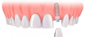
What it is?
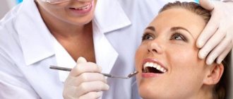
Selective abrasion is an effective technique for treating periodontal disease.
During the procedure, premature contacts are removed by cutting off the top layer within the enamel. The purpose of this procedure is to correct occlusion and articulation, create uniform contacts, relieve excessive loads and tension in dental tissues.
Optimal contact after the procedure
The therapeutic effect is achieved by eliminating the unwanted component of chewing pressure. This step ensures a reduction in vascular damage, corrects the displacement of dental units, and, accordingly, eliminates moments that adversely affect tissue trophism. As a result of reducing the impact on periodontal tissue, blood circulation returns to normal.
The procedure begins only after a diagnosis has been made, analysis taking into account the state of the dental system and examination. Pre-grinding can be carried out on models. Diagnostics provides data on the clinical picture, condition and mechanism of development of the pathology.
The main indication for the procedure is traumatic occlusion - improper closure. In this condition, hyperfunctional tension in individual places occurs. This leads to pathologies such as muscle and mandibular dysfunction, disruption of the structure of periodontal tissue.
The procedure is most often carried out in the first part of the day. Each visit takes no more than 30 minutes. Complete grinding is done on average in 5 sessions, leaving a week between each session. This is necessary for the neuromuscular system and periodontium to fully adapt. After 14 days, the patient comes to check the results and possible correction of the occlusion. In the future, such visits are carried out once every six months.
Ideal bite: closure points: two- and three-point contacts on the supporting cusps
Tools that may be useful for selective sanding:
- waterproof abrasive discs;
- electric drill;
- water-cooled tubular drill;
- set of abrasives;
- diamond coated heads;
- carborubine heads of different shapes;
- hard/soft rubber polishers.
Teeth grinding – what is it and how is it done?
The selective grinding technique is used for periodontal diseases. The procedure is performed to eliminate increased pressure on diseased tissues. Grinding teeth improves nutrition and blood circulation in the periodontium. There are several options for carrying out the technique, but they all have one goal - to eliminate unfavorable factors that interfere with the restoration of diseased tissues.
In what cases is teeth grinding performed?
The main indication for the procedure is periodontal disease, when there are disturbances due to tooth displacement and damage to their ligament. First, the inflammatory process is eliminated. When the symptoms are not acute, treatment and grinding are carried out in parallel. This procedure is performed before surgery, if necessary.
Other indications for the procedure:
- Prevention of periodontal disease in cases where natural tooth wear is absent, which creates a load on the soft tissues and can cause dystrophy.
- Deformation of the dentition, when one or more teeth are missing, which becomes a factor in malocclusion.
- Pathologies of the temporomandibular joint and muscles, when there is a risk of dysfunction due to occlusal obstacles.
- Before implantation for the purpose of correcting the bite in order to avoid overload after installation of the prosthesis, which can cause implant rejection.
Contraindications for grinding
The selective grinding technique also has contraindications:
- Severe inflammatory process of periodontal tissues. This is a relative contraindication. Before grinding, anti-inflammatory therapy is carried out, dental plaque is removed, and foci of the infectious process are eliminated. After this, sanding is performed.
- TMJ pathologies of acute and chronic course, which are accompanied by dysfunction and pain. In this case, grinding can be performed only during the period of subsidence of symptoms, during remission.
- Severe deformations and anomalies of the dental system, when surgical, orthodontic or complex treatment is required.
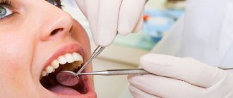
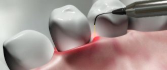
How sanding is carried out
The main method can be started only after a thorough diagnosis and sanitation of the oral cavity. The dentist examines the patient, sends him for x-rays, makes a diagnosis and draws up a treatment plan.
Pre-grinding may be performed first to correct significant occlusal defects. In this way, obvious teeth irregularities are removed. A preliminary procedure is carried out preserving the shape and contour of the teeth, taking into account aesthetic requirements.
Sanding is carried out in several sessions with an interval of 3-7 days, depending on the situation. This period is needed for adaptation. Each procedure lasts no more than 30 minutes.
On the first visit, the dentist records the relationships of the teeth and eliminates supracontacts in the case of posterior and central occlusion, in the second - in the case of lateral and anterior occlusion. On the third visit and subsequent ones, the doctor assesses the condition of the ground crowns and corrects residual contacts.
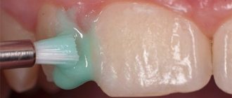
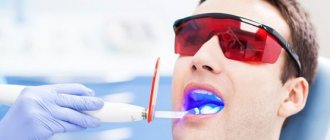
Each visit ends with polishing the contact surfaces. Fluoride-containing preparations can be used to treat the enamel, which will additionally protect the teeth.
After correction, a follow-up examination is carried out after 2 weeks, then after six months.
What could be the consequences?
In case of errors and non-compliance with sanding rules, the following complications may occur:
- displacement of the dentition;
- increased load on periodontal tissue throughout the entire dentition;
- increased sensitivity;
- decrease in interalveolar height;
- increased chewing load on a separate periodontal segment.
To avoid hyperesthesia during grinding, hard tissues are treated with special agents to remineralize the enamel.
The patient is monitored after grinding for 6 months. During this period, adaptation of the TMJ occurs, stabilization of the relationship of the teeth. After orthopedic and surgical treatment, correction and control of contacts are performed.
Over time, the relationship changes due to the natural wear and tear of crowns, fillings and dentures. Therefore, corrections are carried out regularly. This is especially true for patients with severe periodontitis. Then the correction should be performed every 3 years.
The selective grinding technique allows the most gentle way to establish the interaction of the contact surfaces of the teeth to prevent overload of individual areas of the periodontium, while maintaining the normal height of occlusion.
Source: https://dentalzub.com/prishlifovyvanie-zubov
Main directions
All grinding techniques are based on two main directions.
The basic methods include Jenkelson and Schuyler methods:
- Selective grinding according to the Jenkelson method - occlusal relationships are made in a convenient form by the patient himself without the doctor’s efforts. Premature contacts are removed directly in the central/distal occlusion, while anterior/lateral adjustments cannot be made.
- Schuyler method - with this correction method, occlusal contacts are subject to correction, which created interference with the articulation of the rows. They are removed in the following sequence: first distal/central, then anterior, and lastly lateral occlusion. The specialist manually regulates the movement and position of the jaw.
According to Jenkelson, there are 3 classes of unwanted contacts, according to which the procedure is carried out.
The surfaces of the slopes of the mounds are marked with the symbols I, II, III, the surfaces of the antagonists - I a, II a, III a:
- I – vestibular
slopes of the cervical tubercles of the lower 6-8 teeth, 4-5 teeth, the surface of the anterior ones, facing the vestibule of the oral cavity; - I a – oral slopes of the cervical tubercles of the upper 6-8 teeth, 4-5 teeth, the surface of the anterior ones, which faces the vestibule of the oral cavity;
- II – oral slopes of the upper palatine cusps of 6-8 teeth, 4-5 teeth;
- II a – vestibular slopes of the lower 6-8 teeth, 4-5 teeth (lingual cusps);
- III – vestibular slopes of the upper 6-8 teeth, 4-5 teeth (palatal cusps);
- III a – oral slopes of the lower 6-8 teeth, 4-5 teeth (buccal cusps).
Read online Therapeutic dentistry. Textbook page 97. Large and free library
Guided regeneration of periodontal tissues . This operation as a stage of gingivosteoplasty appeared as a result of long-term experimental and clinical studies [Nyman, 1980, Gottow, 1990; Caton, 1992, etc.].
It has been shown that bone and gingival connective tissue do not induce the formation of new connective tissue attachments, and epithelial growth prevents its formation. Granulation tissue arising from the periodontal ligament has the ability to form new connective tissue attachment.
Therefore, it is necessary to promote the proliferation of periodontal ligament cells on the root surface. This is done as follows: after curettage of the pockets and de-epithelialization, before suturing, a Kolapol membrane (KP-2) or an Arisona membrane (USA), or Resolut is applied to the exposed root surface. The operation is completed by applying sutures and a gum bandage.
Trichopolum is prescribed for 5–7 days, maintaining oral hygiene. Non-absorbable membranes are removed no earlier than after 6 weeks. The location of the membrane between the flap and the root surface favors the repopulation of periodontal ligament cells on the root surface.
9.9.4. Physical treatments
Of the physical methods of treatment, the most widely used are electrophoresis, darsonvalization, fluctuarization, microwave therapy, hyperbaric oxygen therapy, laser light, etc.
Physical methods should be used to eliminate or reduce inflammatory phenomena after surgical interventions. The most commonly used are electrophoresis of calcium, vitamin B with lidocaine, fluoride, enzymes and other drugs.
Fluctuation is carried out using the FS-100 apparatus for 20 minutes; There are 6 procedures per course. Darsonvalization is based on the vasomotor effect, it is carried out in long courses, at least 15 procedures.
Diathermic current is widely used for diathermocoagulation of gingival papillae in hypertrophic gingivitis. In the complex treatment of periodontal diseases, various types of massage (vibration, finger) and hydrotherapy are also used.
Recently, laser radiation has been used in periodontology, the action of which on inflamed tissues inhibits the processes of alteration, exudation and activates proliferation with a simultaneous analgesic effect. The procedure is carried out for 3 minutes using a laser physiotherapeutic device “Optodan”, for a course of 5–8 procedures. Indications: ulcerative, catarrhal gingivitis, periodontitis, especially after surgical interventions.
9.9.5. Orthopedic treatment methods
In orthopedic methods of treating periodontal diseases, in order to eliminate the functional traumatic overload of the periodontium, selective grinding of teeth, orthodontic interventions, splinting and rational prosthetics are used [Kalamkarov Kh. A.
, 1995]. Selective grinding of teeth is carried out according to Jenkelson, taking into account the classification of premature contacts (classes I, II, III of supracontacts) or according to the Kalamkarov method.
Orthodontic interventions in adults are used according to strict indications and only in the initial stage of periodontal pathology; Orthodontic treatment ends with splinting and prosthetics.
Splinting can be temporary using composite materials and permanent using removable and non-removable structures, including metal-ceramic prosthetic splints.
Selective grinding of teeth . In case of periodontal diseases, grinding is aimed at eliminating premature occlusal contacts, which lead to horizontal traumatic overload of the teeth.
The possibility of such contacts and periodontal overload increases with periodontitis, since this disease disrupts the physiological process of abrasion of hard dental tissues.
The preserved unworn cusps of small and large molars, as well as the cutting edges of the anterior (frontal) teeth, cause premature contacts in central, anterior and lateral occlusions.
Premature contacts themselves indicate horizontal overload of the corresponding teeth in various phases of articulation. The periodontium of these teeth experiences load directed mesially, distally, vestibularly (to the labial or buccal side), orally (to the lingual or palatal side).
As a result of such a load, with each closure of the dentition, the teeth deviate in the corresponding direction, causing compression of the periodontium and disruption of its trophism, increasing the resorption of bone tissue of the overloaded walls of the sockets.
In addition, premature contacts can lead to parafunctional conditions, which are accompanied by dysfunction of the masticatory and facial muscles and temporomandibular joints.
Several methods of selective tooth grinding have been described in the literature.
According to the method proposed by V. A. Jankelson (1979), premature contacts that appear only with central occlusion are eliminated. Lateral and anterior articulatory movements of the lower jaw are not corrected using this technique.
Jenkelson's technique is called the functional method. During the grinding process, the movements of the lower jaw are not manually controlled or corrected by the doctor.
According to Jenkelson’s classification, class I includes premature contacts on the vestibular slopes of the buccal tubercles of the lower large and small molars and the vestibular surface of the lower anterior teeth, class II includes premature contacts on the oral slopes of the palatal tubercles of the upper large and small molars, class III includes premature contacts on the vestibular slopes of the palatine tubercles of the upper large and small molars.
Premature contacts of classes I and II lead to a displacement of the lower jaw to the lateral side, and the corresponding teeth experience functional overload directed to the oral or vestibular side. Class III contacts result in displacement of the mandible to a mesial eccentric position.
With selective grinding of premature contacts of classes I and II, horizontal overload of the teeth is eliminated. In addition, there is some narrowing of the chewing surface of the teeth, which helps reduce the occlusal load on the periodontium.
At the same time, parafunctional lateral displacements of the lower jaw are eliminated. By eliminating premature contacts of class III, the mesial slopes of the cusps of the upper large and small molars are ground off and thereby prevent the sagittal shift of the lower jaw.
During the grinding process, a decrease in the height of the bite is unacceptable.
Scope and sequence of selective teeth grinding. Selective grinding of teeth is best done in strict sequence. It should begin with the analysis and grinding of premature contacts in the central occlusion.
Premature contacts with central occlusion are easily identified using occludograms. To eliminate diagnostic errors when obtaining occludograms, you need to ask the patient to tightly close the lateral teeth on both sides several times.
Then a wax plate is applied to the lower dentition, and the patient independently (without the help of a doctor) correctly closes the dentition in the position of central occlusion.
The places pressed on the plate through the resulting holes are marked on the teeth with a soft pencil and the wax is removed.
Selective grinding of class I premature contacts from the vestibular surfaces of the buccal cusps of the lower large and small molars is carried out by rounding the wear facets along their periphery and slightly sharpening the cusps of these teeth.
If it is necessary to grind a significant amount, the oral slopes of the buccal cusps of the upper large and small molars can be partially ground (class 1a).
This prevents the appearance of increased sensitivity of the enamel and dentin of the lower teeth (large and small molars).
After eliminating premature contacts in the area of the lateral segments (right and left) of the lower dentition, you can begin to correct the occlusion of the relationship of the anterior teeth in central occlusion.
In this case, it is necessary to grind the vestibular surfaces of the lower incisors and canines, eliminate premature contacts and partially reduce the width of the worn cutting edge.
Grinding should be continued until control occludograms show thinning of the wax plate only at the tops of the tubercles of the small and large molars and along the cutting edge of the front teeth.
Premature contacts of class II are located on the oral surface of the palatal cusps of the large and small molars of the upper jaw. To determine them, you need to apply wax plates to the dentition of the upper jaw in the lateral sections. Depressed areas on the teeth are marked with a soft pencil.
Peripheral areas are corrected along the edge of the occlusal facets of premature contacts. During the process of selective grinding, shortening of the palatal cusps of the upper lateral teeth is unacceptable.
After proper grinding, the peripheral edges of the occlusal facets of premature contacts should be smoothed and rounded, and the palatal cusps of these teeth should be slightly sharpened, the closure with the antagonists should be free, without any obstacles.
Grinding is continued until only the tops of the palatine tubercles leave marks in the wax on the control occludograms.
Source: https://dom-knig.com/read_227749-97
Indications for the procedure
Indications for grinding are the following:
- Disease of a complex of tissues (periodontitis) as a result of tooth displacement. Therapeutic measures and grinding can be carried out simultaneously. In cases of a pronounced inflammatory picture, treatment is first carried out, followed by grinding.
- If there are pockets or if there is pathology of the bone tissue, the procedure is performed before surgery. If pathological mobility of individual dental units or a whole row is observed, then grinding is carried out before/during therapeutic treatment.
- In the absence of natural grinding of chewing surfaces. The procedure is carried out when the jaw is overloaded.
- Repeated preventive measures in the absence of severe symptoms and with intact dentition.
- With slight exposure of the necks of some teeth.
- Correction involving dentures (removable/bridge), crowns, fillings.
- Increased tone of the masticatory muscles.
- Pathological change in a row or displacement due to the loss of a unit.
- Pathological processes/anomalies of the masticatory muscles and the entire dental system.
- Prevention after treatment of diseases of the dental system.
- Correction before implantation if necessary. In this case, grinding is carried out with extreme caution so as not to cause implant rejection.
When prescribing an orthopedic intervention, the following factors should be taken into account:
- patient's age;
- shape, material, sizes of crowns;
- thickness of hard tissue and its condition;
- presence of anomalies – position of teeth, bite, dental arches;
- presence of interdental contacts.
Video lecture from Professor Khvatova:
Selective tooth grinding
Selective tooth grinding is a complex treatment method that is used for periodontal diseases.
The procedure is used to correct occlusion and articulation on natural and artificial teeth, and allows you to evenly distribute the chewing load on the teeth.
This is one of the most common methods in the complex therapy of periodontal diseases, which is used both in the initial and active stages of the pathological process.
Indications
Selective tooth grinding is a complex procedure that requires a strictly individual approach to each patient. In some cases, achieving an ideal ratio is impossible due to the structure of the jaw and individual teeth. In this case, the procedure is carried out in order to get as close as possible to the desired result.
The main purpose of grinding is to create uniform contact throughout the entire dentition, remove excessive load on the jaw and distribute it evenly over all teeth. To do this, hard tissues are ground off from the teeth that come into contact during chewing movements, after which they are aligned with the dentition, on which there are no points of contact.
Main indications for the procedure:
- improper closure of molars;
- increased tone of the masticatory muscles;
- displacement or deformation of the dentition due to injury;
- lack of natural tooth wear;
- abnormalities in the structure of the jaw;
- prevention of periodontal diseases.
In some cases, the grinding method is used to grind down sharp edges of teeth, which injure the tongue and oral mucosa. This saves the patient from minor injuries or damage.
Contraindications
Sanding is not carried out in the following cases:
- During the period of progressive periodontal inflammation. In this case, before the main procedure, the patient is prescribed a course of treatment.
- The patient has been diagnosed with dental anomalies and deformations that require treatment by other specialists (orthopedist or surgeon).
- Acute and chronic diseases of the temporomandibular joint are observed.
The grinding procedure should only be carried out by a qualified specialist. Incorrectly performed correction can lead to displacement of teeth, excessive load on the periodontium due to flattening of the dental tubercle, and removal of one or more teeth from occlusal contact.
How does the procedure work?
The operation is carried out in several stages, with short breaks.
Preparation for the procedure is based on a preliminary visual examination of the patient. Closing the dentition helps to identify points of contact between the dental surface and the arch where this closure is absent. Next, final (more accurate) testing is done using carbon paper to identify all problem areas for further occlusion correction.
Based on the data obtained, zones for grinding are selected. The procedure itself may seem unpleasant, so it is usually carried out under the influence of an anesthetic. A preliminary test for allergic reactions eliminates all negative consequences from the use of painkillers.
The grinding procedure is carried out in three stages:
- At the first stage, a preliminary grinding procedure is carried out in order to eliminate the most pronounced unevenness of the dental surfaces. In case of significant shortening of teeth, mandatory depulpation is carried out.
- Next, the procedure is carried out on other teeth that require correction.
- After final polishing, fluoride-containing polishing pastes are used.
After each stage, treatment is carried out with special preparations that reduce tooth sensitivity.
Rehabilitation
The rehabilitation period can last up to six months, but usually tissue restoration occurs much faster. The dentist's recommendations for this period may vary, depending on each individual clinical case.
If you have chronic dental diseases, you should be especially careful about the condition of your oral cavity and undergo regular examinations by your dentist.
Possible complications
If the rules for the procedure are not followed, the following complications are possible:
- decrease in interalveolar height;
- tooth displacement;
- hyperesthesia of hard tissues;
- excessive load on the periodontium after flattening the cusps of the teeth;
- removal of some teeth from occlusal contact and overload of the periodontium of others.
Therefore, the procedure can only be trusted to a dentist who has undergone special training and has the appropriate qualifications.
Source: https://best-stom.ru/articles/izbiratelnoe-prishlifovyvanie-zuba/
Contraindications for carrying out
In some cases, grinding is contraindicated:
- the presence of acute inflammation of soft tissues, pathological processes of the TMJ - grinding is prescribed at the stage of remission;
- obvious deformation conditions of the dental system, which require different treatment tactics.
The most common mistakes made by periodontists:
- The indications and timing were set incorrectly;
- the volume of work and the sequence of actions are disrupted.
Pathological tooth mobility syndrome
Pathological mobility of teeth is one of the symptoms of periodontal disease. The symptom of tooth mobility sometimes becomes prevalent over other signs (gingivitis, bleeding, osteopathy, periodontal pockets, destruction or atrophy of the bone tissue of the alveolar process, cementopathy) (decalcification, pigmentation, root caries), or these symptoms are summed up, which creates even greater difficulties in achieving positive effect of complex treatment.
The degree of tooth mobility is an objective indicator of the depth of damage to periodontal tissues.
I. Degree – tooth mobility in the vestibular-lingual direction;
II. Degree – tooth mobility in the vestibular-lingual and mediadistal directions;
We suggest you read: Antibiotic for inflammation of a dental cyst
III. Degree – tooth mobility in all directions.
https://www.youtube.com/watch?v=ytdevde
1st degree – slight mobility
2nd degree – horizontal deviation up to 1 mm
3rd degree – tooth mobility in all directions
Today, the dominant assessment scheme in clinical periodontology is the tooth mobility index. According to it, a normal tooth has a physiological minimum deviation, which is designated as zero degree of mobility.
- tooth deviation to the sides up to 1 mm.
-deviation of the tooth to the sides within 1-2mm.
- tooth deviation to the sides is more than 2 mm and vertical mobility.
1) Restoring the lost unity of the dental system and transforming teeth from separately acting elements into a single whole.
2) Redistribution of the functional load over the entire dentition with unloading of teeth with the most weakened periodontium.
3) Protecting teeth from the most dangerous horizontal load for periodontium.
4) In case of defects in the dentition - their replacement with an appropriate prosthesis.
Methods for diagnosing supracontacts
The need for grinding is determined, usually during normal inspection.
If necessary, the following techniques are used:
- An occludogram is a technique in prosthetic dentistry that is used to identify and mark premature contacts on a wax plate. Used to monitor changes in treatment over the entire period. As a rule, the results of the first and last methods are saved.
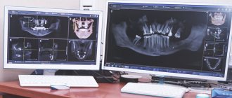
- Diagnostics of jaw models - special impressions of the lower and upper jaws that reproduce its movement. Thanks to this, the doctor receives information about the shape/deformation of the arches, the nature of the deformation, occlusal contacts of the cusps, and sizes. It is carried out before and after sanding to determine the correct result.
- Marking with carbon paper is considered one of the most accurate and at the same time the simplest method for diagnosing supracontacts. A small piece of copy paper is used (approximately 3*4 cm), which is folded into 4 layers. The most suitable method for identifying contacts during mandibular dynamics.
- As an addition, you can use auscultation - a technique for examining the oral cavity, with the help of which certain sound vibrations are determined. In such cases, a bifurcated, dull sound or click is detected.
Functional traumatic overload of the periodontium
This type of treatment is most effective in childhood, when bone tissue is in the process of formation. The use of orthodontic treatment in adolescence and adulthood is possible according to indications, but it takes longer and may require additional measures.
- For bite deformities. This pathology leads to the destruction of periodontal tissue, and in the future may result in tooth loss. In addition, the appearance of the dentition does not look very attractive.
- When there is crowding of teeth: one crown “lies” on top of the other. Such a defect can cause injury to the gums and cheeks; erasing the enamel.
- To eliminate the disproportion between the upper and lower jaws. Such a disorder can occur in infancy, or as a result of bad habits (thumb sucking).
- If there are gaps between the teeth.
Medical Internet conferences
Selective grinding of enamel as a method of creating space in the dentition
Zhiltsova E.S.*, Gogoleva A.V.**
* – dental student
** – orthodontist
Scientific supervisors: ass. Venatovskaya N.V., associate, candidate of medical sciences Petrova A.P.
FSBEI HE "Saratov State Medical University named after. V.I. Razumovsky" of the Ministry of Health of the Russian Federation
Department of Pediatric Dentistry and Orthodontics
Summary. This work contains information about the advantages, disadvantages, complications and techniques for selective grinding of enamel.
Key words: selective grinding of enamel, crowding of teeth, interproximal reduction of enamel, separation.
Corresponding author: Zhiltsova Ekaterina Sergeevna, [email protected] ,ru
Interproximal enamel reduction as a method of obtaining space in the dental arch
Zhiltsova ES*, Gogoleva AV**
* – 4 course, dental faculty
** – orthodontist
Rasumovsky SSMU
Department of Pediatric Dentistry and Orthodontics
Summary. This article contains information about advantages, disadvantages, complications and procedure of interproximal enamel reduction.
Key words: interproximal enamel reduction, enamel stripping, mesiodistal reduction, slenderization.
Introduction. According to the results of numerous studies, the number of dental anomalies is gradually increasing. The abnormal position of individual teeth occurs in 35% of cases of the total number of anomalies of the dental system. At the same time, crowding of teeth ranges from 5.5% to 47.7%, and in 17.2% of cases it is combined with occlusion pathology, and in 72.2% it is an independent pathology [1]. The main methods of obtaining a place in the dentition are as follows: expansion and/or correction of the shape of the dental arch, deviation of the frontal group of teeth into protrusion, distraction osteogenesis, removal, derotation, distalization of teeth, selective grinding of enamel, as well as a combination of various techniques [3, 6, 8 ]. The choice of treatment method in each case depends on the degree of crowding, type of occlusion, Bolton ratio, dental and periodontal condition, facial profile, patient's age and the patient's main complaint [3, 8]. Recently, selective grinding of enamel has been increasingly used in orthodontic practice due to a number of advantages.
Purpose: to study the general provisions and features of selective grinding of enamel as a method of obtaining a place in the dentition.
Tasks:
1) Assess the advantages and disadvantages of selective grinding of enamel.
2) Describe the technique for selective grinding of enamel.
3) Find out the likelihood of complications after selective grinding of enamel.
Materials and methods. An analysis of scientific articles and educational publications was carried out.
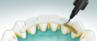
Results and discussion. Selective grinding of enamel (syn. interproximal enamel reduction) is one of the ways to obtain space when there is a deficiency in the dentition. This method involves grinding off part of the enamel from the mesial and/or distal surface of the tooth with abrasive instruments. The first mention of the use of this procedure dates back to 1944 and is associated with the name M.L. Ballard, who proposed the use of interproximal grinding on lower anterior teeth to correct tooth size discrepancies.
Many studies have been conducted to study the normal thickness of enamel and to determine the maximum amount of enamel that can be removed from each tooth surface without harm to the hard and soft tissues of the oral cavity [10]. It was found that the thickness of the hardest tissue in the human body does not depend on the shape (triangular, rectangular, or barrel-shaped) or the size of the tooth (macrodentia, microdentia) [6]. Also, the enamel is the same in thickness in men and women; men’s teeth are larger in size due to the large amount of dentin [10]. On average, the thickness of the enamel ranges from 0.5 to 1.5 mm [11]. However, it is impossible to determine the exact thickness of the enamel for each individual patient, therefore, when drawing up a plan for the procedure, it is necessary to rely on tabulated data of the minimum, and not the average values of enamel thickness for each tooth, obtained in the results of studies by various authors. It is worth noting that some patterns can still be traced. The enamel on the distal surface of the tooth is thinner than on the mesial surface. The only exception is the upper lateral incisors, where the relationship is reversed. Also, in all teeth, the thickness of the enamel increases in the area of the contact point [6].
There is no uniform rule regarding the quantitative possibilities of safe interproximal grinding. But the generally accepted opinion is that more than 50% of the enamel should not be removed from the tooth surface [6]. Thus, when teeth are separated from the mesial surface of the first left molar to the mesial surface of the first right molar, about 9.5 mm of space in the upper jaw and 8.5 mm of space in the lower jaw can be obtained [11]. We observe similar results from other authors.
Indications for interproximal grinding of enamel are: discrepancy in the Bolton ratio, lack of space in the dentition from 4 to 8 mm, increasing the stability of treatment results, improving the aesthetics of the frontal group of teeth by reducing “black triangles”, multiple tortoanomalies, correction of the curve of Spee [10, 6,8,7].
The criteria in favor of carrying out the procedure in a particular patient are: triangular or barrel-shaped teeth, good level of oral hygiene, low CP index, anomalies in the shape of individual teeth, macrodentia, adult patients - 12–14 years old for girls, 15–16 years old for boys and older (lack of possibility of expanding the dentition due to jaw growth), the patient’s categorical refusal to remove teeth in order to create space [8,6,7].
Contraindications are: enamel hypoplasia, dental hypersensitivity, microdentia, the presence of several restorations in the oral cavity, pronounced rotation of the teeth at the time of the procedure, rounded premolars, high KPU index, poor oral hygiene, rectangular teeth, baby teeth (young patients with large pulp chambers), periodontal diseases in the acute stage, lack of space in the dentition of more than 8 mm, patient refusal from this procedure [6,5,7].
The main stages of selective grinding of enamel:
1) Comprehensive planning. At this stage, the possibility of performing interproximal reduction is carefully studied, taking into account the indications and contraindications in this clinical situation, the Bolton ratio is studied in order to determine the zone for separation, and the exact amount of enamel that can be ground down in a particular patient is determined using a diagnostic model and periapical radiographs [4, 6, 9]. It is important that enamel grinding is performed for the first time in this patient to avoid excessive removal of hard tooth tissue from the proximal surfaces. Before carrying out separation, it is necessary to perform preliminary leveling of the teeth in order, firstly, to ultimately avoid the formation of non-physiological contact points and, secondly, to correctly assess the thickness of the enamel on an x-ray [7, 9, 10]. After the leveling stage, there should be no free space between the separated teeth [4]. Attention should be paid to the presence of fillings or crowns in the oral cavity [6].
2) Access to interproximal zones. It is created due to preliminary leveling of teeth [9]. Additional access to the proximal surfaces can be obtained using springs, separators or wedges for 3-4 days before the procedure [4, 9]. The space between teeth obtained using physiological separation must be measured to control the amount of enamel removed [4].
3) Soft tissue protection. JJSheridan recommends the use of a metal ligature to protect the gingival papilla during the separation process. The retractor, metal separator, wedge, and rubber dam also minimize soft tissue damage [7, 9].
4) Actually selective grinding of the enamel. Performed with hand or mechanical tools . There are 3 most common interproximal reduction techniques:
I. using air rotors using fine-grained burs and diamond-coated strips (ARS technology);
II. using diamond-coated discs (manual and for contra-angle);
III. using manual or machine abrasive metal strips [8,5].
The choice of technique depends on the severity of the space deficiency and the segment of the dentition. [8]. There are also a number of general recommendations when carrying out the procedure:
A. It is better to perform the first separating movements manually [6],
b. the enamel should be ground down gradually and symmetrically from both lateral surfaces of the tooth [9];
V. the cusps of canines, premolars and molars should not be subjected to grinding [6];
d. the apex of the interdental papilla and the contact point after the procedure should remain on the same line, perpendicular to the occlusal plane [6];
d. the contact point should remain (or be formed) at a distance of 4.5-5 mm from the upper border of the bone crest [6];
e. the amount of enamel removed must be measured and strictly recorded in writing. For measurements, thickness gauges and a Sheridan meter should be used [6,2];
and. It is not recommended to simultaneously grind off the enamel to the full permissible thickness, as this can result in excess space in the dentition [6];
h. If possible, grinding of enamel from the proximal surfaces of the 2nd and 3rd molars, as well as the distal surface of the 1st molar, should be avoided. This will help preserve the anchorage [7];
And. It is recommended to carry out separation with water or air cooling [8].
5) Recontouring and polishing of enamel. After removing the required amount of enamel, it is necessary to give the treated tooth surface its anatomical shape and form a physiologically correct contact point. These procedures can be performed using a bur, ultra-thin diamond or elastic disc [4]. Polishing of enamel must be carried out using finishing discs or strips and polishing paste [8,4]. Some researchers suggest using 35-37% phosphoric acid in combination with a fine abrasive strip for better effect. However, this statement is disputed [9,4].
6) Local use of fluoride. To enhance the remineralization processes in the enamel after grinding, it is advisable to use fluorine-containing products. On the other hand, B. Zachrisson claims that there is no need to apply fluoride-containing products to healthy patients; in case of increased temperature sensitivity of the teeth, rinsing with a low-concentrated fluoride solution 2 times a day is recommended [9].
Selective grinding of enamel is recommended to be carried out as necessary at three stages of orthodontic treatment: after leveling, immediately after the end of treatment and in the retention period [4]. The procedure can be performed over several visits.
If the principles of the procedure are not followed, excessive separation may cause complications such as: hypersensitivity, irreversible damage to the dental pulp, damage to soft tissues, the formation of false contact points, increased accumulation of plaque, caries and periodontal diseases in the area of ground teeth [8,6]. This can be avoided if you know separation techniques and carry out separation taking into account all the details.
The thinning of the enamel layer and the subsequent roughness of the proximal surfaces of the tooth are a criterion for increasing the risk of developing caries and periodontal diseases, since such a surface becomes a retention point for dental plaque. But with careful polishing, the enamel becomes smoother than even intact enamel [10]. As a result of observations of patients in the post-orthodontic period (subject to all the rules for selective grinding of the enamel in these patients), the following data were obtained:
| Authors of the article | Number of patients | conclusions |
| Zachrisson B.U., Minster L., Ogaard B., Birkhed D. (2011) | 43 | 2.5% of new carious lesions were detected during clinical and radiographic evaluation of 278 mesial or distal surfaces subjected to separation. These lesions were found in 3 of 43 patients |
| Kanoupakis PM, Peneva MD, Moutaftchiev VY (2011) | 53 patients, 18-24 months passed after separation | On 4.7% of ground surfaces, initial enamel changes were detected using the DIAGNOdent device |
| Zachrisson B.U., Nyoygaard L., Mobarak K. (2007) | 61 | 1 patient had a general increased sensitivity of the teeth, 1 patient complained of increased sensitivity of the lower incisors |
| Jarjoura K., Gagnon G., Nieberg L. (2006) | 40 patients, 1 to 6 years passed after separation | New interproximal lesions appeared on 3 of 346 surfaces |
Table. Lapenaite E., Lopatiene K. Interproximal enamel reduction as a part of orthodontic treatment [8].
In orthodontic practice, selective grinding of enamel as a method of obtaining space in the dentition can act as an alternative to tooth extraction for a number of advantages:
- there is no threat of obtaining excess space in the dentition [6,10];
- tooth movement is more gradual, therefore, possible loss of bone and root cement is reduced [6,7];
- tooth movement is more gradual, therefore, the parallelism of the roots is maintained [6];
- smaller anchorage is required [6];
- treatment stability increases [10];
- in case of relapse in the post-orthodontic treatment period, it is possible to obtain additional space [6];
- control over the amount of space obtained makes it possible to achieve better inclination of the incisors (to avoid protrusion and retrusion) [6];
- the total treatment time is reduced [6,10];
- it is possible to correct the shape of teeth [6];
- it is possible to remove “black triangles” [6];
- can be used in patients with contraindications to extraction [7];
- The gingival papilla is more aesthetically pleasing in interdental spaces created by separation than by extraction [7].
Disadvantages of selective enamel grinding:
1) if the technique and principles of the procedure are not followed, serious complications can occur (hypersensitivity, irreversible damage to the dental pulp, damage to soft tissues, the formation of false contact points, caries, periodontal disease) [6, 8];
2) long duration of the procedure [7].
Thus, despite the many details on which the orthodontist must concentrate his attention when performing selective grinding of the enamel, the choice of this procedure when there is a shortage of space in the dentition is indeed advantageous in the treatment of crowding of teeth in a number of clinical cases.
Conclusions:
1) Selective grinding of enamel has many advantages, the main of which are: no threat of obtaining excess space in the dentition, the ability to obtain additional space in case of relapse in the post-orthodontic treatment period, reduction of the overall treatment time, the ability to correct the shape of teeth and remove “black teeth” triangles." The disadvantages are relative, since possible complications can be avoided by following the separation technique, and the long procedure time is compensated by excellent results after orthodontic treatment and a reduction in treatment time in general.
2) The six main stages of separation are as follows: comprehensive planning, access to interproximal zones, protection of soft tissues, selective enamel grinding itself, recontouring and polishing of enamel, topical use of fluoride.
3) If the procedure technique is followed, no complications arise during the procedure. The risk of developing caries of proximal surfaces in the post-orthodontic treatment period is individual. Initial caries affects on average no more than 5% of separated surfaces.