Lump on the lower jaw on the chin
Most often, swelling will be associated with inflammation of the lymph nodes.
There are a lot of them in the neck area, and therefore in the chin area, and they always react to any inflammatory processes in the oral cavity and nasopharynx. Lymphocytes accumulate here, which actively fight infection or other pathogens, and the lymph nodes are the result of their accumulation. Here, lymphocytes are produced and sent by the immune system to the site of inflammation. If pathogens penetrate the lymph node itself, it too begins to become inflamed, called lymphadenitis, defined as a lump under the skin. This lump can form literally overnight: in the evening before bed there was nothing yet, but in the morning the lump has already appeared.
If symptoms do not go away within 2-3 days, this may indicate a purulent process.
and the transition of lymphadenitis to the chronic form, the bumps remain enlarged, but do not hurt. But this does not mean that the node cannot become inflamed and infected again, and then a pain syndrome immediately forms. Lymphadenitis never occurs on its own, it is always the end result of advanced inflammatory diseases of the ENT organs, and below the jaw or on the chin on the right or left it most often occurs with caries. But it must be remembered that lymphadenitis can be the starting point for cancer, so examination and diagnosis by a doctor are necessary.
Lymphadenitis is far from the only reason for the appearance of bumps. A lipoma can also form under the chin on the jaw - elastic, soft, mobile. It is usually asymptomatic, and only when it grows can it compress the nerve endings and then pain appears. And another reason for the appearance of a lump on the lower jaw - on the right or left, or in the middle of the chin - is the formation of an inflamed follicle, which goes through the stage of an internal pimple (like a painful lump under the skin) before appearing on the skin.
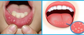
Banal folliculitis occurs as a result of blockage of the sebaceous gland and is the most common. Stomatitis, herpes, atheroma, lipoma, skin cyst, folliculitis can appear in the form of bumps. A subcutaneous ball may appear as a result of facial trauma. In these cases, the formation has clear boundaries and is solid. In the area of the chin on the lower jaw, bumps also often appear due to boils and acne, especially when they become infected.
Lymphoma is always a malignant tumor. It may appear as a painless swelling behind the ear. When palpated, it is defined as a group of lymph nodes that are fused to each other and to the skin and are motionless. Attention is not paid to such formation due to its painlessness. But if a person experiences weight loss over a short period of time, loses interest in life, and doesn’t want anything, they should consult a doctor urgently.
If any lump appears, you should never squeeze it out or heat it, as this can increase inflammation or accelerate the process of malignancy. You cannot lubricate the lump with iodine, rub it, pull it, or expose it to the sun's rays. Folk remedies are also not applicable without a doctor's permission.
An urgent visit to a doctor if a lump appears behind the ear is necessary if:
- lymph nodes enlarge strongly and quickly;
- the lump grows quickly;
- the appearance of the lump is not associated with a cold or other infection;
- the lump begins to change its color and pus appears in it;
- the seal is very sensitive and painful;
- In addition to the lump, some new symptoms appeared.
Prevention
It is much easier to prevent cancer than to cure it later. Rules must be followed:
- treat teeth and oral cavity when necessary;
- monitor oral hygiene;
- stop using tobacco;
- protect the oral cavity from injury;
- limit alcohol consumption to a minimum;
- eat as many healthy foods as possible;
- Avoid prolonged exposure to sunlight.
The rules do not provide a 100% guarantee of not getting sick, but they reduce the chance of the disease occurring.
Causes of bumps
Often, a bump on the cheek appears due to some kind of injury; such a reaction, as a rule, is protective in nature and is considered a completely normal phenomenon. At the same time, it is under no circumstances recommended to touch the tumor in order to allow the wounds to heal properly.
The variety of reasons allows us to talk about their versatility. Despite this, among them we can highlight the main points why such formations most often appear:
- The most common cause is injury. It doesn't have to happen yesterday. Injuries may be old, long forgotten. But at some point, a certain factor gives impetus to the beginning of the appearance of a bone growth. When a bruise occurs, a bruise forms and a swelling forms. In themselves, these manifestations do not pose a danger.
- Another reason is the presence of thrombophlebitis. It causes the formation of blood clots.
- Atheromas (cystic formations on the sebaceous gland) and wen (lipomas), which are benign tumors of adipose tissue, play a certain role in the formation of bone growths.
- The cause may be a situation in which the tendons are stretched or something is constantly pressing on the bone. Most often, with such factors, a lump forms on the wrist bone.
- An abscess is a more serious cause of bone spurs. A similar situation can arise when an infection penetrates into the damaged area of the skin. There is pronounced localized pain with a general change in the condition of the body in the form of a rise in temperature.
- Hereditary factor.
- Varicose veins.
- Bursitis, arthrosis.
- Metabolic disease. In some patients, there may be an increased concentration of uric acid in the blood. This occurs due to a violation of purine metabolism. As a result, crystals are deposited on the surface of the joints and cartilage.
How to distinguish a wen from other formations
To find out how to distinguish a wen from formations of a different nature, you need to know their characteristics and the mechanism of formation. In addition to lipomas, the following formations may appear on the face or other part of the body:
- Atheroma. It is a cyst that formed from the sebaceous gland. The reason for the appearance of such a formation is a violation of the outflow of secretions. This occurs due to mechanical damage to the duct. Most often, atheroma appears on the cheek after self-removal of a pimple or acne;
- Epidermal cyst. It has a special cavity that is filled with sebum and horny masses. It grows slowly, the skin does not change. Appears after an attempt to remove acne on your own or as a result of illiterate treatment;
- Milium. This is the name for epidermal cysts, the size of which does not exceed 2 mm. They are often confused with small lipomas. The most common locations are the cheeks, eyes and forehead. They appear as a result of abrasions, sunburn, dermabrasion or due to genetic predisposition.
You can distinguish a wen on the cheek from an epidermal cyst by palpation. If a cyst has formed on the cheek, then it is characterized by inactivity and the absence of a lobular structure. The wen is mobile and can be easily felt.
Hard lump behind the ear
A lump behind the ear is often a consequence of the same lymphadenitis. It is round in shape, painless, dense and mobile. Does not pose a health hazard. With lymphadenitis, the lump can also be localized under the ear. It may happen that all the symptoms subside on their own and disappear after 1–2 weeks, the lump becomes motionless and dense.
In addition to lymphadenitis, a lump behind the ear can be the result of a blockage or infection of the sebaceous gland, which are present in large quantities here. In addition, the reasons may lie in the following:
- hormonal imbalance and decreased immunity;
- increased sweating;
- a consequence of seborrhea or acne;
- hypothermia of the body;
- lack of hygiene;
- lipoma;
- atheroma;
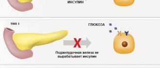
- chronic infections - TBC, diabetes, HIV infection, infectious mononucleosis;
- injuries;
- mumps;
- otitis and dental diseases;
- oncopathology of the lymphatic system.
If it is atheroma (blockage of the sebaceous gland), it appears and grows slowly, over several months, when it does not show itself in any way. Sometimes fat may come out of it, but it is better not to squeeze it out yourself to avoid infection.
its dimensions can be from 5 mm to 5 cm; in this case, the sebaceous gland ceases to function and turns into a seal. Atheroma is a sebaceous gland, stretched due to blockage of its excretory duct, a cystic formation. Its contents are thickened sebum. It can be located behind the ear or under the ear.
Blockage always provokes the formation of a cyst. Its outlines are clear, it is filled with fat, and has a capsule. The skin above it does not fold; upon careful examination, a black dot is visible - a clogged duct, this is its difference from a lipoma. When its size is more than 5 mm, it begins to itch and burn.
Fluctuation can be determined by palpation. Treatment is surgical in the form of removal of the atheroma with the capsule. Atheroma can also be removed with a laser. With good immunity, the lump can open on its own, then all its contents come out: blood, fat, pus. After healing, small scars remain.
Epidemic parotitis, or “mumps,” is an infectious inflammation of the parotid salivary glands. In this case, a rise in temperature, chills, malaise, weakness, pain in the tumor, neck and ears are noted. The disease is contagious and requires isolation of the patient. Treatment is required. The infection can occur in children and adults, in whom it is more severe and has complications.
This proliferation of adipose tissue is a consequence of lipid metabolism disorders, slagging in the body and hereditary predisposition. Discomfort appears only when it is large. In these cases, it is excised. If a lump appears behind the ear and it hurts, this may be a consequence of existing otitis media, eustacheitis and inflammation of the postauricular lymph node.
The lump near the ear may be hard from the very beginning or change in shape. The pathologies will be different. This happens with a lipoma, which at first may be soft, then develop into a malignant tumor. With hyperhidrosis, that is, increased sweating, oily seborrhea, with inflamed acne, secondary atheroma is formed.
A lump in the ear area, if it is of oncological origin, has a flesh-colored or slightly darker color, it is motionless, fused with the surrounding tissue, dense and painful. In benign formations, the tumor is always elastic, mobile and not fused to the underlying tissue. In the last stage of cancer, the lump begins to fester.
Stages of cancer
Symptoms of the disease vary depending on the stage. In medicine, stages are distinguished, distinguished by the size of the malignant formation, the depth of damage to the patient’s body by the disease, as well as the behavior of the cancer cells.
- Stage 1. The tumor is gradually increasing, the nearest tissues are not yet damaged.
- Stage 2. The tumor continues to grow.
- Stage 3. The malignant formation affects nearby lymph nodes, and the affected area grows.
- Stage 4. The process of spreading metastases occurs. They can affect all organs and systems. For example, the palate becomes inflamed. Cancer can also affect the gums. In this case, the gums hurt and bleed.
Factors contributing to the appearance of a lump
Various factors can contribute to the appearance of bumps. These include the following points:
- a person leads a lifestyle in which he is often overworked;
- constant use of certain medications;
- presence of increased physical activity;
- errors in nutrition;
- excess body weight;
- presence of bad habits;
- age factor (people over 30 years of age are most susceptible to the appearance of bumps);
- hormonal disbalance;
- deficiency of certain minerals.
We invite you to familiarize yourself with a sore throat; it hurts to swallow a red spot in the throat.
A compaction under the skin in the form of a ball is a symptom that develops for various reasons. Externally, the soft tissue formation resembles a tubercle. The skin over the bulge may remain normal or become inflamed. In the first case, the lump is a dense ball that usually does not hurt.
You can cope with painful symptoms and prevent or stop the infection process with the help of medications and folk remedies. For pathologies of connective tissues and blood vessels, surgical intervention is required.
Treatment with drugs
Etiological therapy includes drugs that affect the cause of the disease:
- antibiotics - kill bacteria - Augmentin, Amoxicillin, Amoxiclav;
- antiviral drugs - suppress the development of the virus - Amantadine;
- antifungal agents - prevent the development of infection and fight the pathogen - Fluconazole, Amphotericin B.
Pathogenetic therapy means:
- anti-inflammatory drugs in the form of ointments or pastes - Nise, Diclofenac, for internal use - Nimesil, Ibuprofen.
- for insect bites, antihistamines are prescribed - Fenistil gel, Suprastin, Loratadine, Claritin syrup for children;
- antiseptics - Vishnevsky ointment;
- healing drugs – Levomekol
Traditional recipes do not eliminate the causes of the lump, but they help eliminate the symptoms faster.
- Echinacea tincture for lymphadenitis. Combine 50 ml of tincture and 100 ml of water, leave for 30 minutes. Moisten a piece of bandage and apply it to the bump. Apply a layer of cotton wool over the lotion and secure with a bandage or plaster.
- Onions for furunculosis. The vegetable is placed in the oven for a few minutes, cut, and applied to the pimple site.
- If a lump has come out due to a mosquito or midge bite. Catnip oil relieves itching and reduces inflammation.
- Garlic for carbuncles. Grate a few cloves of garlic and squeeze through cheesecloth. Soak a cotton swab in the resulting juice and treat the painful area of the body.
- Lemon juice for a wasp sting. Cut off a slice of lemon, squeeze out the juice and treat the swelling - using the recipe relieves swelling and pain.
If conservative methods do not bring results, surgical treatment is most often used:
- Boils and carbuncles. Opening the purulent focus and installing drainage helps remove pathogenic microbes from the body and prevents further infection.
- Benign processes. Excision is carried out if wandering nodules are present in the thickness of the muscle or adipose tissue. If the diagnosis is confirmed: fibroma, lipoma, chondroma or cyst, surgical treatment is mandatory.
For tumors of vascular origin, an alternative operation may be required - removal with a laser or diathermocoagulator.
If the inflammatory infiltrate is not treated in time, blood poisoning may develop. Formations on the head are especially dangerous - they lead to meningitis and encephalitis.
Blood clots in the veins can be fatal if not treated promptly.
As benign tumors grow, they disrupt the blood flow of adjacent organs. Over time, they can develop into cancer pathologies. If after treatment the lump remains or grows again, a re-examination is required.
Subcutaneous lumps of various etiologies require consultation with a therapist. After collecting anamnesis, palpation and examination, the doctor will refer you to a specialist in a narrow field. Timely diagnosis will help avoid dangerous and fatal complications. In most cases, medication or a simple surgical procedure is sufficient to get rid of the lump.
Skin, nails, hair
Small ball under the skin
A tumor under the skin in the form of a ball, a lump on the abdomen can be either a harmless lipoma or a hernia, especially if it appears near the navel. Therefore, a visit to the surgeon is mandatory. The reasons for such growth are not important, the main thing is to determine what to do with it.
The lump is often a lipoma, formed from excess adipose tissue. In fact, it is harmless and does not cause concern. They feel it, understand the boundaries, the peculiarity of the surface - smooth or bumpy. The skin does not change color, the upper layer of the epidermis is easy to fold with your hand. Occasionally they grow to large sizes, then they should be gotten rid of.
A lump under the skin in the form of a ball on the back sometimes turns out to be atheroma. It cannot be confused with a lipoma, since there is little fatty tissue on the back. In fact, atheroma is another type of subcutaneous cyst that stretches the sebaceous gland, clogging its excretory ducts. Such a cyst is filled with sebum, with a tendency to accumulate, causing the gland to stretch and the atheroma to grow.
A dense, rounded formation has clear boundaries, the skin above it turns blue, and a closed duct can be seen as a dot in the blue. Atheroma tends to become inflamed and fester. Such atheromas must be removed by a surgeon.
A compaction under the skin in the form of a ball under the arm has serious causes - this is the beginning of inflammation of the lymph nodes, leading to lymphadenopathy. The cause may be a cold, to which the lymph nodes respond with inflammation. They are an integral component of the immune system and work as a filter formation to clear interstitial fluid from infection.
Such balls under the armpit decrease slowly after active treatment of a cold with antibiotics. With the onset of purulent inflammation of the lymph nodes, the balls under the armpit are opened by the surgeon to remove the pus and clean the tissue.
When the formation above the lymph node under the armpit becomes dense, the skin does not wrinkle, the node may be damaged by malignant components. These situations require consultation with an oncologist.
Clinical manifestations
The clinic is characterized by diversity, but the following symptoms come to the fore:
- painful sensations in the affected area (naturally, this brings obvious discomfort to the patient);
- development of local edema;
- the skin of the affected area is hyperemic;
- there is stiffness of movement at the site of the pathological focus;
- the skin covering the affected joint thickens;
- general reaction of the body in the form of a rise in temperature.
Other symptoms may also occur, depending on the location and severity of the process.
Diagnostics
Regardless of whether a small ball has appeared under the skin, or a huge protrusion, you need to find out what kind of lump it is.
For this purpose, perform:
- inspection and palpation of the lump - evaluate the color, consistency, contours and size of the formation;
- When inflammatory processes are excluded, an ultrasound is performed.
- All neoplasms are subject to histological examination - it allows you to find out what tissue the lump consists of;
- general blood test, urine test;
- biochemical blood test.
It is not difficult to determine the presence of a lump on a bone in an adult, since it is noticeable, and even the patient himself can do this. It is much more difficult to establish the reason why it appeared. It is necessary, without delay, to pay a visit to the doctor. He will examine the patient, collect anamnesis, and prescribe a number of additional studies.
Dopplerography of the vascular apparatus and x-ray studies will help in making a diagnosis. The results of a general blood test provide some information. An ultrasound of the thyroid gland may be prescribed, since it is the pathology on its part that causes situations in which various seals appear.
Particular attention should be paid to children. If a lump appears and grows on a child’s bone, you should immediately consult a doctor in order to exclude the development of cancer.
Features of treatment
Self-treatment and diagnosis are strictly prohibited. This is the responsibility of the doctor alone.
The greatest effect in treatment is achieved by surgical removal of the lump. If the process is at the very beginning of its development, then attempts can be made to get rid of the lump in a conservative way.
If the lump is on a leg bone, then you can use special orthopedic insoles and arch supports. Conservative treatment methods will not radically eliminate the problem. They can only improve the patient's condition. This will relieve pain and reduce swelling. Along with taking medications, traditional medicine methods are used. Physiotherapy treatment is a good addition. Usually everything is used in combination.
Additional diagnostic methods
To get a complete picture of the disease and determine the method of therapy, a certain number of other examinations are carried out. Among them are:
- Radiography;
- Ultrasound diagnostics;
- Computed tomography.
X-rays allow you to assess the extent of tumor damage to surrounding bone tissue. Since the bones of the skull are in close proximity to the sites of the disease, they are affected first. Radiography allows you to see metastases and assess the extent of damage to the body.
The ultrasound diagnostic method evaluates the cancerous node, its structure and area of damage.
Using computed tomography, it is possible to assess the nature of the tumor: malignant or benign. Computed tomography data are decisive when prescribing a therapy method.
What else can cause neoplasms?
Bumps on the cheek can form simply out of nowhere or can be caused by various factors. Usually occurs in people for a number of the following reasons:
- Due to hormonal imbalance.
- As a result of mechanical injury.
- Against the background of viral infections.
In addition, they can act as symptoms of the following diseases:
- A person has skin fibroids.
- Presence of purulent granuloma.
- Presence of dermatitis or seborrheic keratosis.
- Against the background of soft tissue sarcoma.
- If the patient has a xanthoma or basal cell carcinoma.
In any case, it is extremely difficult to identify on your own what exactly could have influenced the appearance of a lump in the mouth on the cheek, unless it is provoked by visible factors in the form of, for example, insect bites. Now let’s figure out what symptoms accompany the pathology.
A photo of a bump on the cheek is shown below.
Let's look at the main types of bumps that appear on people's cheeks:
- A lipoma is a soft lump, also called a wen, as it is formed from fatty tissue cells. This can occur in the event of a metabolic disorder in the body; it is often small in size and does not cause any inconvenience to its owner. If the size of the wen is insignificant, then drug therapy is prescribed, namely, a special medicinal drug is administered. If it reaches at least three centimeters, surgical excision will be required, which is performed under local anesthesia.
- Presence of atheroma. When the sebaceous duct is blocked, the secretion accumulates under the skin, against which a lump appears on the cheek in the form of a round and colorless compaction with a diameter of one to ten centimeters. If you palpate, you will notice the mobility of the atheroma; as for pain, it is completely absent. Such a tumor will require careful study and treatment, since in advanced cases side effects may occur in the form of pain, accumulation of pus, inflammation, increased body temperature, and the like. Even if at first a person does not feel discomfort except for a violation of aesthetics, it is still highly recommended not to delay therapy. As a rule, patients are prescribed a surgical operation in the form of excision of the lump and capsule, and there is a possibility of scarring of the operated area.
- Treatment of mucocele. This is the name of a cyst that occurs in the oral cavity on the buccal mucosa. It can be characterized by a bluish color, lack of pain, and during palpation the homogeneity of the structure can be felt. There is also sometimes a cloudy white liquid inside. The opening can occur spontaneously, otherwise it is possible that excision of the bladder will be required. The likely causes are injuries to the salivary glands along with constant biting of the mucous membrane. Often such a lump occurs in young people.
- With cancer of the salivary gland, a lump may appear in the mouth near the ear on the inside of the cheek; the nature of the formation will be malignant. As a result, compression of the branches of the trigeminal nerves may occur, and the patient may feel numbness in the face. Against the background of all this, muscle tone may deteriorate, and pain will be present in the tonsil area. In addition to a biopsy, in this case it is necessary to carry out a diagnosis using computed tomography, and based on the results obtained, a specialist will prescribe therapy.
We suggest you read: Pain in the periosteum after tooth extraction - ProfiMed
Phases of cancer tumor development
Small formations and ulcers can transform into complex forms of the disease
The life cycle of malignant tumors consists of several phases:
- Primary;
- Active;
- Launched.
At the initial stage, small lumps or ulcers appear on the cheek. They cause a certain discomfort to a person. As a rule, no one pays much attention to the first signs. However, diagnosing the disease in this phase significantly increases the success of treatment measures.
The active phase is characterized by the appearance of painful formations. The ulcers turn into cracks. The person becomes lethargic, suffers from severe headaches, and rapid weight loss occurs. At this stage, mucosal cancer becomes obvious, and any qualified dentist will pay attention to this.
Traditional medicine in the treatment of cones
It must be clearly remembered that alternative medicine methods cannot be classified as the main treatment. They just complement it. You can use the following folk remedies.
Soap and iodine will help in treating bumps. These simple remedies are great for getting rid of bone bumps. You need to take a small piece of soap, grind it with a grater and add a little water. The resulting composition should be rubbed into the affected area, using light massage movements.
You can apply camphor oil to the sore spot, followed by iodine. Before this, the application area is well steamed.
It is possible to treat cones using the “grandmother’s” method. It uses potatoes and propolis. Treatment is carried out with potato baths. The following lineup is being prepared for them. Potato peelings are placed in a saucepan. Water is poured into it and boiled. The resulting solution is poured into a basin. The hot composition is used for baths. Warm up the affected area for at least 30 minutes.
You can apply propolis to the sore spot. It must first be softened. Propolis is fixed on the sore spot with a bandage. For this purpose, you can use alcohol tincture of propolis, which can be purchased at a regular pharmacy.
Using a regular chicken egg and vinegar, you can prepare an ointment. She has excellent treatment for hallux valgus deformity. The egg is poured with vinegar and left in a dark place to infuse. It should stay there for 2-3 weeks. The shell dissolves and the shell must be removed. It is necessary to add a little rendered lard or turpentine in the amount of 10 g. The ointment is applied to the affected area every other day. In the intervals between applications, a mesh of iodine is performed.
Which doctor should I contact?
As a rule, such a neoplasm has a high growth rate. It can develop up to two centimeters in diameter in just a week, and sometimes even more.
An additional unpleasant symptom is that the bump on the cheek causes a person extremely painful sensations, starting to bleed and acquiring a convex shape. Against the background of all this, the color of the mucous membrane can vary from bright red to a pronounced purple hue.
After a biopsy, such patients may be prescribed the following types of therapy:
- Carrying out surgical treatment of a bump on the cheek under the skin. For this, electronic scissors are used, and immediately after removing the tumor, the affected area is cauterized. Therapy is continued with a course of antibiotics.
- Injection treatment. It involves special injections containing alcohol, which are given directly to the area of the granuloma.
- Performing laser treatment.
- Local injections using aletretin gel.
So, a bump appeared on my cheek. Regardless of the size of the pathological growth, one should not delay going to a specialist, this way a person can quickly feel comfortable and avoid all sorts of complications.
Many patients are not only unaware of how to treat a lump on the inside of the cheek, they are not even aware of which doctor to consult. You should make an appointment with the following specialists:
- First of all, you should go to a dermatologist. This specialist will certainly help you cope with such seals, be it a wart, lichen, papilloma, and so on.
- Often, a surgeon is consulted for the removal of a benign tumor and for the treatment of purulent inflammation.
- To an oncologist. After examination and receiving test results, the doctor will be able to exclude the presence of a malignant tumor.
But first, it would be better to consult a therapist. After the initial examination, the specialist will write a referral to a doctor of a more narrow specialization.
If a lump appears on the cheek in the mouth, treatment should be timely and comprehensive.
With formations such as hygromas, bumps on the finger, or painful nodules on the cheekbones in men, you need to contact a general practitioner - a therapist, family doctor.
If a nodule appears inside the skin after surgery or opening a pimple, a consultation with a surgeon is necessary. The same applies to dark blood bumps.
Nodules under the skin of the penis are a reason to consult a urologist. Protrusions on the genitals in women may indicate a viral infection - the problem is dealt with by gynecologists.
If necessary, the patient is referred to an infectious disease specialist.
“Lump” or “ball” near the ear are, of course, non-medical terms. These words mean a voluminous formation, the nature of which remains to be determined. And to clarify the pathological process, the doctor needs to conduct a clinical examination: questioning, examination, palpation.
Furuncle
A lump on the cheekbone next to the ear may be a boil. This is an acute purulent inflammation of the hair follicle. At first it looks like a small focus of infiltration with the following manifestations:
- Redness.
- Swelling.
- Soreness.
Next, the boil takes on a cone-shaped appearance, and a necrotic core begins to mature in it. This process is accompanied by an increase in local symptoms. The pain becomes more intense and can radiate to the ear or eye. With severe inflammation, the temperature rises and signs of intoxication appear.
A yellow spot soon forms at the top of the swelling - this is the accumulated pus trying to come out. The breakthrough of the boil is marked by the release of greenish contents with necrotic masses, which leads to an improvement in the general condition and a decrease in inflammatory symptoms. During the healing period, the wound is filled with granulating tissue, and a small scar remains on the surface.
Lymphadenitis
When the lymph nodes are involved in the inflammatory process, swelling also forms. Most often this occurs in the corner of the lower jaw, in front of or behind the ear. Lymph nodes react to any inflammatory process in the area of their functional activity: otitis and mastoiditis, tonsillitis, periodontitis, sinusitis, skin infections, etc.
Signs of lymphadenitis are mainly local in nature. These include the usual signs of inflammation such as swelling, redness and tenderness. The affected lymph nodes increase in size, and the skin over them becomes hotter. During an acute purulent process, an abscess forms in the thickness of the tissue, which leads to an increase in local symptoms and a deterioration in the general condition.
Sialadenitis
The parotid glands become inflamed quite often, which does not allow sialadenitis to be completely excluded. In such cases, a compaction appears, which becomes sensitive to palpation and is accompanied by pain. The latter intensify with movements:
- Chewing.
- Opening the mouth (talking, yawning).
- Turn your head.
The pain radiates to the ear, lower jaw, and temple. The function of the salivary gland is also impaired, which is manifested by hyposalivation (decreased secretion) and the appearance of pathological inclusions (mucus, pus, flakes). Acute sialadenitis is accompanied by fever and a disturbance in general health. On palpation, the gland feels dense; with suppuration, fluctuation (trembling) is determined in the center of the swelling. A complicated course of the disease is indicated by abscess formation, the appearance of fistulas, or stenosis of the salivary ducts.
Mastoiditis
Swelling behind the ear can indicate mastoiditis. This is an inflammatory process that affects the cave (antrum) and the cells of the mastoid process of the temporal bone. It is characterized by pain (including ear pain), swelling and sensitivity of the skin. Additional signs of pathology include:
- Headache.
- Fever.
- Hearing loss.
- Discharge from the ears.
There may also be a protrusion of the auricle anteriorly, and upon examination, pus is visible in the ear canal. The latter can break through the skin with the development of an abscess. This leads to increased swelling, redness and pain.
Lipoma or atheroma
A painless ball in front of the ears occurs with benign formations, for example, lipoma or atheroma. The fatty tissue is localized under the skin, is not fused to it, and has an elastic consistency. Its size is usually small, so it does not cause subjective discomfort (besides aesthetics).
We invite you to familiarize yourself with Dental Implantation for Oncological Diseases
Atheroma is formed in cases where the sebaceous gland duct is blocked. The secretion accumulates, stretching the walls, which leads to the appearance of a limited, mobile swelling. If the atheroma becomes inflamed, then signs characteristic of a boil or other purulent process in the skin appear: redness, pain, swelling, fever. The formed microabscess breaks out with the discharge of pus and thick sebaceous secretion.
Swelling of traumatic origin will be diffuse. It occurs after mechanical damage to tissue (usually a bruise) and is accompanied by pain, abrasions, and hematoma. Nosebleeds may also occur, and with severe injuries, patients experience dizziness, tinnitus, and headaches.
Insect bites are considered a commonplace situation, but it is necessary to exclude an allergic reaction or infectious consequences (ehrlichiosis, borreliosis, malaria, tick-borne encephalitis). If we are talking about signs of hypersensitivity, for example, after a bee sting, then, along with local swelling and redness, hives and itching will be a concern. More negative consequences are also possible, for example, Quincke's edema, bronchospasm, anaphylaxis.
Sometimes a lump near the ear should be considered a malignant process. Skin cancer can manifest itself in various ways:
- Lumpy wart.
- Pigmented spot.
- Flaky seal.
- A plaque that becomes crusty.
- Ulcer with foul-smelling discharge.
The malignant neoplasm is characterized by intensive growth, unclear boundaries, adhesion to the underlying tissues, and enlargement of regional lymph nodes. If the cancer becomes widespread, pain and signs of intoxication appear: weakness, emaciation, pallor, loss of appetite, low-grade fever. If isolated metastases appear, the function of the affected organs is also impaired.
Cheek cancer: symptoms, photos of the initial stage, types and treatment
Cheek cancer is a cancer of the mouth.
Rarely seen. It often manifests itself as a secondary disease; the first signs are the same as other diseases of the oral cavity. Sometimes it appears against the background of leukoplakia. According to ICD-10 it has code C06.0. Cancer affects different parts: tongue, buccal mucosa, palate, lip, alveolar process.
Causes and risk factors
Men over fifty years of age are most susceptible. The disease is more common in Asians. Women are five times less likely to get sick. In exceptional episodes, children. Causes of occurrence and risk factors:
- smoking and use of tobacco products;
- alcohol addiction;
- leukoplakia;
- chronic gum diseases;
- poor oral care;
- injuries to teeth and mucous membranes;
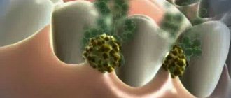
- the presence of viruses such as herpes and papillomas;
- lack of vitamins in food;
- contact with harmful substances at work.
Reasons and factors:
- smoking and use of tobacco products;
- alcohol addiction;
- precancerous condition of the oral mucosa;
- chronic gum disease;
- insufficient oral care;
- unfavorable heredity;
- injuries;
- the presence of papilloma and herpes;
- food with insufficient vitamin content;
- frequent contact with harmful substances at work.
Kinds
The initial stage is asymptomatic and, accordingly, does not attract the patient’s attention.
Shapes:
- Ulcerative. Tiny sores appear inside the mouth. They do not last for a long time and cause inconvenience. The severity is that the swelling is located close to the blood vessels. When it enters the bloodstream, it instantly spreads throughout the body. It is more difficult to stop the oncological process in such a situation.
- Papillary. The form is easy to treat and does not leave swelling. Looks like growths.
- Nodal. The swelling has the appearance of a rough-to-the-touch compaction covered with pale spots. Compaction with clear, fairly visible boundaries. This type of tumor is characterized by rapid development.
Symptoms of the disease
- A painful lump appears on the inside of the cheek. The appearance of swelling is determined visually. Formations of unknown origin must not be ignored.
- The presence of long-term non-healing wounds.
- Bleeding from affected areas.
- Having problems swallowing.
- The appearance of periodontal disease and the transition to a chronic form.
- Decreased sensation in the mouth.
- Significant size of lymph nodes.
- Swelling and decreased tongue mobility.
- Unhealthy feelings during movement of the lower jaw.
- The mouth may be lined with spots. At later stages, bad breath may occur, which is associated with decay of cancer cells.
Cancerous lump on the inside of the cheek
Formation cycles
The development of malignant neoplasms goes through a number of periods: initial, active, neglected. Each one manifests itself differently.
- During the first phase, a painful lump appears on the inside of the cheek. There are no other symptoms.
- The active phase is characterized by the transformation of ulcers into cracks. The patient develops lethargy and a feeling of apathy, and his head often hurts. Sometimes weight drops sharply. At the described stage, cheek cancer is easy to detect. As a rule, dentists pay attention to such symptoms. The likelihood of successful treatment at this stage is high.
- During the advanced phase, metastases spread throughout the body. Treatment no longer helps.
Stages of cancer
Symptoms of the disease vary depending on the stage. In medicine, stages are distinguished, distinguished by the size of the malignant formation, the depth of damage to the patient’s body by the disease, as well as the behavior of the cancer cells.
- Stage 1. The tumor is gradually increasing, the nearest tissues are not yet damaged.
- Stage 2. The tumor continues to grow.
- Stage 3. The malignant formation affects nearby lymph nodes, and the affected area grows.
- Stage 4. The process of spreading metastases occurs. They can affect all organs and systems. For example, the palate becomes inflamed. Cancer can also affect the gums. In this case, the gums hurt and bleed.
Diagnostics
As a rule, the initial diagnosis is carried out by a dentist. He can see the neoplasm upon visual examination. If one is detected, an anamnesis is collected. Upon completion of the collection of information, a referral to oncology is issued.
After an appointment with an oncologist, a biopsy of the damaged tissue is performed, and a cytological examination is also performed. Some forms of cancer can be detected by the conclusion of a laboratory test, which also reveals the stage and how far the disease has spread.
Do not forget - the correct diagnosis is established only after receiving the results of additional studies:
- X-ray. Allows you to determine which bone tissues are involved in the process. Detects the presence of metastases and the degree of damage to the body.
- Ultrasound. Allows you to study the surface area of the tumor and its structure.
- CT scan can determine the shape and size of a malignant formation.
However, the first step to determining the presence of cancer is the result of a self-examination. Detection of any abnormalities should prompt a visit to a doctor.
Cancer Treatment and Side Effects
Treatment often depends on the location where the swelling is found and the area of spread. In the treatment of cheek cancer, a complex method is often used. Main methods:
- Surgical intervention. The tumor is removed by surgery. Regional lymph nodes located on one side with swelling are also subject to removal.
- Chemotherapy. A method is used to reduce swelling before surgery. It is often carried out in conjunction with radial therapy and surgery.
- Radial therapy. Calculated doses of radiation are used to destroy cancer cells.
- Multimodal method. A method that combines two or more other methods.
- Long-term monitoring. The bottom line is systematic medical examinations. They allow you to prevent relapse of the disease in time.
There are many methods, but surgical intervention is still considered one of the main ones in the fight against cancer. The use of surgery involves, in some cases, the removal of part of the bone tissue. Then the person needs plastic surgery to recreate the appearance of his face. Removal of lymph nodes can lead to facial numbness and improper functioning of the facial muscles due to rupture.
Types of radiation during radial therapy:
- Remote. Irradiation is performed away from the cheek.
- Contact. The device is applied to the cheek from the outside.
- Interior. The substance is directly injected into cancer cells. Radial therapy is used with caution due to the presence of side effects. The patient may experience loss of voice, pain when swallowing, and loss of appetite. To relieve symptoms, doctors recommend following a diet and drinking water more often.
For chemotherapy, various drugs are used, divided according to the degree of toxicity into red, yellow, blue, white.
Taking red medicine is considered the most toxic and can aggravate the patient’s condition. The big disadvantage of this method is that the substances affect all cells and destroy healthy tissue along with the diseased ones in the process.
It is carried out before and after surgery to prevent recurrence of the disease.
Among all the methods, chemotherapy has a strong effect on the body; the procedure is accompanied by baldness, a feeling of nausea, and numbness of the limbs.
Other complications:
- tooth loss after radiation therapy;
- the need for prosthetics;
- scar formation after surgery;
- increased susceptibility to infections.
- mucous secretion is not produced properly.
- The jaw moves worse.
Traditional methods
It is permissible to use these methods of treatment only under the supervision of a doctor! They are often treated with herbs and herbs.
Few people know that annual wormwood is used as an anti-cancer agent. It has a lot of useful properties, but at the same time it also has contraindications. Do not forget that the grass is poisonous.
If you consume more than the norm, unwanted side reactions are possible, some of them life-threatening.
Scientists have found that the tumor requires a significant amount of iron to increase its size. An extract prepared from wormwood can organize a chemical reaction, the result will be oxidation of iron, and the cancer cell will die.
This is the effect of the substance. The product is undergoing clinical trials. If the result is positive, the medicine will be classified as a drug that kills diseased cells without affecting healthy tissue. In this case, wormwood does not require much time.
In addition to the already known tinctures and preparations based on wormwood, an extract that can overcome cancer is being created. The only type of wormwood suitable for the production of medicine is annual wormwood.
It differs from the others in appearance and chemical composition. This wormwood exhibits medicinal properties and is harmful to cancer. The effect occurs due to the content of the substance artemisinin in the leaves. Together with iron it can destroy cancer.
In addition to cheek cancer, wormwood can cure leukemia and adenoma.
Hemlock as a cure for cancer
The plant has many names, but it remains poisonous. The composition contains necrotic poison. After entering the bloodstream, it poisons the body. This is precisely what the mechanism of action on cancer is based on. The body tries to cleanse itself of toxins by mobilizing all resources. As a result, the immune system begins to fight the poison and at the same time destroy cancer cells.
Affected cells absorb much more poison than healthy ones. As a result, a temporary improvement in the patient's condition occurs.
The effect of hemlock on the body:
- has an effect similar to chemotherapy, affecting all cells;
- activates the immune system;
- relieves pain by blocking nerve endings.
Medicinal properties of birch chaga mushroom
Chaga mushroom is used to treat cancer. The plant contains many useful microelements and is distinguished by:
- anti-inflammatory effect;
- antibacterial property;
- diuretic effect;
- removes spasms;
- protects.
Chaga for the treatment of disease
How does the mushroom help:
- increases immunity;
- removes toxins;
- reduces blood sugar levels;
- normalizes metabolism;
- normalizes blood pressure;
- removes inflammation;
- heals wounds;
- stops the growth of cancer cells;
- inhibits the spread of metastases;
- improves well-being;
- relieves pain.
- the area of inflammation decreases.
Gives a positive effect when combined with other herbs.
The beneficial properties of plants are undeniable, but you cannot rely only on traditional methods! It is necessary to combine folk remedies with traditional ones. Then the treatment will become comprehensive.
Prevention
It is much easier to prevent cancer than to cure it later. Rules must be followed:
- treat teeth and oral cavity when necessary;
- monitor oral hygiene;
- stop using tobacco;
- protect the oral cavity from injury;
- limit alcohol consumption to a minimum;
- eat as many healthy foods as possible;
- Avoid prolonged exposure to sunlight.
The rules do not provide a 100% guarantee of not getting sick, but they reduce the chance of the disease occurring.
Forecast
Early diagnosis ensures a favorable treatment outcome. Late detection of cancer can guarantee survival in 60% of cases.
It is easy to conclude that a person should take care of his health on his own. Any malignant process is dangerous. Do not neglect self-examination of the oral cavity. You should visit a doctor if you have any suspicions. The earlier the disease is diagnosed, the easier it is to cure. Complications are possible during treatment, but they can be managed.
Source: https://onko.guru/organ/rak-shheki.html
Preventive actions
No one is immune from the appearance of lumps on bones. For some reason, everyone thinks that this will not affect them. But at some point they still appear. To prevent this from happening, you need to find time to take preventive measures:
- Shoes for everyday wear should be convenient and comfortable, not restrict movement when walking. High heels should only be worn on special occasions.
- Shoes should be worn only from natural materials of high quality.
- It is necessary to perform simple exercises for the feet every day.
- In the summer, you need to find time to walk around barefoot a little.
- You should say goodbye to bad habits forever.
- It is useful to adhere to the regime, without deviating from it, even on holidays and weekends.
- Get enough sleep.
- Balanced diet.
- Constant dosed physical activity.
By following these simple rules, you can avoid the appearance of bumps on the bones.
Papillomas as one of the varieties
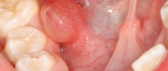
This growth occurs mainly on the inside of the cheek and looks like a bump on a stalk. The structure of the lump is soft, usually pink in color and looks like a wart, which has a rough surface and does not cause pain. Such a formation can be isolated, but sometimes it can cover a significant area. Localization sites include not only the cheeks, but also the tongue, gums, larynx and palate.
As a result of regular interaction of the papilloma with various irritants, especially when eating, it can become injured. When a wound occurs, bacteria accumulate in the affected area, causing inflammation. The human papillomavirus, which is the source of the above-mentioned formation, if a person’s immunity is strong, may not make itself known at all, but when the body’s defenses weaken, the likelihood of such an illness increases sharply.
For removal, surgical techniques or exposure to formations using liquid nitrogen are used. The use of chemicals is not recommended, as this can lead to the degeneration of the tumor into malignant forms.
Lipoma
In the case of a lipoma, it is often removed only in situations where they cause cosmetic discomfort to their owners along with a feeling of self-doubt. If an abscess occurs on the skin, it is treated only surgically, and the surrounding tissue is often also removed (if oncology is present).
It is worth noting that formations of various seals on the cheeks are not so uncommon. It is worth knowing that not all of them are considered dangerous, and some may even go away on their own. But if a person has a lump on his cheek, then you should not hesitate and need to see a specialist as soon as possible.
However, is it possible to get rid of a bump on the inside of the cheek at home? Let's talk about this in more detail.
Tumor forms
Mucosal cancer at the initial stage, as a rule, does not attract much attention. Often, a person only learns that he may have a malignant tumor when he visits the dentist.
The following forms of the disease are distinguished:
| Ulcerative |
| Papillary |
| Knotty |
The most common form of oncology of the mucous membranes is ulcerative. It consists of small ulcers that do not heal for a very long time and cause a lot of inconvenience to the patient. The main danger is that the cancerous node is localized in close proximity to blood vessels and spreads very quickly throughout the body. Consultation with an Israeli specialist
The papillary form is considered the easiest to treat. The growths hanging towards the gums can be effectively treated and do not allow the disease to go beyond the boundaries of the formation.
The ulcerative form of cheek cancer, the photo of which shows small compactions on the mucous membrane, is characterized by rapid growth. At the same time, the surface of the cheek itself may not change its color or become just a little whitish.
Home therapy
Treatment can be started at home in the case of a single manifestation, when the tendency for the lump to grow in size does not persist. Here are a few recipes that can help you cope with this disease on your own without seeking medical help:
- If the manifestation is completely fresh, then you should try using raw chicken egg white several times a day.
- You should take a small piece of cotton wool and soak it in castor oil, then apply it to the bump in the cheek area.
- The procedure should be repeated twice a day.
- It is necessary to cut a clove of garlic into slices, and then lubricate the growths with them three times.
- You need to prepare a tincture, for which you will need the peel along with walnut leaves. You should pour some alcohol into the dry ingredient and let it sit for two weeks, and then lubricate the formation with it once or twice daily.
Doctors' advice and recovery period
According to the advice of experts, after excision of the affected area (in the event that the internal bump on the cheek was removed surgically), the patient should be very careful and take his condition seriously. For example, you should completely review your diet, eliminating roughage from it.
At first, if a bandage has been applied to the patient, you must be extremely careful not to touch the seam; it is important to allow it to tighten efficiently and painlessly. In addition, in case of excision of areas on the cheeks, the risk of scar formation remains.
We must not forget that the operation itself is additional stress for the body; after it, it is very important to strengthen all your strength from the inside. To do this, you will need to eat right, in addition, good sleep is important along with giving up bad habits, and if possible, you need to avoid any worries and stress. You should not self-medicate; seeking medical help will help you quickly cope with the disease.
Primary diagnosis
In order to identify the problem at the initial stage of development, it is very important to conduct self-diagnosis. A careful examination of the mucous membrane will allow you to notice changes on the inner surface of the cheek in time and seek medical help.
Self-visual examination of the oral cavity should be carried out regularly
The specialist will conduct a visual examination and assessment of the condition of the oral cavity. The first examination is carried out by a dentist, who, if a malignant tumor is suspected, refers to a consultation with an oncologist.
The oncologist uses palpation to determine the degree of penetration of the disease into the soft tissues. Next, a cytological examination of the affected tissue is performed - a biopsy. A biopsy also allows you to determine the stage of the disease and the affected area.
Attention! Only based on the results of this study can a diagnosis of cheek cancer be made.
To clarify the diagnosis, a number of other studies are carried out.