How and why is baby teeth removed?
Milk teeth begin to appear in children even before reaching the first year of life and serve faithfully until the age of 6-7 years. When the time comes, the ligaments begin to gradually dissolve and the teeth begin to fall out. However, there are situations when it is advisable to seek the help of a pediatric dentist and speed up the process of natural change. However, the need may arise earlier if the condition of the teeth is very poor and treatment is impossible.
The procedure for removing a child's baby tooth is an exciting event for both parents and the child. Modern dentistry offers a lot of opportunities to ensure its painlessness, as well as the psychological comfort of the little patient.
Indications for the procedure
Indications for removal may be as follows:
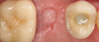
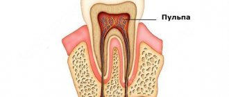
- deep caries, pulpitis, periodontitis, periostitis: the doctor will conduct therapeutic treatment, do everything possible for recovery, but if this is not possible, he may decide to remove it. Here it would be advisable to remind parents that baby teeth succumb to the destructive effects of bacteria much faster than adults. In this case, the baby may not show any concern, the process will proceed painlessly until the appearance of pulpitis and periostitis. To avoid the problem and not think about further prosthetics, you need to show your child to the dentist 3-4 times a year,
- cyst, root granuloma, fistula on the gum: the problem is that the inflammatory process on the root can most directly threaten the health and formation of the rudiments of the permanent element,
- loosening: if a baby tooth does not fall out on its own, but is very loose and interferes with proper nutrition, speaking and, in general, just living, then the procedure for its removal is unlikely to be avoided,
- keeping a baby tooth longer than expected: in this case, it may interfere with the eruption of the permanent unit and therefore must be extracted,
- serious injury: deep crack, root fracture as a result of a fall, mechanical impact,
- the presence of a supernumerary or additional tooth,
- the need to correct malocclusions using plates and other removable structures,
- eruption disorders: a milk element that has not fully erupted or is growing in the wrong direction will have to be removed if it threatens the health of the “neighbors”, the formation of the bite, causes pain and discomfort, or injures the mucous membrane.
Indications
Removal is indicated as a last resort when therapeutic treatment is not possible and there is a significant risk to the patient's health. The operation is performed when:
- purulent inflammatory process in periapical tissues;
- destruction of the root system;
- osteomyelitis;
- abscess;
- phlegmon;
- the impossibility of conservative treatment of periodontitis and cysts on the root tips;
- units located in the fracture gap;
- planned orthodontic treatment;
- high degree of tooth mobility due to periodontitis or periodontal disease;
- hyperdontia;
- the presence of impacted or dystopic structures.
Tooth extraction is prescribed as a last resort when therapeutic treatment is impossible.
Important! If the patient doubts the need for extraction, it is better to consult 2–3 dentists. Sometimes surgery is prescribed when the doctor does not have the necessary equipment for complex treatment.
Removal technique and steps
Removing baby teeth in children requires the professionalism of a dentist, since the procedure has some features associated not only with the structure of the child’s dental system, but also with a psychological component.
Important! The first tooth extraction procedure should be carried out for the baby in the most comfortable conditions. It is the absence of fear and emotional stress that will largely determine his future relationships with dentists and will become the key to oral health. It is better if your child is already familiar with the dental chair, and does not find himself in it for the first time in his life right before the procedure.
The process is carried out very carefully so as not to injure the rudiments of the permanent element. It is carried out in several stages:
- preparation: performing anesthesia - about which anesthesia is better and safer to choose for a child, read on,
- applying forceps to the crown: after fixing the crown part, the doctor carefully dislocates it from the jaw. Next, the tooth is removed from the socket,
- X-ray diagnostics: this measure is necessary in order to completely eliminate the entry of root fragments into the hole and reduce the risk of developing a further inflammatory process,
- treating the wound area with antiseptics.
“At the age of 4, my son had to have a tooth removed - it was badly damaged due to caries. The doctor applied topical anesthesia (applied it with gel), the child didn’t make a sound, didn’t even understand what happened. But the doctor was excellent, he spoke to my son before the procedures, everything went in a playful way. I advise everyone: choose special dentistry, for children, for these purposes, the dentists there have a psychological approach to children, and everything is beautifully decorated, like in cartoons. My opinion is this: it’s better to overpay for the service than to leave your child with a lifelong injury and fear of going to the doctors.”
Irene, fragment of a message from the site Otzovik.com
TECHNIQUES FOR TEETH REMOVAL WITH FORCES
The operation begins with the separation of the circular ligament from the neck of the tooth and the gum from the edge of the alveoli. It is best to do this with a smoothing iron or a narrow flat rasp. Careful separation of the circular ligament and gums facilitates the advancement of the cheeks of the forceps under the gums and prevents rupture of the mucous membrane during the intervention.
Tooth extraction consists of a number of techniques carried out in a certain sequence: 1) application of forceps; 2) moving the cheeks of the forceps under the gum; 3) closing the forceps (fixation);
4) tooth dislocation (luxation or rotation); 5) extraction of the tooth from the socket (traction). The success of the surgical intervention depends on the precise and consistent implementation of these techniques.
Forceps delivery. Having chosen the forceps according to the tooth to be removed, they are held in the hand according to one of the methods (see Fig. 31.1, II). Then the cheeks of the forceps are opened so that the crown of the tooth can fit between them.
One cheek of the forceps is applied to the tooth from the outer (vestibular) side, the other - from the inner (oral). The axis of the cheeks of the forceps must coincide with the axis of the tooth (Fig. 33, a). Incorrect application of forceps leads to a root fracture during tooth dislocation (Fig. 33, b).
Advancing the cheeks of the forceps. Pressing on the forceps, move the cheeks under the gum. On the upper jaw, this is done by moving the hand holding the forceps; on the lower jaw, by pressing on the locking area with the first finger of the left hand. Make sure that the axis of the cheeks of the forceps coincides with the axis of the tooth.
The cheeks of the forceps are advanced until
neck of the tooth, further advancement is hampered by the edge of the alveolus (Fig. 34, a). When the bone around the root of the tooth is reabsorbed, it is possible to move the forceps deeper into the upper part of the root. To obtain good fixation of the forceps when removing a tooth with a completely destroyed crown, apply the cheeks of the forceps to the edges of the alveolus (Fig. 34, b). During tooth extraction, these sections of bone break off.
Closing the forceps. It should be such that the tooth to be removed is firmly fixed in the forceps (Fig. 34, c). The tooth and forceps form a common lever arm. When moving the forceps, the tooth should also move at the same time. If the closure of the forceps is not strong enough, they move along the tooth or slip off it. If the fixation of the forceps is weak, the tooth cannot be removed. Compressing the handles of the forceps with excessive force leads to crushing of the crown or root, especially when their strength is reduced as a result of the carious process.
Tooth dislocation. When a tooth is dislocated, the periodontal fibers that connect its root to the walls of the socket are torn. At the same time, the walls of the hole shift or break. The tooth is dislocated in two ways: 1) by rocking (luxation) outward and inward, moving the forceps along with the tooth alternately to the vestibular and oral sides (Fig. 34, d); 2) rotation (rotation) around the tooth axis by 20-25°, first in one direction, then in the other direction (Fig. 34, e).
Luxation and rotation should be done gradually, without rough movements and jerks. Rocking the tooth should begin in the direction of least resistance, where the wall of the socket is thinner and, therefore, most pliable. The first swinging movement of the tooth is made weak, then the amplitude of movements is gradually increased. During dislocation, the forceps must be constantly closed and hold the tooth tightly.
In the upper jaw, the outer (vestibular) wall of the alveoli is thinner than the inner (palatal) wall. Therefore, the first dislocation
movement when removing teeth of the upper jaw should be made outward. The exception is the first large molar, in the area of which the outer wall of the alveolus thickens due to the zygomaticalveolar ridge.
On the lower jaw, the thickness of the compact layer of alveoli in the area of the incisors, canines and small molars on the outside is less than on the inside (lingual). In the area of large molars, the thickness of the compact layer of bone increases due to the bone ridge passing here (oblique line). The compact bone on the outside of the second and third molars is especially thick, while on the inside, on the contrary, it is thin. In the first large molar, the thickness of the alveolar walls on the outer and inner sides is the same. The first dislocation movement when removing these teeth is made to the internal (lingual) side, when removing the remaining teeth of the lower jaw - to the external (vestibular) side.
Rotational movements - rotation - are performed when removing teeth that have one root, resembling a cone in shape. Such teeth are the incisors and canines of the upper jaw and the separated roots of the first small molar and large molars of the upper jaw. However, it is not always possible to remove these teeth and roots using rotational movements alone. Then rotation has to be combined with luxury.
Extracting a tooth from the socket (traction). After the roots of the tooth have lost contact with the alveolus and the movement of the forceps together with the tooth has become free, they begin to remove the tooth from the socket and from the oral cavity. This is done smoothly, without jerking, often outward, up or down (depending on which jaw the tooth is being removed from) (Fig. 34, f).
If they begin to remove the tooth from the socket before losing connection with al-| veola and apply force at the same time, then at the moment of ligamentous rupture;
dental apparatus, forceps can forcefully hit the teeth of the opposite jaw and damage them or injure the mucous membrane.
The success of a tooth extraction operation does not depend on the physical strength of the doctor, but on the correct and consistent execution of all stages of the operation.
Position of the patient and the doctor during tooth extraction. The tooth extraction operation is performed in a dental chair. The outcome of the operation largely depends on the correct position of the patient and the doctor during this intervention.
Typically, tooth extraction is carried out in a sitting or semi-lying position. In patients experiencing severe fear and anxiety in connection with the intervention, as well as in persons with severe concurrent diseases, acute vascular insufficiency (fainting, collapse) often develops during surgery. To avoid these complications, before removing a tooth, it is advisable to recline the back of the chair to a horizontal position and raise the headrest.
The position of the patient in the chair should be such that one rational field is clearly visible and there are favorable
nik0^^ execution ^chom
all “Reems of the operation.
Headrests^™ durable Ф^^Р0^. did
not move during the operation.
When removing the teeth of the upper jaw, the patient sits in a chair with a slightly reclined back and headrest. Chair^lift^u^
s^st^1″06^ removed ^ was at the level of the shoulder
SS ?5 T4^ ra4nahod™ ^P3^ and in front of the ball fis. J5, a). This position of the patient and the doctor contributes to the successful completion of the operation.
When removing the teeth of the lower jaw, the chair is lowered, its back and headrest are moved so that the patient’s torso and head are in an upright position or the head is tilted forward, and the lower jaw is located at the level of the elbow .
the knee joint of the doctor's lowered hand. During the removal of small and large molars on the right side of the lower jaw, the doctor stands to the right and somewhat posterior to the patient (Fig. 35, b). When removing all the teeth on the left and the front teeth on the right, the doctor is positioned slightly ahead and to the right of the patient (Fig. 35, c).
Features of the use of anesthesia
I would like to dwell in more detail on the issue of pain relief during the removal of baby teeth. Anesthesia is not needed in all cases. So, when extracting a very loose temporary tooth, you don’t have to resort to it, since its root is already in a very mobile state, and the procedure will not cause pain. Sometimes, in order to carry out manipulations, the doctor uses superficial anesthesia - he pre-treats the area of influence with a special gel, which is most preferable for children psychologically.
Contraindications for removal
There is a list of relative contraindications that will require postponing the removal procedure until better times. Experts include diseases of the central nervous system, exacerbation of chronic diseases, fever, acute inflammatory phenomena in the oral cavity, infectious diseases (whooping cough, ARVI, influenza, mononucleosis, etc.), location of the root near a tumor or plexus of blood vessels. However, in emergency cases, removal can be carried out immediately - this is especially true for cases accompanied by a high risk of tissue infection and blood poisoning.
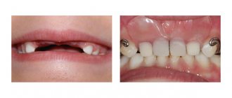
Consequences of early removal of primary occlusion elements
Early removal of primary teeth is associated with certain risks: the presence of free space in the row will lead to displacement of the “neighbors,” which may result in permanent dentition problems1.
Important! Early tooth extraction can be considered to be removal of a tooth more than a year before a permanent one appears in its place.
The absence of a chewing tooth can affect the characteristics of digestion, which is associated with insufficiently good chewing of food, the front one can cause speech defects and psychological discomfort in the child, difficulties in communication and low self-esteem. And do not forget that even at a young age, if one tooth is lost, the best option to solve the problem is temporary prosthetics.
Complications of tooth extraction surgery
Tooth extraction may be accompanied by general and local complications. Common complications include fainting, collapse, and anaphylactic shock. Local complications occur during surgery and in the postoperative period. During tooth extraction, a fracture of the crown or root, injury to an adjacent tooth or antagonist tooth, fracture of the lower jaw, separation of the tubercle of the upper jaw, damage to soft tissues, bleeding from the socket of the extracted tooth, perforation and pushing of the root into the maxillary sinus or nasal cavity occur.
These complications most often arise due to improper tooth extraction technique, and some of them are serious mistakes. The postoperative period may be accompanied by early and late alveolar pain, complicated by osteomyelitis of the alveolar process, and damage to the alveolar nerves. Early alveolar pain, caused by damage to the alveolar nerves, usually occurs after the anesthetic wears off.
The appearance of the alveoli and adjacent tissues, as well as the intensity of pain, correspond to the nature of the surgical intervention. In some cases, early socket pain is the result of significant trauma to the socket tissue. Such a complication can occur when the mouth of the hole does not correspond to highly branched roots.
Late socket pain (alveolitis) occurs 24–48 hours after tooth extraction as a result of excessive tissue damage and infection, as well as improper wound care. With alveolitis, there is no blood clot, the walls and bottom of the socket are covered with a gray mass consisting of fibrin, white and red blood cells in a state of decay, microbes and their decay products, as well as the remains of decomposing food. The tissues adjacent to the socket and regional lymph nodes are painful on palpation.
What to do after removal: rules for quick rehabilitation
To prevent the consequences of baby tooth extraction, it is important to strictly follow the doctor’s recommendations for further care:
- refrain from eating and drinking for 2 hours after visiting the dentist,
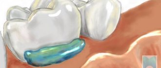
- do not give up oral hygiene, but carry it out extremely carefully on the first day: you cannot actively rinse your mouth; to clean and disinfect the wound surface, you just need to do oral baths with herbal decoctions or medications prescribed by a doctor,
- The first meals should be very careful if local anesthesia has taken place: some areas may have reduced sensitivity, it is easy to bite them and not notice the wounds. Instruct your child to chew food on the opposite side of the intervention. Make sure that it is soft, mushy, moderately warm, but in no case cold or hot, not salty or spicy,
- Limit your baby’s physical activity for a couple of days, stay away from beaches, bathhouses, swimming pools,
- Make sure that the child does not touch the socket with his fingers or put foreign objects in his mouth. Also explain to the baby that you can’t touch it with your tongue - this will make the healing go faster.
The parents' task is to assess the child's condition in the first days after the intervention. Thus, a high temperature in a child after removal or malaise is a reason for a second visit to the doctor.
Important! Fever 1-2 days after surgery is a possible symptom of inflammation and infection in the wound. It is definitely worth visiting a specialist if there is swelling after the removal of a baby tooth, which after 2-3 days not only does not subside, but becomes larger. All this may be accompanied by bleeding, pain, and the baby’s refusal to eat.
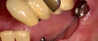
It is also very important to consult a pediatric orthodontist after the socket has healed. In order to avoid orthodontic complications after the removal of a baby tooth, you need to think about prosthetic options. This will prevent bite defects, ensure uniform distribution of the load on the remaining teeth and prevent complications in the form of their displacement, loosening, changes in the direction of growth and shape.
Removal technique
All situations can be divided into two groups:
- Extraction of teeth with the crown portion preserved.
- Removal of teeth with damaged crowns (roots).
When saving the crown, the following stages of the operation are distinguished:
- Peeling of the circular ligament of the tooth (for better grip on the tooth).
- Forceps delivery.
- Promotion.
- Fixation.
- Tooth dislocation.
- Extraction from the hole.
Read more about tooth extraction options in the corresponding article.
Removing roots has its own characteristics. Thus, the roots of the upper jaw are removed with bayonet-shaped forceps, sequentially carrying out the above steps. In some cases, elevators are used.
The roots of the lower jaw are often removed using straight elevators. At the same time, its concave surface is turned towards the tooth, and its convex surface is turned towards the wall of the alveoli. Moving the elevator between the tooth and the bone, they begin to rotate the handle of the instrument, thereby dislocating the tooth (using the lever method).
Read a detailed article about methods for removing tooth roots.
Also look at techniques for removing individual teeth:
- Dairy
- Impacted
- Dystopic
- Supernumerary
- Wisdom teeth
Can I solve the problem myself?
If the tooth is sick, then, of course, you need to see a doctor. There can be no talk of any independent treatment. But if a tooth becomes loose, many parents do not rush to the dentist, trying to remove it themselves. But it is undesirable to do this, because unlike a doctor, adults will not be able to accurately foresee situations and vouch for the safety of the child’s health.
However, if the period for the loss of the deciduous unit of the dentition has come, it is wobbly and literally “hanging by a thread,” you can try to remove it yourself. The main thing is the child’s positive attitude and the absence of concomitant oral diseases. To do this, you can give the baby, for example, an apple or a carrot - perhaps, during the process of mechanical action, the baby tooth will fall out on its own, unable to withstand the load. Care after a home removal is similar to what your doctor recommends after an in-office procedure.
Video on the topic
1 Alshimbaeva A. Causes and consequences of early removal of primary teeth, 2011.