What does enamel consist of?
Tooth enamel is the hardest tissue of the human body. It is low in water (2-3%) and organic matter (1-2%). The main share (95-97%) falls on minerals - mainly calcium phosphate in the form of hydroxyapatite crystals, as well as magnesium, fluorine, carbon and some other elements.
Beneath the outer covering is a more porous layer of dentin, covering the pulp chamber, which is filled with loose connective tissue containing blood vessels and nerve fibers (pulp).
The thickness of the enamel layer can reach 2 mm in the chewing plane of the tooth and thins to 0.01 mm in the cervical zone.
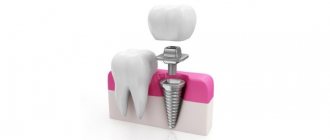
The structure of human teeth
Crown
Crown (lat. corona dentis) is the part of the tooth protruding above the gum. The crown is covered with enamel - hard tissue, 95% consisting of inorganic substances and subject to the most powerful mechanical stress.
There is a cavity in the crown - dentin (hard tissue 2-6 mm thick) comes closer to the surface, then pulp, filling both part of the crown and the root part of the tooth. The pulp contains blood vessels and nerves. Cleaning and removal of dental plaque is carried out specifically from the crowns of teeth.
Tooth neck
Neck (lat. collum dentis) is the part of the tooth between the crown and root, covered by the gum.
Roots
Root (lat. radix dentis) is the part of the tooth located in the dental alveolus.
Fissure
On the chewing surface of the back teeth, between the cusps there are grooves and grooves - fissures. The fissures can be narrow and very deep. The relief of the fissures is individual for each of us, but dental plaque gets stuck in the fissures of everyone.
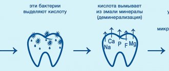
It is almost impossible to clean the fissures with a toothbrush. Bacteria in the oral cavity, processing plaque, form acid, which dissolves tissue, forming caries. Even good oral hygiene is sometimes not enough. In this regard, fissure sealing has been successfully used throughout the world for 20 years.
Enamel
Tooth enamel (or simply enamel, Latin enamelum) is the outer protective shell of the coronal part.
Enamel is the hardest tissue in the human body, which is explained by the high content of inorganic substances - up to 97%. There is less water in tooth enamel than in other organs, 2-3%.
Hardness reaches 397.6 kg/mm² (250-800 Vickers). The thickness of the enamel layer differs in different areas of the crown part and can reach 2.0 mm, and disappears at the neck of the tooth.
Proper care of tooth enamel is one of the key aspects of human personal hygiene.
Dentine
Dentin (dentinum, LNH; lat. dens, dentis - tooth) is the hard tissue of the tooth, constituting its main part. The coronal part is covered with enamel, the root part of the dentin is covered with cement. Consists of 72% inorganic substances and 28% organic substances. Consists mainly of hydroxyapatite (70% by weight), organic material (20%) and water (10%), permeated with dentinal tubules and collagen fibers.
Serves as the foundation of the tooth and supports tooth enamel. The thickness of the dentin layer ranges from 2 to 6 mm. Dentin hardness reaches 58.9 kgf/mm².
There are peripulpal (internal) and mantle (external) dentin. In peripulpal dentin, collagen fibers are located predominantly condensally and are called Ebner fibers. In mantle dentin, collagen fibers are arranged radially and are called Korff fibers.
Dentin is divided into primary, secondary (replacement) and tertiary (irregular).
Primary dentin is formed during the development of the tooth, before its eruption. Secondary (replacement) dentin is formed throughout a person’s life. It differs from the primary by a slower pace of development, a less systemic arrangement of dentinal tubules, a larger number of erythroglobular spaces, a larger amount of organic substances, higher permeability and less mineralization. Tertiary dentin (irregular) is formed during tooth trauma, preparation, caries and other pathological processes, as a response to external irritation.
Dental pulp
Pulp (lat. pulpis dentis) is loose fibrous connective tissue that fills the tooth cavity, with a large number of nerve endings, blood and lymphatic vessels.
Along the periphery of the pulp, odontoblasts are located in several layers, the processes of which are located in the dentinal tubules throughout the entire thickness of the dentin, performing a trophic function. The processes of odontoblasts include nerve formations that conduct pain sensations during mechanical, physical and chemical influences on dentin.
Blood circulation and innervation of the pulp are carried out thanks to dental arterioles and venules, the nerve branches of the corresponding arteries and nerves of the jaws. Penetrating into the dental cavity through the apical opening of the root canal, the neurovascular bundle breaks up into smaller branches of capillaries and nerves.
The pulp helps stimulate regenerative processes, which manifest themselves in the formation of replacement dentin during the carious process. In addition, the pulp is a biological barrier that prevents the penetration of microorganisms from the carious cavity through the root canal beyond the tooth into the periodontium.
The nerve formations of the pulp regulate the nutrition of the tooth, as well as the perception of various irritations, including pain. The narrow apical opening and the abundance of vessels and nerve formations contribute to the rapid increase in inflammatory edema in acute pulpitis and compression of the nerve formations by the edema, which causes severe pain.
Tooth cavity
(lat. cavitas dentis) The space inside, formed from the cavity of the crown and root canals. This cavity is filled with pulp.
Cavity of the tooth crown
(lat. cavitas coronae) Part of the tooth cavity, located under the crown and repeating its internal contours.
Tooth root canals
Root canal (lat. canalis radicis dentis) is an anatomical space inside the root of a tooth. This natural space within the coronal part of the tooth consists of a pulp chamber, which is connected by one or more main canals, as well as more complex anatomical branches that can connect the root canals to each other or to the surface of the tooth root.
Nerves
(lat. nervae) Neuron processes passing through the apex of the tooth and filling its pulp. The nerves regulate the nutrition of the tooth and conduct pain impulses.
Arteries
(lat. arteriae) Blood vessels through which blood from the heart flows to all other organs, in this case to the pulp. Arteries nourish dental tissues.
Vienna
(lat. venae) Blood vessels through which blood returns from organs back to the heart. The veins enter the canals and penetrate the pulp.
Cement
Cement (lat. - cementum) is a specific bone tissue covering the root and neck of the tooth. Serves to firmly secure the tooth in the bone alveolus. Cement consists of 68-70% inorganic components and 30-32% organic substances.
Cementum is divided into acellular (primary) and cellular (secondary).
Primary cement is adjacent to the dentin and covers the lateral surfaces of the root.
Secondary cement covers the apical third of the root and the bifurcation area of multi-rooted teeth.
Root tips
(lat. apex radicis dentis) The lowest points of the teeth, located on their roots. At the tops there are openings through which nerve and vascular fibers pass.
Apical foramina
(lat. foramen apices dentis) Places of entry of vascular and nerve plexuses into the dental canals. The apical foramina are located at the apex of the tooth roots.
Alveolus (alveolar socket)
(alveolar socket) (lat. alveolus dentalis) A notch in the jaw bone into which the roots enter. The walls of the alveoli form strong bone plates impregnated with mineral salts and organic substances.
Alveolar neurovascular bundle
(lat. aa., vv. et nn alveolares) A plexus of blood vessels and nerve processes passing under the alveolus of the tooth. The alveolar neurovascular bundle is enclosed in an elastic tube.
Periodontium
Periodontium (lat. Periodontium) is a complex of tissues located in the slit-like space between the cementum of the tooth root and the alveolar plate. Its average width is 0.20-0.25 mm. The narrowest section of the periodontium is located in the middle part of the tooth root, and in the apical and marginal sections its width is slightly greater.
The development of periodontal tissue is closely related to embryogenesis and teething. The process begins in parallel with the formation of the root. The growth of periodontal fibers occurs both from the side of the root cement and from the side of the alveolar bone, towards each other. From the very beginning of their development, the fibers have an oblique course and are located at an angle to the tissues of the alveoli and cementum. The final development of the periodontal complex occurs after tooth eruption. At the same time, the periodontal tissues themselves are involved in this process.
It should be noted that, despite the mesodermal origin of the constituent components of the periodontium, the ectodermal epithelial root sheath takes part in its normal formation.
Gingival grooves
(lat. sulcus gingivalis) Crevices that form where the crown of the tooth adheres to the gums. The gingival grooves run along the line between the free and attached parts of the gum.
Gum
Gums (lat. Gingiva) is a mucous membrane that covers the alveolar process of the upper jaw and the alveolar part of the lower jaw and covers the teeth in the cervical area. From a clinical and physiological point of view, the gums are divided into interdental (gingival) papilla, marginal gum or gingival margin (free part), alveolar gum (attached part), mobile gum.
Histologically, the gum consists of stratified squamous epithelium and the lamina propria. There are oral epithelium, junctional epithelium, and sulcal epithelium. The epithelium of the interdental papillae and attached gingiva is thicker and can become keratinized. In this layer, there are basal, spinous, granular and stratum corneum. The basal layer consists of cylindrical cells, the spinous layer consists of polygonal cells, the granular layer consists of flattened cells, and the stratum corneum is represented by several rows of completely keratinized and nucleated cells that are constantly exfoliated.
Mucous papillae
(lat. papilla gingivalis) Fragments of gums located at their elevation in the area between adjacent teeth. The gingival papillae are in contact with the surface of the dental crowns.
Jaws
(lat. maxilla - upper jaw, mandibula - lower jaw) Bony structures that are the basis of the face and the largest bones of the skull. The jaws form the mouth opening and determine the shape of the face.
Source: CreateSmile.ru
When is it time to strengthen
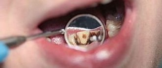
It is necessary to carefully monitor the condition of your teeth. If you experience frequent inflammation of the mucous membranes of the oral cavity, think about how to strengthen your gums. Otherwise, there is a risk of developing cervical caries, since the thickness of the enamel at the point of contact between the gum and the crown is minimal.
It’s time to strengthen teeth enamel if one or more signs appear.
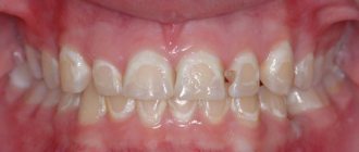
- The color has changed. As the outer covering wears away, the yellowish layer of dentin begins to show through.
- The shape has changed. Wear and tear can cause teeth to become flatter, shorter, and rounder, making it more difficult to bite and chew food.
- A white chalky spot has appeared - the initial stage of caries, at which it is easiest to deal with it. The destruction of the layer is just beginning, so its surface remains even and smooth.
- Increased sensitivity (hyperesthesia) has appeared - a painful reaction when brushing with a toothbrush, eating sour, sweet, cold or hot foods.
- The teeth began to crumble.
The structure of human teeth
Crown
Crown (lat. corona dentis) is the part of the tooth protruding above the gum. The crown is covered with enamel - hard tissue, 95% consisting of inorganic substances and subject to the most powerful mechanical stress.
There is a cavity in the crown - dentin (hard tissue 2-6 mm thick) comes closer to the surface, then pulp, filling both part of the crown and the root part of the tooth. The pulp contains blood vessels and nerves. Cleaning and removal of dental plaque is carried out specifically from the crowns of teeth.
Tooth neck
Neck (lat. collum dentis) is the part of the tooth between the crown and root, covered by the gum.
Roots
Root (lat. radix dentis) is the part of the tooth located in the dental alveolus.
Fissure
On the chewing surface of the back teeth, between the cusps there are grooves and grooves - fissures. The fissures can be narrow and very deep. The relief of the fissures is individual for each of us, but dental plaque gets stuck in the fissures of everyone.
It is almost impossible to clean the fissures with a toothbrush. Bacteria in the oral cavity, processing plaque, form acid, which dissolves tissue, forming caries. Even good oral hygiene is sometimes not enough. In this regard, fissure sealing has been successfully used throughout the world for 20 years.
Enamel
Tooth enamel (or simply enamel, Latin enamelum) is the outer protective shell of the coronal part.
Enamel is the hardest tissue in the human body, which is explained by the high content of inorganic substances - up to 97%. There is less water in tooth enamel than in other organs, 2-3%.
Hardness reaches 397.6 kg/mm² (250-800 Vickers). The thickness of the enamel layer differs in different areas of the crown part and can reach 2.0 mm, and disappears at the neck of the tooth.
Proper care of tooth enamel is one of the key aspects of human personal hygiene.
Dentine
Dentin (dentinum, LNH; lat. dens, dentis - tooth) is the hard tissue of the tooth, constituting its main part. The coronal part is covered with enamel, the root part of the dentin is covered with cement. Consists of 72% inorganic substances and 28% organic substances. Consists mainly of hydroxyapatite (70% by weight), organic material (20%) and water (10%), permeated with dentinal tubules and collagen fibers.
Serves as the foundation of the tooth and supports tooth enamel. The thickness of the dentin layer ranges from 2 to 6 mm. Dentin hardness reaches 58.9 kgf/mm².
There are peripulpal (internal) and mantle (external) dentin. In peripulpal dentin, collagen fibers are located predominantly condensally and are called Ebner fibers. In mantle dentin, collagen fibers are arranged radially and are called Korff fibers.
Dentin is divided into primary, secondary (replacement) and tertiary (irregular).
Primary dentin is formed during the development of the tooth, before its eruption. Secondary (replacement) dentin is formed throughout a person’s life. It differs from the primary by a slower pace of development, a less systemic arrangement of dentinal tubules, a larger number of erythroglobular spaces, a larger amount of organic substances, higher permeability and less mineralization. Tertiary dentin (irregular) is formed during tooth trauma, preparation, caries and other pathological processes, as a response to external irritation.
Dental pulp
Pulp (lat. pulpis dentis) is loose fibrous connective tissue that fills the tooth cavity, with a large number of nerve endings, blood and lymphatic vessels.
Along the periphery of the pulp, odontoblasts are located in several layers, the processes of which are located in the dentinal tubules throughout the entire thickness of the dentin, performing a trophic function. The processes of odontoblasts include nerve formations that conduct pain sensations during mechanical, physical and chemical influences on dentin.
Blood circulation and innervation of the pulp are carried out thanks to dental arterioles and venules, the nerve branches of the corresponding arteries and nerves of the jaws. Penetrating into the dental cavity through the apical opening of the root canal, the neurovascular bundle breaks up into smaller branches of capillaries and nerves.
The pulp helps stimulate regenerative processes, which manifest themselves in the formation of replacement dentin during the carious process. In addition, the pulp is a biological barrier that prevents the penetration of microorganisms from the carious cavity through the root canal beyond the tooth into the periodontium.
The nerve formations of the pulp regulate the nutrition of the tooth, as well as the perception of various irritations, including pain. The narrow apical opening and the abundance of vessels and nerve formations contribute to the rapid increase in inflammatory edema in acute pulpitis and compression of the nerve formations by the edema, which causes severe pain.
Tooth cavity
(lat. cavitas dentis) The space inside, formed from the cavity of the crown and root canals. This cavity is filled with pulp.
Cavity of the tooth crown
(lat. cavitas coronae) Part of the tooth cavity, located under the crown and repeating its internal contours.
Tooth root canals
Root canal (lat. canalis radicis dentis) is an anatomical space inside the root of a tooth. This natural space within the coronal part of the tooth consists of a pulp chamber, which is connected by one or more main canals, as well as more complex anatomical branches that can connect the root canals to each other or to the surface of the tooth root.
Nerves
(lat. nervae) Neuron processes passing through the apex of the tooth and filling its pulp. The nerves regulate the nutrition of the tooth and conduct pain impulses.
Arteries
(lat. arteriae) Blood vessels through which blood from the heart flows to all other organs, in this case to the pulp. Arteries nourish dental tissues.
Vienna
(lat. venae) Blood vessels through which blood returns from organs back to the heart. The veins enter the canals and penetrate the pulp.
Cement
Cement (lat. - cementum) is a specific bone tissue covering the root and neck of the tooth. Serves to firmly secure the tooth in the bone alveolus. Cement consists of 68-70% inorganic components and 30-32% organic substances.
Cementum is divided into acellular (primary) and cellular (secondary).
Primary cement is adjacent to the dentin and covers the lateral surfaces of the root.
Secondary cement covers the apical third of the root and the bifurcation area of multi-rooted teeth.
Root tips
(lat. apex radicis dentis) The lowest points of the teeth, located on their roots. At the tops there are openings through which nerve and vascular fibers pass.
Apical foramina
(lat. foramen apices dentis) Places of entry of vascular and nerve plexuses into the dental canals. The apical foramina are located at the apex of the tooth roots.
Alveolus (alveolar socket)
(alveolar socket) (lat. alveolus dentalis) A notch in the jaw bone into which the roots enter. The walls of the alveoli form strong bone plates impregnated with mineral salts and organic substances.
Alveolar neurovascular bundle
(lat. aa., vv. et nn alveolares) A plexus of blood vessels and nerve processes passing under the alveolus of the tooth. The alveolar neurovascular bundle is enclosed in an elastic tube.
Periodontium
Periodontium (lat. Periodontium) is a complex of tissues located in the slit-like space between the cementum of the tooth root and the alveolar plate. Its average width is 0.20-0.25 mm. The narrowest section of the periodontium is located in the middle part of the tooth root, and in the apical and marginal sections its width is slightly greater.
The development of periodontal tissue is closely related to embryogenesis and teething. The process begins in parallel with the formation of the root. The growth of periodontal fibers occurs both from the side of the root cement and from the side of the alveolar bone, towards each other. From the very beginning of their development, the fibers have an oblique course and are located at an angle to the tissues of the alveoli and cementum. The final development of the periodontal complex occurs after tooth eruption. At the same time, the periodontal tissues themselves are involved in this process.
It should be noted that, despite the mesodermal origin of the constituent components of the periodontium, the ectodermal epithelial root sheath takes part in its normal formation.
Gingival grooves
(lat. sulcus gingivalis) Crevices that form where the crown of the tooth adheres to the gums. The gingival grooves run along the line between the free and attached parts of the gum.
Gum
Gums (lat. Gingiva) is a mucous membrane that covers the alveolar process of the upper jaw and the alveolar part of the lower jaw and covers the teeth in the cervical area. From a clinical and physiological point of view, the gums are divided into interdental (gingival) papilla, marginal gum or gingival margin (free part), alveolar gum (attached part), mobile gum.
Histologically, the gum consists of stratified squamous epithelium and the lamina propria. There are oral epithelium, junctional epithelium, and sulcal epithelium. The epithelium of the interdental papillae and attached gingiva is thicker and can become keratinized. In this layer, there are basal, spinous, granular and stratum corneum. The basal layer consists of cylindrical cells, the spinous layer consists of polygonal cells, the granular layer consists of flattened cells, and the stratum corneum is represented by several rows of completely keratinized and nucleated cells that are constantly exfoliated.
Mucous papillae
(lat. papilla gingivalis) Fragments of gums located at their elevation in the area between adjacent teeth. The gingival papillae are in contact with the surface of the dental crowns.
Jaws
(lat. maxilla - upper jaw, mandibula - lower jaw) Bony structures that are the basis of the face and the largest bones of the skull. The jaws form the mouth opening and determine the shape of the face.
Source: CreateSmile.ru
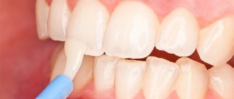
How to strengthen tooth enamel
At the first sign of decay, visit the dental clinic. The doctor will conduct an examination and determine the reason why your teeth are crumbling. He will advise you on how to treat tooth sensitivity.
In dental practice, there are several methods aimed at strengthening enamel and protecting against demineralization.
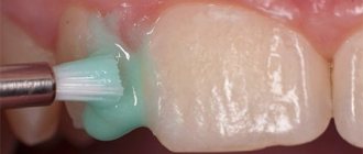
The most common is fluoridation. The effectiveness of this procedure has been confirmed by practice. It involves applying a special fluoride-containing composition to the surface of the teeth. Fluoride penetrates deep into the enamel and promotes better absorption of phosphorus and calcium.
As a result of fluoridation:
- the permeability of the top layer decreases;
- the smallest cracks are sealed;
- the structure of the outer shell becomes smooth and uniform;
- a natural healthy shine appears;
- resistance to pathogenic microflora and acidic environment increases;
- caries is eliminated at the stain stage;
- tooth sensitivity decreases.
Cosmetic defects in the outer covering can be hidden by installing veneers and lumineers or polishing teeth.
Prevention of tooth enamel destruction
Simple recommendations will help slow down the destruction of the enamel layer and keep teeth healthy.
- Visit your dentist at least once a year. Even in the absence of discomfort in the oral cavity, the doctor will see the initial stages of the disease and begin timely treatment.
- Brush your teeth at least twice a day, morning and evening, and use dental floss to remove food debris from your mouth.
- After eating, rinse your mouth with clean water and take your medication with plenty of water.
- Eat foods containing sugar less often. Each consumption of sugar leads to the growth of bacteria in the mouth.
- Limit your consumption of sugary sodas and juices - some of the acids in them are more corrosive than battery acid.
Do not forget that oral care should be carried out regularly and comprehensively throughout your life. Following one rule will only slightly reduce the risk of developing pathology. Oral care should become a habit and become a lifestyle.