Computed tomography is a modern and accurate examination method that helps specialists working in the dental field, as well as in the field of maxillofacial surgery. This method, despite being completely non-invasive, is completely safe, since the X-ray radiation used in the process is supplied in very small quantities. Unfortunately, for certain groups of people even this can be dangerous, but contraindications will be discussed a little later. In this material, let's give answers to questions about where to get a CT scan of the jaw, what this examination shows, as well as how it is carried out.
Ready images are given to patients in various planes, which is very important for proper treatment. It is also worth noting that special equipment helps to implement the technology for constructing a three-dimensional model of a given area of the face. Computed tomography of the jaw space will help you take the most accurate picture of the jaw, and the specialist will even be able to obtain data on the height and width of all elements. It will even be possible to locate the mandibular canal, as well as determine the condition of the maxillary sinus. The method will allow a specialist, using one computer tomogram, to identify the most dangerous diseases, such as tumors, as well as inflammatory processes, cysts and pathologies in the early stages. It is worth mentioning the possibility of controlling implantation.
Computed tomography of the upper and lower jaw is performed for a child in the same way as for adults.
Indications for dental CT
CT (computed tomography of teeth) has its own indications for use. A 3D photograph of teeth is not prescribed for the treatment of ordinary dental caries or pulpitis; here it will be enough to take a regular x-ray. CT scanning is performed before treatment of more complex dental diseases and before surgical operations.
Let's take a closer look at the full range of indications for CT scanning of the jaw:
- 1. A CT scan is required before surgical operations on the jaw - bone grafting, implantation.
- 2. CT scan is prescribed before prosthetics and allows you to correctly select dentures in the most difficult clinical situations.
- 3. A 3D photograph of the teeth must be taken before orthodontic treatment, which involves wearing braces for a long time. In this case, CT data will allow you to assess the condition of the tissues of the mouth and teeth, choose the right braces and eliminate possible contraindications to their use.
- 4. A CT scan is also performed in case of any injuries to the jaw, or when a person experiences strange pain in the jaw, the cause of which cannot be determined during a visual examination.
- 5. Computed tomography is prescribed if the presence of neoplasms in the soft and hard tissues of the oral cavity is suspected.
- 6. CT can be used to monitor the quality of root canal treatment.
The need for a CT scan to obtain a 3D image of the teeth is determined by the doctor during the patient’s first appointment.
Carrying out tomography
Dental tomography equipment comes in different sizes and shapes. During startup, it rotates clockwise around the patient's head, while it takes pictures. The study lasts 1 – 2 minutes and during this time the device manages to take more than 500 pictures. After this, the computer converts the images into digital format. The scanning takes place very quickly and does not cause any discomfort to the patient. A CT scan can be performed lying down, sitting or standing.
Computed tomography of teeth is carried out according to the following scheme :
- The tomograph table is moved apart and the patient lies down on it. His head is secured with soft straps, and a protective vest is put on his body. If the procedure is performed while sitting, the patient places his head on a special stand of the device, and if standing, he presses his face against the equipment stand.
- If there is a need to improve the quality of the study, a contrast agent is administered intravenously to the patient. In this case, soft tissues and blood vessels are visualized much more clearly.
- Once the patient is ready, the device is turned on. The table begins to move smoothly, as a result of which the head ends up in the ring of the device. In this case, the noise of the tomograph occurs; it is quite natural and you should not be afraid of it.
- When rotating, the tomograph takes a series of layer-by-layer images of the required area in various projections. The device's monitor records these images.
The patient may experience discomfort due to the fact that he has to remain motionless for several minutes. In addition, the use of a contrast agent may cause discomfort. In this case, the patient feels nauseous, has slight dizziness or an unpleasant bite in the mouth. If his health worsens, he must immediately notify the doctor using a special communication system.
Is CT scanning of teeth harmful?
During this procedure, a whole series of layer-by-layer images is taken. In this case, the patient receives a small dose of radiation. The tomograph tube contains emitters and scanners. The larger the surface area to be examined and the number of images, the higher the dose of radioactive radiation. For example, the radiation dose for examining the pelvis, chest or abdominal cavity is 6 – 11 m3v. If the radiation dose is more than 50 m3v, this can provoke the formation of cancerous tumors.
Dental tomography is considered the most gentle study in terms of obtaining a radioactive dose, since it is only 1 - 3 m3v. Due to its minimal harmfulness and high efficiency, tomography is beginning to increasingly replace x-rays.
Thus, dental computed tomography is a very effective and completely safe diagnostic method that allows us to identify various dental diseases. This method is becoming increasingly popular and has virtually no contraindications.
CT scan of teeth before dental implantation
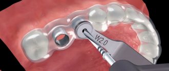
A dental CT scan must be done before dental implantation. Based on the computed tomography data, the operation will be planned in detail; based on the 3D image of the teeth, implants and the method of their implantation will be selected; contraindications to this method of dental restoration will be excluded.
TAKE NOTE: The accuracy of diagnosis is of fundamental importance for the quality of implantation. The risks of complications after implantation, the healing time and service life of installed dental implants will depend on how thoroughly the diagnostic measures are carried out. Implantation can only be planned using a CT scan of the jaw - all other diagnostic studies do not provide the necessary information before such a serious surgical intervention.
Using CT data, it is possible to plan implantation in detail and give a prognosis based on its results. Computed tomography will not only allow you to correctly select dental implants and determine the area for their installation: computer modeling is used to demonstrate the future results of implantation and is a good motivation for people who are worried about how their smile and teeth will look after surgery.
Also, CT scanning of the jaw serves as the basis for creating special surgical templates used in dental implantation. Implantation templates help install implants into the bone without causing serious injury to the soft and hard tissues of the jaw, and this has a positive effect on the recovery time after surgery.
A CT scan before implantation is also important because it allows the doctor to obtain the most detailed data on the condition of the bone tissue, assess its height, volume and density. If a CT scan of the jaw shows that the bone is loose or has a height that is not enough for high-quality implantation, it will be necessary to undergo bone grafting surgery before implantation.
Contraindications
An absolute contraindication to performing a dental CT scan is pregnancy at any stage. For breastfeeding women, computed tomography can be done only if after such a procedure it is possible not to breastfeed the baby for 24 hours. Tomography is not prescribed for preschool children.
Since a contrast agent is often used during the procedure, in this case it is contraindicated for:
- renal or liver failure;
- diabetes mellitus;
- allergies to iodine.
For neuroses and claustrophobia, tomography may be accompanied by the use of light tranquilizers.
Types of equipment for CT jaw
Various devices are used for CT scanning of teeth, and therefore, when planning to undergo a study in any clinic in St. Petersburg or any other city, you must find out which specific device is used in a particular dentistry. Why is this so important? It’s simple: not all models of dental CT devices have the required accuracy, and in addition, there are devices that, when taking pictures, will have too much radiation exposure on your body.
Below we will look at the most commonly used CT devices in clinics and indicate their pros and cons.
Types of CT devices
In dentistries in St. Petersburg, the following types of devices for CT scans of teeth and jaws are most often found:
1. Units for step-by-step dental CT. They appeared and are used for diagnostics quite a long time ago and are considered obsolete. The use of devices of this type does not allow obtaining accurate data on the condition of bone tissue; the quality of the images is quite low. But the main disadvantage of step-by-step CT machines is that when using them, the body is exposed to strong and harmful radiation.
2. Spiral devices for CT. The level of safety when using them will be higher, but still the use of such units for CT scans of teeth is undesirable, since the radiation dose remains high. The quality of images of spiral devices for CT of the jaw also leaves much to be desired, since they do not allow one to see and evaluate the structure of the bone tissue.
3. Cone beam tomographs. These devices are considered the most modern, accurate and informative. A CT scan performed with a cone beam device will allow you to see clear images and evaluate:
- The condition of the root canals of the tooth, the tooth itself and surrounding tissues;
- Condition of the bone and soft tissues of the jaw.
Based on CT data performed using cone-beam tomographs, it is possible to successfully plan prosthetics, implantation, bite correction with braces, and assess the quality of endodontic treatment. In this case, the radiation dose during CT will be minimal, no more than 90 µV, which allows, if necessary and with complete safety for the patient, to conduct a repeat study.
TAKE NOTE: The cost of a CT scan in St. Petersburg to a certain extent depends on the device with which the study will be carried out. But we categorically do not advise you to save money on a CT scan of the jaw: CT scanning on outdated devices is not informative enough and, moreover, is harmful to your health.
In our dentistry in St. Petersburg - “New line Dent”, modern and high-precision devices are used for CT scanning of the teeth and jaw - GALILEOS Sirona (Germany) and Vatech (South Korea). The diagnostic data that we obtain using these high-tech and modern devices helps us successfully plan and carry out dental treatment of any complexity!
Where is it used in dentistry?
When contacting a specialist, a visual examination of the teeth and oral cavity is not enough. It is not always possible to understand and study the problem in detail in the simplest way of our ancestors, so the specialist prescribes additional diagnostics for the patient.
It is impossible to see the condition of a tooth from the inside by visually assessing it. Previously, dental diseases were diagnosed using X-rays, however, thanks to progress, the CT method has become widespread.
Using the CT method, it is possible to obtain a three-dimensional (3D) image of all parts of the dentofacial system. This innovation allows dentists to timely identify pathology, make a diagnosis and prescribe effective treatment.
Modern equipment for CT examination makes it possible to identify and diagnose the inflammatory process in the first stages of development, as well as to recognize the nature of jaw injuries and diseases of certain dental units.
Thanks to the three-dimensional image produced by computed tomography, the dentist can carefully examine all the smallest details of the disease and correctly make a diagnosis.
Based on the results obtained, a specialist can find out the anatomical features of an individual person in 2-3 minutes:
- number and location of bone organs of the oral cavity;
- the characteristic shape of the dental unit and its structure;
- feature of the root canals;
- internal and external anatomy of the movable jaw canal.
Also, a universal device is used in the presence of a number of problems and to improve the quality of individual events:
- injury;
- congenital malformations of the jaws and bone organs, disorders;
- localization of infection in a specific organ of the oral cavity, acquiring a chronic form;
- planned surgical procedure, titanium root implantation;
- complex tooth extraction;
- prosthetics of bone organs;
- diagnostic manipulations to prescribe effective treatment.
The CT machine has found wide application in various fields of dentistry and has practically replaced x-rays.
Orthodontics
The diagnostic device in CT is a dental tomograph. In the field of orthodontics, the device is used before and after surgery, but no more than 6 times in total.
Examination using a standing tomograph has proven itself well among young patients.
The child is placed along a vertical stand. A special stand is placed under the lower part of the face. A special apron is worn as protection against radiation.
To correctly direct the beam, a laser is used. It regulates the source of electromagnetic waves before gamma radiation. The shooting lasts half a minute. During this interval, the patient should not breathe or swallow saliva.
The clarity of the image will depend on the number of scanned sections and the quality of their resolution.
This method is most often used in the following cases:
- analysis of the causes of the problem;
- establishing diagnosis;
- preventive research;
- treatment of all kinds of deviations from the normal condition of teeth and other bone tissues of the oral cavity.
Indications for the operation of resection of the apex of the tooth root and the procedure for its implementation. Here we will discuss contraindications to the use of the method of one-stage dental implantation.
Follow the link https://dr-zubov.ru/lechenie/desny/periodontit/vse-o-kovarstve-i-slozhnosti-zabolevaniya.html if you are interested in the symptoms of chronic periodontitis.
Implantation
It is very difficult to reliably install an implant without a CT scan. The specialist will have to perform complex and painstaking manipulations, which in most cases do not give positive results due to the lack of information about the anatomy of a particular patient.
That is why in the USA the survival rate of artificial roots is much higher than in our country. The essence of the reason lies in the lack of CT equipment in a number of dental clinics in the Russian Federation.
The three-dimensional image allows you to select a high-quality implant and fastening elements and guarantees a successful operation.
Watch the video about computed tomography when planning dental implantation.
Surgery
In the surgical field, CT is an indispensable study. With its help, you can reliably determine the characteristics of the damage.
The method is used to carry out the following operations:
- implantation into the jawbone;
- osteoplasty;
- diagnostics;
- analysis of tumor formations.
Treatment
The task of endodontic therapy is to assess the condition of the problematic dental unit. The specialist must perform canal therapy and filling in such a way that the restored tooth no longer causes trouble for the patient.
Thanks to a digital computed tomograph, a specialist can analyze a high-definition image. The thickness and height of the hard tissues of the dental unit are determined accurately.
The examination allows you to determine the condition of not only the oral cavity and bone organs, but also the paired paranasal sinuses.
In therapeutic practice, CT scanning of bone organs is carried out to control the correct and uniform consistency of the filling material, as well as its uniform distribution in the carious cavity and canals.
This method allows you to plan treatment and fully control the results of all its stages.
The principle of the formation of tartar and the reasons that are the source of its occurrence. Come here to read the instructions for use of Stomatofit.
Let's look at this address https://dr-zubov.ru/lechenie/zuby/karies-z/etiologiya-i-metodiki-ustraneniya-prisheechnogo.html tactics for treating cervical caries.
Take a 3D CT scan of teeth: what advantages does the technique give to patients?
Compared to other diagnostic methods, CT of the jaw has a number of advantages, including:
- 1. Speed of implementation. A CT scan can be done in just a few minutes and also quickly obtain all the data necessary for analysis and treatment planning.
- 2. CT eliminates the possibility of obtaining inaccurate and blurry images.
- 3. The CT method is absolutely safe for the patient’s health (provided that modern tomographs are used in the examination, with minimal radiation doses).
- 4. You can do a CT scan for one or two jaws at once, a separate area of the oral cavity. The ability to select the area of study is convenient for both doctors and patients.
All data obtained after a dental CT scan is easily stored on any information medium and this will allow them to be transferred to another specialist if additional consultation is necessary.
Is preparation necessary for a dental CT scan and what should it be?
No special or complex preparation is required before a CT scan of the jaw: before the study, you can lead your usual lifestyle without any restrictions. Immediately before a CT scan at the clinic, you may be asked to remove all jewelry, metal accessories, and dentures. Metal products on the body during computed tomography make the examination difficult and negatively affect its reliability.
If you have dentures and metal-ceramic crowns installed in your mouth, warn the specialist about this before the CT scan. Then the radiologist will be able to adjust the equipment so that the presence of metal-ceramic prostheses does not negatively affect the accuracy of the diagnostic results. Pregnant women planning to undergo a CT scan should notify the diagnostician about their situation, and also tell him the exact stage of pregnancy.
TAKE NOTE: during the CT procedure, special protective aprons are used, which are important to apply correctly. If this is not done, the patient may receive an increased dose of radiation, and the images will be blurry and inaccurate.
How a CT scan is done in the clinic: a detailed description of the procedure
Depending on the type of machine that will be used for CT in a particular clinic in St. Petersburg, you will be laid on a couch or placed in front of the device in a certain position. The diagnostician must pay the closest attention to choosing the position of the patient during CT: to obtain high-quality images, the beams from the unit must accurately hit a certain area of the face. After preparing for the examination, the specialist will definitely ask you not to move during the entire time required for a dental CT scan.
But there is no need to worry: you will need to remain motionless for a very short time. An image of even two jaws is completed in less than one minute, and the doctor can receive about 200 clear and high-quality images of the jaw, on the basis of which the computer will create a three-dimensional model.
The most popular questions about CT scans in St. Petersburg
How often can I have a CT scan?
Radiation doses when using modern tomographs are minimal, so if you need a repeat CT scan, you do not have to worry that the procedure will cause harm to your health.
Is it painful to have a CT scan?
Computed tomography is performed quickly and does not cause any pain or discomfort during the procedure.
Is it possible to do a CT scan during pregnancy?
There is no exact data on the effect of CT on the fetus during pregnancy, so pregnant women can undergo this study only in cases of extreme necessity and only after agreeing on the procedure with the doctor monitoring the pregnancy. It is definitely not recommended to undergo a CT scan in the first trimester of pregnancy.
Where to get a dental CT scan in St. Petersburg?
The CT service is offered by different clinics in St. Petersburg, and when choosing a dentist for diagnostics, it is important to find out what equipment will be used in the procedure. Choose those clinics in St. Petersburg that have modern and high-tech equipment that allows you to conduct accurate research!
How to choose a clinic? It’s simple - enter a request on the Internet - do a CT scan of teeth in St. Petersburg, addresses and prices, and the system will give you a list of dentists offering the service. You will need to call clinics and find out what equipment is used to perform CT scans.
Where can I get a dental CT scan in St. Petersburg inexpensively?
The desire to save on dental services is quite understandable, but you should not chase excessively low prices for CT scans in St. Petersburg. An underestimated cost of a service may indicate that the research is being conducted on outdated equipment. Therefore, it is more correct not to ask the question about the price of CT scans in St. Petersburg, but to look for a clinic where the study will be carried out with the highest quality and safety for your health.
Get a CT scan of your teeth in St. Petersburg: price of the service
The cost of computed tomography of teeth in dentistry in St. Petersburg may vary, because the price of the service will depend on:
- On the type of equipment used in the study;
- The price of the service will also be affected by the size of the jaw area that needs to be examined;
- The need for printing the resulting images will increase the price of CT scanning.
It is worth considering the fact that the price of a CT clinic may include various services, but usually it includes the examination itself, recording of finished images on a digital medium and a specialist’s report, which is handed to the patient in writing.
Below we give approximate prices for different types of CT scans in St. Petersburg:
- The price of a dental CT scan will be 1,500 rubles;
- The average price for a CT scan of the jaw will start in the northern capital from an amount of 3,000 rubles;
- The price of a comprehensive diagnostic (image of two jaws and maxillary sinuses) is from 6,000 rubles.
You can get a CT scan of your teeth and jaw at a competitive price and using modern, ultra-precise equipment at our dentistry in St. Petersburg - “New Line Dent”!
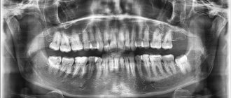
What does it show?
Dental computed tomography visualizes the nasal cavity and sinuses, teeth, lower and upper jaw. The following pathologies are noted:
- Inflammatory disease of the salivary gland;
- Tumor in the dentofacial region;
- Inflammatory periodontal disease;
- Root canal pathologies;
- Various diseases of the temporomandibular region;
- Injuries;
- Infections;
- Abscess;
- Necrosis;
- Dental-gingival pockets;
- Anomalies of teeth and their growth;
- Deviated septum;
- Unerupted teeth.