The average person may not know that they have teeth in their mouth called molars. Much more often these teeth are simply called molars. And with a big mistake they believe that they grow in place of dairy ones. Surprisingly, the first molars – molars – are already present in the primary dentition. They are chewable and have the largest crown area. On the upper jaw, their shape resembles a diamond, and the shape of the lower jaw is similar to a cube. This feature allows these teeth to perform the main function of chewing food.
Location and number of molars in the oral cavity in children
Children with have 8 molars, 4 on each jaw. They are located symmetrically, 2 on the left and right sides of the jaw. If you count from the front incisors, they occupy positions 4 and 5 in the dentition. The first molars appear on the lower jaw at the age, according to various sources, of 13-18 months, and a month later they appear on the upper jaw. Then the fangs erupt. The second molars complete the formation of the primary occlusion at the age of 2-2.5 years. Almost at the same time on both jaws. However, the timing of teething is now quite arbitrary, since the appearance of teeth individually depends on many conditions:
- genetic predisposition,
- child health,
- the presence of congenital characteristics,
- climatic living conditions,
- completeness of nutrition.
Chewing teeth are very important; they can have quite deep fissures where food gets into. Therefore, hygiene and thorough cleaning of the surface contributes to the health and safety of teeth until they are replaced with permanent ones.
Molars in humans
Molars are the fourth and fifth teeth in the order of eruption in the primary row or the sixth to eighth teeth in the permanent row of teeth on the right and left sides of the jaw. They are located behind the premolars. Very rarely, so-called additional, fourth, .
Typically, molars have four canals and three roots in the upper jaw, and three canals and two roots in the lower jaw. Upper first molars may have three canals in 7%, four canals in 90%, and five canals in only 3% of cases. In 40% of cases, the upper second teeth may have four canals. Upper third molars may have two, four, or five roots.
The roots of the molars extend from the cavity of the tooth in the form of thin branches. On the line that connects the mouth of the palatal canal and the mouth of the mesiobuccal canal, the mouths of the fourth upper first and second molars are also located.
On the lower jaw, the first molar is larger in size than the second and third .
On the human lower jaw, the first molar has five cusps on the chewing surface - two oral and three vestibular (distal, medial vestibular). The second one has four tubercles - two oral and two vestibular (distal, medial vestibular).
The difference between premolars and molars
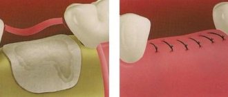
When baby molars fall out, it is not the molars that erupt, but the premolars. Many parents cannot understand the reason for this order of teething. This is explained by the fact that in all children the oral cavity increases in size, and the molars erupt behind the primary molars. Premolars are smaller in size than molars, and they are located behind the canines.
The first premolar has two roots, all the others have one. There are eight premolars in the human oral cavity (four premolars on each jaw). In the primary dentition there are no premolars, unlike molars. After all, a small child’s jaw has not yet grown to a size that can accommodate so many teeth. Despite the fact that premolars are considered the smallest molars, they are not very small in size. Premolars also perform the functions of chewing and grinding food . In their shape they are similar to fangs, only their crown is much wider than that of fangs. There are only two cusps on the crown of the premolar.
The appearance of baby teeth molars
Surely every parent remembers that period in the life of their baby when his molars began to erupt. After all, cutting through them is more painful than others. The only exception in this case are fangs. Molars erupt earlier than canines, although in their location they are behind them.
The first molars begin to appear when the child has already reached 12 months of age . They erupt like all others - in pairs. The molar appears first on the lower jaw, then on the upper jaw. Ideally, the first primary molars should appear before the baby is 18-20 months old. During this same period of a child’s life, the most painful teeth, the fangs, may begin to come out. That is why the age of up to 24 months is considered the most active age for the eruption of painful teeth.
Now, as for the second molars of the deciduous type, they appear at the age of 2 years. Sometimes they can erupt either earlier or a little later. But when the child is 30 months old, he should already have both first and second molars. Don't worry if your baby has any deviations from the due dates. This may not always be a pathology. This may also be due to genetics and heredity.
Eruption of permanent molars
The replacement of temporary teeth with permanent ones begins with the appearance of molars, which are not present in the primary dentition. They will be the first in the root and occupy the 6th position in the row. This process lasts for two years and begins when the child reaches 5-8 years of age. During this period, the resorption of the roots of temporary teeth begins, they gradually become loose and fall out. The age at which teeth change, as well as their appearance, is individual and depends on a number of factors. Including place of residence and gender. It has been established that boys become the owners of permanent teeth later than girls.
Next, at 10-12 years old, the first and second premolars are replaced. In this situation, they are the first and second molars of the primary occlusion. The second molars, whose serial number is seven, erupt in about a year. And the third molars complete the formation of the dentition. They are called wisdom teeth.
Structure
These teeth are even different from each other. The structure of the molars of the upper and lower jaws has significant differences. The first molar tooth is the largest. The rest are smaller than the first, the sizes decrease from the first to the third. The upper molar has a more powerful root than the lower molar: the upper has 3 roots, while the lower row has 2 roots. The second molar tooth is significantly smaller in crown area than the first. But nevertheless, all 3 molars on each dental arch have a powerful crown, as they are intended for chewing and grinding food.
On the crown of the molars of both the upper and lower rows there are tubercles: normally there are from 3 to 5 of them on each tooth. The cusps of the upper molars are sharper and more prominent, especially the buccal cusps. The lingual ones are more rounded in shape. And on the lower molars, lower and blunt tubercles can be noted. True, in contrast to the upper molars, the lingual cusps of the lower ones are actually more pointed and protruding compared to the buccal cusps.
As for the size of the teeth, the molars of the lower row are larger than the similar teeth of the upper row. Only wisdom teeth can differ in shape and structure. These molars may have 2 or 3 roots. And the shape of the crown can be varied. This is what distinguishes wisdom teeth from all others: they are not permanent, and it is impossible to predict what shape they will be.
Molar placement
The molar large units are located on the back of the jaw. Each jaw has six of them. They decrease in size from front to back. That is:
- the first rudiment is the largest;
- the second one is smaller;
- the third tooth is small;
The molar units are located most posteriorly in the arc. They are also called extreme. The number eight, that is, the wise tooth, grows in the period from 18 to 35 years. It happens that he does not appear at all. Row anomalies and bite defects can form in the absence of individual units or groups of rudiments. Often this concerns the end organs. Remember that normally there should be 28 units without eights, and with them 32 organs. The primary dentition has eight outermost units, the permanent dentition has twelve. In the molar group, the sixth units are key, and the 7s are variable, the most variable being the eights.
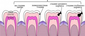
If caries treatment is carried out in a timely manner, the chewing organs will last a long time. The health of the whole body, the functioning of the gastrointestinal tract, liver and kidneys depends on this. Therapy should begin at the white spot stage, while the cavity has not yet formed. Prevention of caries is of great importance. To do this, you need to visit the dentist at least once every 6 months, carefully care for your oral cavity, and follow the dentist’s recommendations. Modern medicine has technologies the use of which can save even a dilapidated organ. Therefore, the key to health is timely treatment after a complete diagnosis and correct diagnosis.
It is important from childhood to teach a child to brush his teeth and treat the oral cavity. Children should understand that the main cause of caries is pathogenic bacteria living in dirty plaque. Therefore, trips to the hygienist will not allow the active proliferation of pathogenic microbes, subject to regular preventive cleanings.
Structural features
Those who looked at their teeth in the mirror noticed that they all had a different design. The structure of the crowns depends on:
- from the arrangement of organs in a row;
- depending on the shape of the unit;
- depending on the size of the rudiment;
The anatomical structure of the mammary organs is basically the same as the permanent units. True, temporary rudiments have distinctive features.
- They are smaller in size;
- The width of their crown prevails over the height;
- In the cervical area, the enamel layer is thickened;
- The shade of the enamel is bluish;
- The roots are widely spaced and short;
In addition, in the alveolar arch the temporal units are arranged steeply vertically. This is influenced by the fact that behind the roots there are already rudiments of permanent teeth. The upper units have 3 roots, one of them is lingual and 2 are buccal. The crowns have a cuboid shape, on the chewing surface of which there are 3 or more tubercles. The groove that separates the tubercles has two parallel lines and one perpendicular. The crowns of the rudiments on the lower jaw are somewhat elongated, and the fissures are located almost cross-shaped. These chewing organs have 2 roots. In eights, the roots often merge, the shape of such a root is conical. It happens that wise teeth grow with 4-5 roots. Since the upper and lower eights are close to each other, occasionally they can grow together.
To keep the chewing organs healthy, dentists may prescribe dental remineralization. This procedure restores the enamel layer well due to its saturation with deficient elements. The enamel ceases to be porous, acids do not penetrate its pores and do not contribute to the leaching of useful minerals. This event prevents and eliminates caries at an early stage, improves the aesthetics of crowns with fluorosis, reduces the sensitivity of units, brightens crowns and normalizes the microflora of the oral cavity.
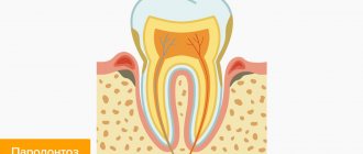
Teeth structure
All teeth consist of three main tissues, these are:
- Enamel. This is the hard outer layer. It contains the largest amount of minerals. On top it is covered with pellicle. This casing is acid resistant. Without proper care, the enamel is affected by caries.
- Dentine. Located under the enamel.
- Pulp. The softest part of the tooth. It contains nerves and blood vessels. When caries reaches the pulp, a person begins to experience pain. In this case, it is necessary to fill the root canals.
The top part of the tooth is called the crown. Between it and the root there is a neck. It is thinner than a crown. The root is located inside the alveoli. In the area of the neck, the thickness of the enamel is 0.01 mm, and on the masticatory tubercles it is 1.7 mm.
The root is attached to the hole using connective tissue fibers. They are called periodontium. They also ensure tooth mobility under load.
Milk teeth differ not only in shape, but also in the composition of the enamel. The latter contains less minerals, so caries is observed much more often in children. The pulp takes up more space than permanent teeth. The roots of the teeth have a rounded shape and wide channels.
Large molars - molars
(dentes molares) occupy the 6th to 8th positions in the dental arch; these teeth are located after the small molars. A person has 12 large molars (molars): 1st, 2nd, 3rd molars of the upper jaw (right, left), 1st, 2nd, 3rd molars of the lower jaw (right, left) . A common feature of the structure of large molars is the presence of several tubercles on the chewing surface of the crown and several roots. The molars of the lower jaw have two of them - mesial and distal (smaller than mesial), the molars of the upper jaw have three roots - one lingual (palatal) and two vestibular, one of which is mesial, and the second is distal (smaller than mesial).
The 1st molar of the upper jaw is the largest of all molars. The height of the tooth varies from 17.0 to 27.4 mm, the height of the crown is 6.3–9.6 mm, the height of the lingual root is 10.6–17.5 mm, the mesial vestibular root is 8.5–18.8 mm, vestibular distal - 8.9-15.5 mm. Its crown is prismatic in shape. In the lingual norm, the contact contours of the crown are convex; protrusions are defined on the crown, which are separated by a vertical groove. A vertical groove divides the lingual surface of the tooth into two parts, different in size. The mesial part of the crown is larger than the distal part. The apices of both lingual cusps are less sharp than those of the vestibular cusps.
In the vestibular norm, the contact contours of the crown of the 1st large molar of the upper jaw converge towards the neck of the tooth. Along the edges of the vestibular surface there are protrusions of enamel in the form of vertically located ridges, between which there is a groove. Of the two vestibular roots, the mesial one is the longest and widest. The roots of the tooth are often curved.
On the chewing surface of the 1st large molar of the upper jaw (occlusal norm), four tubercles are identified: vestibular-mesial (paracone), vestibular-distal (metacone), lingual-mesial (protocone), lingual-distal (hypocone). Each tubercle has a centrally located triangular ridge, along the edges of which there are less pronounced marginal ridges. Transverse ridges are more pronounced along the mesial edge of the chewing surface, which has a diamond shape. The tubercles are separated from each other by the mesial-vestibular and ulcer-distal grooves, which are connected in the middle by the deepest central groove or fossa, which separates the paracone and metacone. The furrows form the letter "H". The root of the tooth in a horizontal section in the cervical area has the appearance of an irregular quadrangle. The vestibular contours are flattened.
In the mesial norm, the 1st large molar has a smooth enamel-cement border, the vestibular-mesial tubercle is sharper than the lingual-mesial tubercle; the roots are cone-shaped, the longest of them being the lingual root. In the distal normal, this tooth has visible contours of all the cusps; the enamel-cement border forms an almost straight line. From the distal surface, two roots are visible (lingual and vestibular-distal). The crown cavity of the 1st large molar of the upper jaw corresponds in shape to the outline of the crown; there are four upper depressions (pulp horns) corresponding to the tubercles of the chewing surface. The lower wall of the pulp cavity of the crown is smooth and triangular in shape. Of the tooth root canals, the most direct is the lingual canal, which deviates slightly vestibularly in the apical part of the root. The vestibular canals are narrower and more curved than the lingual canal.
The 1st large molar may have additional cusps. In the dental cavity, the vestibular-mesial root canal is most variable.
The 2nd molar of the upper jaw is smaller in size than the 1st molar of this jaw. The height of the 2nd tooth varies from 16.0 to 26.2 mm, the crown - 6.1-9.4 mm, the lingual root - 10.0-18.8 mm, the vestibular-mesial root - from 9.0 to 18 .2 mm, vestibular-distal root - from 9.0 to 16.3 mm. The crown is narrower in the transverse (mesio-distal) direction. In the lingual norm, the vestibular tubercles are taller and less rounded than the lingual ones
tubercles. The lingual tubercles on the side of the lingual surface are separated by a shallow groove. The lingual root has a cone-shaped shape, larger than the vestibular roots.
In the occlusal norm, tubercles similar to the 1st upper molar are revealed. The 2nd molar has the largest lingual-mesial cusp, and the lingual-distal one has the smallest. The shape of the chewing surface is irregular quadrangular. The vestibular distal tubercle is separated from the lingual mesial tubercle by a central groove. In cross section, the lingual root is larger than the vestibular root.
In the mesial norm, in the 2nd large molar of the upper jaw, the vestibular mesial cusps are higher and sharper than the lingual mesial cusps. The apex at the roots is curved towards the longitudinal axis of the tooth.
In the distal normal, both vestibular cusps of this tooth are higher and sharper than the lingual distal cusp. The enamel-cementum border is slightly curved, with its convexity directed towards the lingual surface of the tooth. The vestibular-lingual size of the crown is larger than the mesio-distal size. In the distal norm, the lingual root is the largest; of the vestibular roots, the mesial one is larger.
The cavity of the crown of the large molar of the upper jaw corresponds to the external contours of the crown; the deepest corresponds to the vestibular mesial tubercle. The lower wall of the crown cavity forms a convexity directed towards the chewing surface. The cavity of the root canals is slit-like in shape, the largest lumen is at the lingual canal.
In the 2nd large molar of the upper jaw, the size and number of tubercles on the chewing surface vary; the roots and their sizes vary. There may be four roots.
The 3rd large molar of the upper jaw is the most variable in shape and size. It is often difficult to distinguish between right and left teeth. In the vestibular and lingual norms, the crown of this tooth is narrower and lower compared to the other large molars of the upper jaw. There are 3-5 tubercles on the chewing surface, the grooves on this surface have different configurations, and the roots are often not separated.
The 1st molar of the lower jaw is the largest tooth. The mesio-distal size of the crown is larger (10-13 mm) than the vestibular-lingual size (9-12 mm). The tooth has a mesial root measuring 14-16 mm and a distal root measuring 13.4-14.6 mm. In the vestibular norm, the mesial contour of the tooth is longer than the distal one. On the vestibular surface there are three vertically located ridges, decreasing in diameter towards the neck of the tooth. There are two grooves between the ridges, the depth of which increases towards the chewing surface. Both roots form a bend directed distally.
In the lingual norm, a vertically directed groove is determined on the surface of the tooth, passing between the lingual tubercles. The groove gradually disappears at the level of the middle third of the crown.
The chewing surface has a pentagonal irregular shape - a five-tubercle tooth. On the chewing surface, vestibular-mesial (protoconid), vestibular-distal (hypoconid), distal (mesoconid), lingual-mesial (metaconid), lingual-distal (entoconid) tubercles are identified. The metaconid is considered the highest. The vestibular-distal tubercle is smaller than the vestibular-mesial one. The lingual-distal tubercle is less pronounced than the lingual-mesial one.
Rice. 271. Variants of roots of the first lower molar in the vestibular norm. The dotted line and solid line show possible deviations of the tooth root. The lines on the surfaces of the crowns indicate their relief. Scheme.
1 - mesial root, 2 - buccal-mesial tubercle, 3 - lingual-mesial tubercle, 4 - buccal-distal tubercle, 5 - lingual-distal tubercle, 6 - distal tubercle, 7 - distal root,
D - distal surface, M - mesial surface.
The relief of the grooves on this surface is complex; The most pronounced groove is the one that separates the vestibular tubercles from the lingual ones. A groove is also identified that separates the mesial tubercles (vestibular and lingual) from the rest. The deepest place on the chewing surface (central fossa) is formed at the intersection of these grooves.
In the mesial norm, the 1st large molar of the lower jaw has a noticeable inclination in the lingual direction of the vestibular contour. The mesial root has a cone shape, a wide base; the distal contour is almost invisible. In the distal norm, the vestibular and lingual contours of the crown are convex; the distal root has a conical shape.
The cavity of the crown of the 1st large tooth has depressions corresponding to the tubercles. When viewed from the chewing surface, the cavity of the crown has a trapezoidal shape. The lower wall is convex in the direction from the roots of the tooth. On the lower wall there are holes leading into the root canals. The mesial root corresponds to two openings leading to the mesial-vestibular and mesial-lingual canals, the distal root has one canal. The mouth of the mesial vestibular canal corresponds to the location of the mesial vestibular canal. The mouth of the mesial-lingual canal is located near the mesial-lingual tubercle, the distal canal is located near the central fossa. The mesial-lingual canal has a large diameter and a straight direction, in contrast to the mesial-vestibular canal.
The 1st large molar of the lower jaw may have additional tubercles, the location and shape of the enamel-cementum border is variable, the enamel in some places can reach the middle of the tooth root.
The 2nd large molar of the lower jaw has a smaller crown and roots that are more closely spaced relative to each other compared to the 1st molar of the lower jaw. The height of the tooth ranges from 15 to 25.5 mm, the crown - 6-10 mm, the mesial root - 9.5-18 mm, the distal root - 8.5-18 mm. In the vestibular norm, in the 2nd large molar of the lower jaw, the contact contours of the crown are rounded, and two vertical ridges are defined. The vestibular mesial tubercle is larger than the vestibular distal one. The enamel-cementum boundary does not have a constant shape.
In the lingual norm, in the 2nd large molar of the lower jaw, the lingual cusps are lower and less pointed, the vertical groove between the vertical ridges is less deep than in the 1st lower molar. The 2nd large molar of the lower jaw has two vestibular (mesial and distal) and two lingual (mesial and distal) tubercles on the chewing surface. The vestibular-mesial tubercle is the largest, the vestibular-distal tubercle is the lowest. Between the tubercles, intertubercular (mesio-distal and vestibulo-lingual) grooves in the form of a cross are defined. The mesial and distal roots in the occlusal norm are flattened in the mesio-distal direction.
In the mesial norm, the vestibular contour of the crown of the 2nd large molar of the lower jaw in the direction of the chewing surface deviates to the lingual side. In the distal norm, the vestibular and lingual contours of the crown are convex; the most prominent points occur in the middle third of the crown. In the distal norm, the distal root, cone-shaped in shape, is smaller in size than the mesial one.
The cavity of the crown of the 2nd large molar of the lower jaw, when viewed from the chewing surface, has a quadrangular shape with rounded corners and contains recesses that protrude towards the tubercles. On the lower wall of the crown cavity there are holes leading into the root canals. Two holes belong to the mesial root. The mouth of the mesial-vestibular canal corresponds to the mesial-vestibular tubercle, the mouth of the mesial-vestibular canal is located next to this tubercle. The mouth of the distal canal is located near the central fossa of the chewing surface.
The 2nd large molar of the lower jaw, on its chewing surface, may have from 3 to 6 tubercles. The roots of the tooth are often not separated and have bends of different shapes. The number of root canals in a tooth varies from 1 to 4.
The 3rd molar of the lower jaw is very variable in shape and size. The height of the tooth varies from 15 to 22 mm, the crown - from 6 to 9 mm, the mesial root - from 7 to 14.5 mm, the distal - from 5 to 14 mm. Its crown size is smaller than that of other mandibular molars. In the vestibular norm, the shape of the crown is similar to the rest of the large molars of the lower jaw. There are usually 4 tubercles on the chewing surface. Sometimes multidirectional small grooves are revealed on the chewing surface. The roots are usually located closer together compared to the 2nd molar of the lower jaw.
The position of the roots of permanent teeth relative to the lower wall of the nasal cavity, maxillary sinus, and mandibular canal is different.
The roots of the upper medial incisor in people with a rounded head and a wide face and a small height of the alveolar process are usually separated from the nasal cavity by a thin plate of compact substance involved in the formation of the hard palate. In people with an elongated head, a narrow face, and a significant alveolar process, the apex of the root of the upper medial incisor is located, on average, at a distance of 1 cm from the nasal cavity.
The root of the upper lateral incisor usually does not fit close to the nasal cavity. The apex of the root of the upper canine with a flattened hard palate (in people with a wide face) often reaches the lower wall of the nasal cavity near the nasal notch. With significant development of the maxillary sinus, the root of the 1st premolar of the upper jaw comes close to its lower wall, and the apex of the root of the 2nd premolar is separated from the sinus only
Rice. 276. The relationship of the roots of the teeth of the upper jaw with the maxillary sinus and the lower wall of the nasal cavity and the lower teeth with the canal of the lower jaw. Right view.
1 - orbit, 2 - maxillary sinus, 3 - lower wall of the maxillary sinus, 4 - nasal cavity, 5 - roots of the teeth of the upper jaw, 6 - roots of the teeth of the lower jaw, 7 - lower jaw, 8 - canal of the lower jaw, 9 - angle of the lower jaw jaws, 10 - ramus of the mandible, 11 - coronoid process of the mandible, 12 - condylar process of the mandible, 13 - external auditory canal, 14 - mandibular fossa, 15 - zygomatic process of the temporal bone.
mucous membrane. With strong development of the maxillary sinus, the roots of the molars can protrude into its lumen, separated from it only by the mucous membrane covering the roots.
The apex of the root of the 1st lower premolar with a short lower jaw passes very close to the mandibular canal. The roots of the 2nd and 3rd lower molars may come close to its wall.
Related posts:
- Krasnoyarsk surgeons performed a unique...
- The Russian Federation was forced to pay 99 thousand euros...
- Leeches on the spine: why put them on...
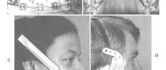
- Bite correction methods
- Video-computer psychodiagnostics
- Inguinal hernia: treatment
Diagnosis and treatment of maxillary second molars with two palatal roots
In addition to ideal knowledge of the morphology of the root system, every doctor during endodontic treatment must also know the prevalence of individual aberrations deviating from normal anatomy in order to identify all changes and features of the root structure before treatment begins.
In clinical practice, conventional radiography and an operating microscope are, in principle, sufficient for an objective assessment of the anatomy of the root canal, but it is quite difficult to achieve any more rigorous detail using these methods. It is for this purpose that endodontics uses cone beam computed tomography (CBCT), which is simply a critically necessary diagnostic tool that allows for a comprehensive assessment of all the features of root morphology (most often the molars of the upper jaw need this). One of the most unusual and rare forms of anatomical aberration appears to be the case of a maxillary second molar with two separated palatal roots. This article presents a clinical case of treatment of a patient with the above-mentioned features of the root system. Clinical case A
26-year-old patient sought help at the Department of Therapeutic Dentistry and Endodontics of the University of Belgrade with the following symptoms, which had bothered him for several weeks:
- spontaneous attacks of pain in the area of the left maxillary molar;
- moderate sensation of pain when eating solid food.
From the anamnesis it was also possible to find out that:
- no other symptoms were observed, there was no irradiation of pain;
- this tooth underwent endodontic treatment several years ago;
- two teeth on the offending side of the upper jaw were removed at least ten years ago.
During the clinical study it was possible to establish that:
- on the left side of the upper jaw there was only a second molar (tooth 27) with a huge amalgam restoration;
- the patient showed moderate sensitivity to vertical percussion of the buccal tubercle and a painful reaction to percussion of the mesiopalatal tubercle;
- no sensitivity to palpation of the vestibular or palatal sides was detected near the disturbing tooth;
- tests for hot and cold stimuli, as well as electroodontometry showed negative results;
- No signs of pathological tooth mobility were found.
A diagnostic periapical radiograph revealed:
- partially obturated palatal and mesiobuccal root canals and unobturated distal buccal root canal;
- an area of slight radiolucency around the apex of the palatine root; lack of a distinctive border with the surrounding bony structure of the maxillary sinus.
The need for repeated endodontic treatment was explained to the patient, to which he immediately agreed, after which a plan for subsequent visits was drawn up.
Treatment procedure
The old amalgam restoration and phosphate cement liner were completely removed and the cavity walls were further prepared to provide clear visibility and straightforward access to all root canal orifices. The openings of the palatal and mesiobuccal root canals were obturated with phosphate-iodine cement material and gutta-percha pins. Approximately 3 mm distal to the orifice of the obturated palatal root canal, an oval foramen with perforation was found. Further assessment of the pulp chamber was carried out using magnifying loupes (4.5X) and an Endodontic Probe Orifice Opener (DENTSPLY Maillefer). Using a probe and a size 10 K-file, a second palatal canal (distal palatal canal) was identified in the area of the foramen ovale. The orifice of the distobuccal root canal was hidden under brown deposits of tertiary dentin and was located approximately 2 mm distal to the obturated orifice of the mesiobuccal canal and approximately 2 mm more buccal to the orifice of the distalopalatine canal. The orifice of the distobuccal canal was slightly widened with an instrument to make it easy to identify later. The second mesiobuccal root canal was not found even under the control of loupes after applying a decalcifying solution (17% EDTA).
After consultations and obtaining the patient’s consent, it was decided to carry out the entire procedure in two stages. First, using rotating nickel-titanium files, ProTapers D1, D2 and D3 (DENTSPLY Maillefer) and manual H-files (DENTSPLY Maillefer), root fillings were removed from the mesiobuccal and mesiopalatal root canals. Further instrumental processing of these canals was carried out with reciprocating movements using WaveOne files (DENTSPLY Maillefer): the mesiopalatal canal with a tool with black markings (# 40), the mesiobuccal canal with a red marking (# 25). The working length was determined and checked several times during the treatment using an electronic apex locator (RomiApex A-15, Romidan).
The distal palatal root canal was completed to approximately 1–1.5 mm from the apical foramen using size 10 and 15 K-files. The coronal part of the canal was expanded using Gates–Glidden burs sizes 3 and 2. The same procedure was performed in the distobuccal root canal. Thus, it was possible to achieve direct access and visibility of all four channels (photo 1).
Photo 1. Straight-line access to all four canals of the 27th tooth.
X-rays were taken at two different horizontal angles with K-files in all four canals, but only one image was able to differentiate all four of them (photo 2) with vague contours of the apical parts of the roots. The mesiobuccal canal was filled with calcium hydroxide, and a paper point soaked in 2% chlorhexidine solution (R4, Septodont) was left in the mesiobuccal root canal. A cotton swab soaked in chlorhexidine was left in the pulp chamber, and the cavity was closed with a temporary filling material. After two weeks, the distal-palatal and distal-buccal root canals were carefully prepared using the same WaveOne technique as for the MP and MB root canals: the distal-palatal canal with a black-marked instrument (#40) and the distal-buccal with a red (#25). The working length was determined and checked in the same way.
Throughout the endodontic treatment, solutions of 2.2% sodium hypochlorite and 10% citric acid were used as irrigants in all four canals. Each of the four canals was finally irrigated with 40 ml of 2.2% NaOCl, dried and obturated with Acroseal (Septodont) with one gutta-percha point of appropriate taper (DENTSPLY Maillefer; Figure 3).
Photo 2. Identification of all four canals of tooth 27 on a targeted radiograph.
Photo 3. Obturated orifices of the root canals of the 27th tooth.
The results of post-treatment intraoral retro-alveolar radiography were of relatively poor quality due to overlap and interference of the lower zygomatic arch and adjacent bony structures, making it extremely difficult to objectively assess the condition of the periapical tissues (Figure 4). With the patient's consent, a CBCT scan was performed, which made it possible not only to verify the result of the treatment, but also to document an extremely rare clinical case. With the formation of a small field of view (50 x 50 mm) and using the SCANORA 3Dx device (Soredex), treatment results were recorded immediately after treatment and at a six-month follow-up period. The edited images (OnDemand3D, Cybermed) clearly visualized the two separate palatal roots, as well as their position in relation to the two buccal roots and adjacent anatomical structures, but most importantly, it was possible to objectively assess the quality of obturation of all four root canals (photos 5-9).
Photo 4. Intraoral radiography of the 27th tooth did not provide objective information regarding the quality of treatment.
Photo 5. Axial cut at the mid-length of the roots.
Photo 6. Axial section in the apical region.
Photo 7. Sagittal section of the 27th tooth.
Photo 8. Detailed view of tooth 27 after treatment
Photo 9. Volumetric rendering and multiplanar reformats of tooth 27 after treatment.
Conclusions and Key Points
Careful evaluation of the internal anatomy of the pulp chamber is critical to identifying all root canals. An upper second molar with two separate palatal roots is a rare anatomical aberration, and to our knowledge such a clinical case is encountered only once every ten years. CBCT images provide more accurate and reliable information about roots and root system morphology compared to conventional radiographs. In addition, the use of CBCT images provides more predictable and successful endodontic treatment results.
Author: Katarina Beljic-Ivanovic, Assistant Profesor, Serbia