Stitching |
Posted at 14:45h in Services by dobro
In their work, surgeons use surgical sutures, there are different types; this is one of the most common methods used to connect biological tissues: the walls of internal organs, wound edges, and others. They also help stop bleeding and the flow of bile, all thanks to properly selected suture material.
Recently, the main principle for creating any type of suture is considered to be careful treatment of each edge of the wound, regardless of its type. The suture should be placed so that the edges of the wound and each of the layers of the internal organ that requires suture are accurately matched. Today these principles are combined under the term "precision".
Depending on what tool is used to create the seam, as well as the execution technique, two types can be distinguished: manual and mechanical seams.
To apply a suture manually, ordinary and traumatic needles, needle holders, tweezers and other devices are used.
Absorbable threads of synthetic or biological origin, metal wire or other materials can be selected for stitching.
The mechanical seam is applied with a special apparatus that uses metal staples.
When stitching wounds and forming anastomoses, the doctor can apply sutures either in one row - single-row, or layer-by-layer - in two or even four rows. Along with the fact that the sutures connect the edges of the wound together, they also perfectly stop the bleeding. But what types of suture material exist today?
Classification of surgical sutures
As we have already said, seams can be either manual or mechanical, but there are several more classes of their division:
- according to the technique of their application, they can be nodal or continuous;
- if you divide them by shape - simple, knotted, in the shape of the letter P or Z, purse-string, 8-shaped;
- according to their functionality, they can be divided into hemostatic and screw-in;
- by the number of rows - from one to four;
- according to the length of time they remain inside the fabric - removable and submerged; in the first case, the seams are removed after a certain time, and in the second case they remain in the human body forever.
It is also worth mentioning that surgical sutures, their types are divided depending on the material used: they can be absorbable if they use catgut - this is a biological type and vicryl, dexon - these are synthetic. Erupting into the lumen of an organ - this type of suture is applied to hollow organs. Permanent sutures are those types of sutures that are not removed; they remain in the body forever and are surrounded by a connective tissue capsule.
Types of raw materials for suture
Suture material includes various materials used for ligating blood vessels using surgical sutures. The types of material for suturing tissue and skin changed greatly every year depending on how surgery developed. What surgeons did not use to connect the tissues of internal organs and skin:
But, thanks to modern developments, synthetic threads have now become popular. There are also cases when metal ones can also be used.
- high strength;
- Smooth surface;
- elasticity;
- moderate stretchability;
- high level of slip on fabrics.
But one of the important criteria for suture material is compatibility with the tissues of the human body. The currently known materials used for sutures have antigenic and reactogenic properties. There are no absolute types for these characteristics, but their degree of expression should be minimal.
It is also very important that the suture material can be sterilized and retain it for as long as possible, while its main characteristics must remain original. Suture thread can consist of one or more fibers that are connected together by twisting, knitting or braiding, and to ensure a smooth surface, they are coated with wax, silicone or Teflon.
Currently, absorbable and non-absorbable types of suture material are used in surgery.
Classification of surgical sutures, most of it involves the use of absorbable threads - catgut, which is made from the muscular lining of the small intestine of a sheep, and the submucosal layer can also be used to create it. Today there are 13 sizes of catgut, which differ in diameter.
The strength of the seam material increases with size. So, for example, the strength of the three-zero type is about 1400 g, but the sixth size is 11500 g. This type of thread can dissolve from 7 to 30 days.
Non-absorbable suture material in surgery uses threads made of silk, cotton, flax and horsehair.
Types of seams
When applying a suture to the skin, the doctor must take into account how deeply the wound is cut or torn, its length and how much its edges have diverged. The location of the injury is also taken into account. The most popular surgical sutures are considered to be the following; the photos in the article will show what they look like:
- subcutaneous continuous;
- subcutaneous nodular;
- cutaneous nodules;
- continuous multi-row applied inside the skin;
- continuous in one row, applied inside the skin.
This will help to understand which methods of applying surgical sutures are most often used when suturing an external wound.
Continuous intradermal type
It has recently been used most often, providing the best cosmetic results.
Its main advantage is excellent adaptation of the wound edges, excellent cosmetic effect and minimal disruption of microcirculation when compared with other types of sutures.
The thread for stitching is carried out in the layer of the skin plane itself parallel to it. However, for easier threading, it is better to use monofilament material.
After the primary surgical treatment of wounds is carried out, different types of sutures can be chosen, but often doctors give preference to absorbable suture material: biosin, monocryl, polysorb, dexon and others. And for threads that do not dissolve, monofilament polyamide or polypropylene are ideal.
Interrupted seam
This is another popular type of outer seam. When creating it, the skin is best pierced with a cutting needle. If you use it, the puncture looks like a triangle, the base of which is directed towards the wound.
This form of puncture allows you to securely hold the suture material.
The needle is inserted into the epithelial layer as close as possible to the edge of the wound, retreating only 4 mm, after which it is carried out obliquely in the subcutaneous tissue, while moving slightly away from the edge, as far as possible.
After reaching the same level as the edge of the wound, the needle is turned towards the midline and injected into the deepest point of the wound. In this case, the needle passes strictly symmetrically into the tissue on the other side of the wound, only in this case the same amount of tissue will enter the seam.
Horizontal and vertical mattress seam
The types of surgical sutures and knots are selected by the surgeon depending on the severity of the wound; if there are slight difficulties in matching the edges of the wound, it is recommended to use a U-shaped mattress suture running horizontally.
If an interrupted primary surgical suture is applied to a deep wound, then in this case a residual cavity can be left. It can accumulate what is separated by the wound and leads to suppuration. This can be avoided by applying a seam in several layers.
This method of suturing is possible for both nodal and continuous types.
In addition, the Donatti suture (vertical mattress suture) is often used. When performing it, the first puncture is made 2 cm from the edge of the wound. The puncture is made on the opposite side and at the same distance.
The next time the injection and puncture are carried out, the distance from the edge of the wound is already 0.5 cm. The threads are tied only after all the sutures have been applied, thus making it easier to manipulate in the very depths of the wound.
The use of the Donatti suture makes it possible to suture wounds with large diastasis.
In order for the result to be cosmetic, during any operation, primary surgical treatment of wounds must be carefully carried out, and the types of sutures must be selected correctly. If you do not carefully align the edges of the wound, this will result in a rough scar. If you apply excessive force when tightening the first knot, ugly transverse stripes will appear along the entire length of the scar.
As for tying knots, all silk threads are tied with two knots, and synthetic and catgut threads with three.
Types of surgical sutures and methods of applying them
When applying any type of suture, and many are used in surgery, it is extremely important to strictly follow the technique. How to apply a knotted suture correctly?
Using a needle on a needle holder, first pierce the edges at a distance of 1 centimeter, holding with tweezers. All injections are carried out one opposite the other. The needle is allowed to be passed through both edges at once, but it can be passed alternately, first through one, then through the other.
After completion, the end of the thread is held with tweezers and the needle is removed, and the thread is tied, while the edges of the wound should be brought one to the other as close as possible. The rest of the stitches are done in this manner until the wound is completely stitched up. Each seam should be 1-2 cm apart.
In some cases, knots can be tied when all the stitches have already been applied.
How to tie a knot correctly
Most often, surgeons use a simple knot to tie the suture material. And they do it like this: after the suture material is threaded into the edges of the wound, the ends are brought together and a knot is tied, and another one above it.
A surgical knot can be made in another way: they also thread a thread into the wound, take one end with one hand, and the other with the other, and, bringing the edges of the wound closer together, make a double knot, and then a simple knot above it. The ends of the thread are cut at a distance of 1 cm from the knot.
How to properly stitch a wound using metal staples
The types of surgical sutures and methods of their application can be different, which is determined by the location of the wound. One option would be stitching using metal staples.
Staples are metal plates, several mm wide and about a centimeter long, but can be longer. Both of their ends are presented in the form of rings, and from the inside they have a tip that penetrates the tissue and prevents the staples from slipping.
To apply staples to a wound, you should grab its edges with special tweezers, bring them together, place them well, holding it with one hand, and with the other you need to take the staple with another tweezers. After this, apply it to the seam line, squeezing the ends and applying force. As a result of such manipulation, the staple bends and wraps around the edges of the wound. Apply at a distance of 1 cm from each other.
The staples, like the sutures, are removed 7-8 days after they are applied. For this, a hook and special tweezers are used. Once removed, the staples can be straightened, sterilized, and used again to close wounds.
Types of seams in cosmetology
A cosmetic surgical suture can be made with any of the existing suture materials: silk, catgut, linen thread, fine wire, Michel staples, or horsehair. Among all these materials, only catgut is absorbable, and all the others are not. Seams can be embedded or removable.
According to the application technique in cosmetology, continuous and knotted sutures are used, the latter can also be divided into several types: marine, ordinary female or surgical.
The nodular type has one main advantage over the continuous one: it securely holds the edges of the wound. But a continuous suture is in demand because it is applied faster and is more economical in the quality of the material used. The following types can be used in cosmetology:
- mattress;
- continuous Reverden suture;
- continuous furrier;
- tailoring (magical);
- subcutaneous (American Halsted suture).
In cases where the patient has strong tissue tension, the doctor can use plate or lead-plate sutures, as well as a suture with rollers, which makes it possible to close large defects and securely hold the tissue in one place.
In plastic surgery, the doctor may also sometimes use an apodactyl suture. Its essence lies in the fact that it is applied and tied only with the help of a special tool: a needle holder, tweezers and a torsion pean.
Horsehair is the best suture material. The types of surgical sutures and knots that exist in cosmetology can be created well with its help. It is often used during ENT operations, because it practically does not become infected, does not irritate the skin and tissues, and there is no suppuration or scars in the places where it is applied. Horsehair is elastic, so unlike silk, it will not cut into the skin.
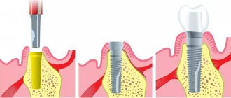
Use of sutures in dentistry
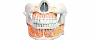
Dentists also use different types of sutures to stop bleeding or hold the edges of a large wound together. All types of sutures in surgical dentistry are very similar to those that we have already described, the only thing is that there are slight differences in the types of instruments. To apply a suture in the oral cavity, the following are most often used:
- needle holder;
- eye surgical tweezers;
- small two-prong hook;
- eye scissors.
It can be difficult to perform operations in the oral cavity, and only a professional will be able to perform this work efficiently, because not only high-quality primary treatment of wounds is important here. It is also important to choose the right type of sutures in dentistry, but most often it is a simple interrupted suture. And it is applied like this:
- It is necessary to sequentially pierce both sides of the wound at a sufficient distance from one another, the thread must be pulled out as much as possible, leaving only a small end - 1-2 cm.
- The long end of the thread and the needle are held in the left hand, after which they need to wrap the needle holder clockwise 2 times.
- Using a needle holder, grab the short tip and pull it through the loop formed - this is the first part of the knot, carefully tighten it, slowly bringing the edges of the wound closer together.
- Also, while holding the loop, you need to do the same manipulations, only scroll counterclockwise once.
- Tighten the already fully formed knot, be sure to monitor the uniform tension of the thread.
- Move the knot from the cutting line, trim the end of the thread, that's all, the seam is ready.
Source: https://dobromedbutovo.ru/lechenie/nalozhenie-shvov/
Never grab the edges of a wound with tweezers, especially anatomical ones!!!!!
The needle should be inserted at a sufficient distance from the edge of the incision.
When the edges of the wound are brought closer together, excessive tension should not be allowed in the suture area; the edges of the wound should only touch each other tightly, and at the same level. Please note that overlapping one edge of the wound over the other is absolutely not permissible; in this place there will be a rough scar or even divergence of the sutures. In case of a deep wound, it is necessary to apply sutures in layers; therefore, in the deep layer, it is necessary to apply sutures made of “absorbable” material.
It is most convenient to fix the edge of the wound with a two-pronged hook; the puncture is made between the “teeth” of the hook. It is best to sew according to the “puncture” pattern,
those. It is undesirable to pierce both edges of the wound at once - in this case, excessive tension of the tissues occurs and, accordingly, the risk of injury and subsequent cutting of the seam. Sutures should be tied using instruments; this is especially important in the oral cavity. Another advantage is the extremely low consumption of suture material, which is important in our time. I'll tell you in more detail how this is done.
1. Having punctured both edges of the wound sequentially, you pull the thread to the point where a short end 1-1.5 cm long remains
2. Holding the long end, with the needle, in your left hand, you wrap it around the needle holder 2 times clockwise
3. Grasping the short end of the thread with the needle holder, you pull it through the loop already formed on the needle holder and get the first part of the knot
4. Carefully tighten the knot, bringing the edges of the wound closer together; the double loop will not allow it to come apart
5. Repeat position 2, taking into account that you need to wrap the long thread once and now counterclockwise.
6. Finally tighten the knot, ensuring that the edges of the wound are brought together evenly and “evenly”
7. Move the knot so that it is not located above the incision
8. Cut the end of the thread to the required length, the seam is ready...
It is advisable to apply sutures evenly and not too often, starting from the middle of the wound. Frequent sutures often lead to problems with the blood supply to the wound edges and poor healing. It is a good idea to leave glove drainage between the seams for 2-3 days, especially if the wound was traumatic.
Well, that's all. I hope this article will remind non-surgeons how to properly close a wound with a simple interrupted suture. I do not consider other options for sutures, since they are practically inapplicable in the oral cavity. But by the way, if anyone is interested, write 
Classification of dental suture materials and their areas of application
834
Not a single serious dental procedure is complete without suturing. The range of suture materials today is impressive.
Based on the complexity of clinical cases, the dental surgeon has the opportunity to select the appropriate version of the surgical thread in terms of absorption rate, structure and diameter.
General concept
In dentistry, suture material is a surgical thread used during surgery in the mouth; it is used to connect dissected tissues and activate the process of their regeneration upon completion of the operation.
Reliability, biological compatibility with oral tissues and non-traumatic properties certainly affect the outcome of the operation and the course of the postoperative period, and also provides the required cosmetic effect.
Today, surgical thread is presented in a wide range on the market. Each variety has certain qualities and characteristics that suit its characteristics and tasks.
Requirements
Surgical operations performed in the oral cavity require careful selection of not only the method of performing them, but also the materials used, including threads for suturing tissue.
It should have the following indicators:
- Biocompatibility with oral tissues , i.e. There must be no teratogenic, toxic, allergic or carcinogenic effects on the body. Ideally, there should be no tissue reaction.
- Durability and preservation before scar formation , i.e. resistance to destruction and change in original shape.
- Reliability in the knot (i.e. have minimal slip).
- Versatility in use - suitable for most types of surgical procedures.
- Simplicity in sterilization , namely, easy to disinfect without loss of original characteristics.
- Sliding structure (no sawing effect).
- With gradual biodegradation , which means: its resorption period must exceed the time required for tissue scarring.
- Atraumatic , which means there should be no effects of “tearing” and “sawing” of tissues when pulling a thread through them.
- Resistance (resistance) to infection.
- Ease of use , namely good handling qualities: optimal elasticity (the length should not increase by more than 20% of the original value), softness, flexibility.
- Passivity to wicking and capillarity , which means it does not absorb liquid from the puncture channels and does not pass it between the fibers.
In addition, the packaging must be clearly and completely labeled, intact and easy to open.
Important! There is no universal surgical thread that meets all the listed criteria at once. The option is usually selected in accordance with the purpose of the operation and the characteristics of the tissue at the site of the manipulation.
Classification
The products differ among themselves in structure (number of fibers in the thread), speed of biodissolution (resorption, etc.).
By material
Classified:
- Natural when flax, catgut, silk, or natural cellulose are used in its production.
- Natural inorganic is fibers made from nichrome and platinum.
- Synthetic - threads made from homopolymers, polyesters, polyolefins, polybutesters and fluoropolymers.
By structure
Surgical threads are divided into:
- Homogeneous - ethylone, maxon, prolene.
- Multi-fiber - dexon, surgilon.
- Combined - vicryl, polysorb, terylene, tikron.
According to the rate of biodegradation
According to the speed of resorption, there is the following division.
Absorbable
This group includes derivatives of ε-caprolactone, polydioxanone, polyglycolic acid, i.e. all synthetic threads and catgut.
There are threads with a short resorption period (when the tissue holding time is 8-10 days), an average period (this figure reaches 4 weeks), and a long resorption period (the holding time is 2 months).
Biodegradation occurs under the influence of proteases (proteolytic enzymes) and hydrolysis (when liquids enter the fibers and destroy their structure).
In addition, the process is significantly influenced by individual indicators of the body, such as body temperature, protein volume, etc. Resorption in most cases is associated with a tissue reaction at the site of the operation, inflammation, and suppuration.
Conditionally absorbable
This includes polyurethane and polyamide threads, as well as some types of natural suture products. The process of biodestruction in such threads does not completely end, i.e. there is only a loss of initial strength.
Non-absorbable
These are polyolefins, polyesters, polypropylenes, fluoropolymers. They are removed by the dentist after the wound area has healed. There is practically no tissue reaction to this type.
Threads are the most popular group because they have the best handling properties , are non-traumatic and have the lowest risk of tissue infection.
It is recommended to remove the conditionally absorbable and even resorbable type, after scarring of the tissues in order to prevent their infection. The need for this action is explained by the loss of the thread of its original strength; moreover, upon contact with the external environment, it becomes a substrate for the development of microorganisms.
Important! When there is simply no particular difference in what material to use, it is better to give preference to threads that cause less reaction from the body.
In the video, a specialist will talk about absorbable and non-absorbable suture materials.
By design
Based on their design, surgical threads are divided into:
- Monofilament (homogeneous).
- Polyfilament (multi-filament).
- Combined.
The first group includes products made from solid homogeneous fiber with a smooth surface , which is why it is less traumatic on the one hand, and on the other hand, it slips in the hands and is not so reliable in the knot. Such threads also have good elasticity and hydrophobicity, are non-wicking, non-capillary, do not “grow” to the tissues, and do not cause irritation and inflammation.
The second group includes threads consisting of a cross-section of several fibers that provide additional strength. They can be twisted (obtained by axial twisting of several filaments) and braided (obtained by weaving many filaments together in a rope).
The threads have reliability in knots, good handling characteristics, increased strength and ductility. The disadvantages include wicking and sawing properties, which can cause purulent inflammation in the tissues.
The last group, as a rule, consists of braided threads that are coated or impregnated with a polymer composition. The coating reduces the “sawing” effect. The material also has excellent handling characteristics and does not injure tissue. Disadvantages - high cost and the possibility of premature loss of initial indicators.
Important! For operations in the oral cavity, it is more advisable to use homogeneous (monofilament) threads, because they are smaller than multi-filament threads and contribute to the accumulation of pathogenic microorganisms.
Main types
The assortment is extensive. Let's consider the options for those threads that are used only when performing operations in the mouth.
Traditional
The sutures commonly used in most dental procedures are silk and catgut.
The “standard” for manipulation properties in traditional surgery, and dentistry in particular, is silk. This type has softness, elasticity, the necessary strength, and can be used to tie several knots.
The structure is a complex of several polyfilament fibers of different thicknesses, characterized by increased strength and sufficient flexibility.
Silk is classified in this group because it retains its original properties in tissues for up to six months. Produced with a thread length of up to 2 meters in sealed ampoules in a braided or twisted version. To increase strength, a little cotton fiber is added to them.
The material has significant disadvantages. Thus, being an organic fiber, it can provoke an aseptic reaction in soft tissues. In addition, its high hygroscopicity can cause inflammation. Sawing and wicking properties appear.
Important! Silk has a slow absorption rate, which is why it is not recommended for use in prosthetics.
The most popular in dentistry, rather than silk, is catgut. It is a multifilament product made from the submucosa of the mammalian intestine.
The following indicators are considered advantages:
- ability to withstand excessive loads;
- excellent handling properties;
- the ability to form strong nodules.
Cons of catgut:
- high allergenicity;
- pronounced absorption;
- reactogenicity;
- insufficient tensile strength.
The duration of catgut biodestruction varies from 3 days to 2 weeks, which may be insufficient or excessive time for scar formation.
Non-absorbable
This group includes threads made from polyolefins, polyesters, polypropylene and fluoropolymers. Commonly used in dentistry:
- Nylon (multifilament or monofilament). It is distinguished by the memory of the original parameters, and is subject to degradation with the loss of the original strength indicator.
- Polyester (multifilament only). It is not subject to changes in parameters; it is encapsulated by fibrous tissue.
- Polyethylene (monofilament only). It undergoes degradation with a change in strength, which always ends in thread rupture. Has shape memory.
- Polypropylene (monofilament).
It is considered a good non-absorbable material, since it is passive in relation to body tissues, does not have high resistance to infections, and is easily removed. Minus (relative disadvantage) - requires the doctor to have suture skills, because the tip of the thread is quite sharp. - Polytetrafluoroethylene (monofilament). It is the best option for this type of suture thread. It has only one drawback - the high price.
Polysorb
These are new generation products. It is a fiber consisting of several combined polyglycolic acid braided fibers coated with a polymer.
Comparative characteristics of Polysorb:
- It is easily pulled through fabrics as a homogeneous thread.
- In terms of handling properties, it is not inferior to natural fiber (for example, silk).
- It has sufficient strength (80% of its strength remains for 2 weeks).
- It is distinguished by the reliability of its nodes.
- The biodestruction period is 70 days.
- Produced in two color options - colorless and purple.
- Sold at an affordable price.
conclusions
Medicine, and in particular surgical dentistry, is constantly developing, which means that existing technologies and materials are being improved, and new ones are appearing.
Constant updating and proper use of new products allows us to cope with any complex pathological conditions, and thereby improve the quality of life of patients.
Reviews
Modern suture materials are used in all areas of surgery, and dentistry is no exception. The correct selection of threads largely determines the outcome of the operation, affects the comfort of the recovery period, the quality and speed of tissue fusion.
Source: https://www.vash-dentist.ru/hirurgiya/udalenie-zubov/stomatologicheskogo-shovnogo-materiala.html
Suture materials used in dental surgery
267
Any type of surgical therapy will have a positive result if the postoperative wound heals quickly, without causing complications and scar changes.
Therefore, during dental operations, as in conventional surgery, suture products are used, which accelerate the healing process and help restore the integrity of damaged tissues.
Overview characteristics
The suture material resembles a thread that is used after surgical therapy to tighten the edges of a surgical wound in order to form a scar or epithelialize.
Great attention and special requirements are placed on it, since if a low-quality product is used, the following negative consequences may occur:
- failure of the suture can cause subsequent suppuration (abscess formation);
- the risk of bleeding increases;
- a poorly executed suture causes the appearance of fistula tracts and granulomas;
- in some cases, a hernial protrusion occurs.
Suture thread is made in various ways. Synthetic, animal and plant components are used as raw materials for its production.
Thus, to obtain catgut, the muscle layer from the small intestine of sheep is used. Some threads (occelon, cacelon) are made from cellulose, while vicryl and dexon are a synthetic product. Sometimes metal wire or staples are used to connect the edges of the wound.
According to the materials used
This type of classification is based on the type of raw materials used . Based on this, the suture product comes in the following types:
- Biological. For its production, components such as animal tendon fascia, sheep wool, horse hair, cattle meninges or cellulose derivatives are used. Using the latest technologies, using these raw materials, we obtain catgut, occelon, silk, cacelon, rimin, etc.
- Inorganic. These include metal wire made of steel, platinum or titanium.
- Artificial (synthetic or polymer). Produced using polyesters, polymer compounds and acids (polyglycolic, lactic). The final product is lavsan, dexon, prolene, mersilene, fluorlon.
By biodestruction
Biodegradation is the process of dissolution in tissues. Based on this ability, suture products are classified into types:
- absorbable (differently adsorbable). These include catgut threads, polysorb, bioxin, rimin and polyurethane.
- conditionally absorbable - these are types such as nylon, silk, treated with silicone.
- non-absorbable suture products are made using polyesters (lavsan, marsilene), polyolefins (surgipro, prolene, mersilene, polypropylene), fluoropolymers (vitafon, fluorlon, fluorlin, fluorex).
In the video, a specialist will talk about absorbable and non-absorbable suture materials.
Product Types
Various suture products are used to solve surgical problems. They have different characteristic features that allow the products to be used in certain cases.
Reviews, opinions, comments
Practicing specialists constantly monitor innovations in the field of surgical dentistry. Polysorb suture material has become especially relevant in use.
With its help, it becomes possible to solve many surgical problems, which minimizes the chance of developing complications in the postoperative period.
You can leave your opinions and comments at this point in this article.
, please select a piece of text and press Ctrl+Enter.
Source: https://zubovv.ru/hirurgiya/udalenie-zubov/stomatologicheskogo-shovnogo-materiala.html
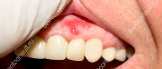
Stitches in the mouth
The need for suturing the oral mucosa arises after surgical operations. The procedure is performed to reduce the area of the wound surface, speed up the healing process, and prevent the risk of bleeding and wound infection.
When is suturing necessary in dentistry?
Surgical intervention is performed if:
- cystectomy – removal of a medium to large cyst that extends over several dental units. Resection of the apex of the previously sealed roots occurs;
- cystomy - partial removal of the cyst in order to reduce the pressure inside the formation and its gradual flattening;
- eliminating certain inflammatory processes - opening abscesses, treating osteomyelitis, periodontitis;
- gingivectomy – excision of overgrown gum tissue;
- gingivoplasty – tissue transplants to improve the aesthetics of the gums;
- removal of dental units - resection of a wisdom tooth is especially often accompanied by suturing;
- preparation for prosthetics – alveoloplasty, sinus lift, bone block transplantation.
A deep incision in the soft tissues of the oral cavity always requires sutures.
Types of suture material
Material for suturing is divided into two large groups - absorbable and non-absorbable.
Absorbable suture materials include:
- catgut is a thread obtained from purified connective tissue; when left in the oral cavity for a long time, the possibility of an inflammatory process arises due to rejection of the material;
- vicryl - synthetic threads containing polyglycomic acid, polyclatin, do not provoke the development of inflammatory processes, which makes it possible to use them where there is a high risk of postoperative complications.
Non-absorbable suture material:
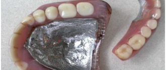
- monofilament - a synthetic thread with a rigid structure that can injure the mucous membrane;
- silk is an elastic, durable material, but requires timely removal of sutures (after 10 days it causes inflammation of the soft tissues);
- polyster is a durable woven thread that requires special care when fixing; in order to make it more smooth, it is often coated with silicone.
Main types of sutures in dentistry
The most commonly used seams are:
- single – used for minor incisions in the soft tissues of the patient’s oral cavity;
- nodal - a reliable way to fix the edges of the wound;
- supporting – used when flaps (buccal, lingual) are located at different levels;
- continuous – its advantage is good adaptation of the wound edges and is highly aesthetic.
Instruments used for suturing in dentistry:
- needle holder - there are many models of the instrument, the choice depends on the preferences of the surgeon;
- two-pronged hook, eye surgical tweezers - necessary for maintaining the edges of the wound;
- eye scissors – needed for cutting suture material;
- suture needles - made of medical stainless steel, having different sizes, shapes, diameters.
Prevention of complications after suturing
It is important to complete the medication prescribed by your dentist.
The following will help speed up and relieve pain from wound healing:
- applying cold on the first day for 10–15 minutes helps relieve swelling and reduces the risk of bleeding;
- baths with antiseptics - rinses, herbal decoctions with antimicrobial, anti-inflammatory properties;
- Three days after surgery, you can clean the suture material with a soft toothbrush.
After surgery it is prohibited:
- take aspirin and other anticoagulants - they thin the blood and can cause bleeding;
- eating hard, spicy foods can damage soft tissues and cause irritation;
- carry out warming procedures - heat compresses, visiting saunas, baths leads to inflammatory processes;
- rinse your mouth - active rinsing of the mouth is prohibited until the stitches are removed;
- active physical activity.
A visit to a dental surgeon is a prerequisite for preventing and timely eliminating the causes that contribute to the occurrence of postoperative complications.
How does the procedure work?
The suture should be applied from the side of the moving edge of the wound towards the fixed one. The free edges of the tissue flap must be carefully held with special anatomical tweezers. In the process of suturing the skin and mucous membranes, the needle grasps the flap from the edge of the wound by about 3 mm, and then the wound is tightened along the edges with the ends of the suture material, tightening the loops.
It is very important that this procedure is done professionally; mistakes and carelessness are unacceptable. For example, you should not tighten the knot too much, as this disrupts the blood supply at the edges of the wound, as a result of which the tissue quickly turns pale at the suture site. However, it is also unacceptable for the seam to be too loose.
Seam removal and post-processing
If the wound healing process proceeds normally, then already on the seventh day it is possible to perform an operation to remove the sutures. This is a simple procedure; special surgical threads are simply removed using surgical tweezers, sharp scissors, or a special suture cutter.
You need to grab the knot with tweezers, and then simply cut one side of the seam. Carefully remove the suture material from the tissue. In modern dentistry, materials are often used that dissolve themselves and do not need to be removed at all. Some remnants of materials from the wound can also be removed later using a chlorhexidine solution (if necessary).
Source: https://best-stom.ru/articles/nalozhenie-shvov-v-stomatologii/
Surgical treatment of non-gunshot injuries to soft tissues of the maxillofacial area
Primary surgical treatment is the first surgical operation performed on a patient regarding a wound under aseptic conditions and pain relief. The main types of primary surgical treatment: Early primary surgical treatment - performed up to 24 hours from the moment of wound infliction. Usually ends with the application of primary sutures. The peculiarity of the timing of early surgical treatment of facial wounds is that it can be carried out within 48 hours. The possibility of performing primary surgical treatment of a wound at a later date on the face is associated with good blood supply and innervation. Delayed primary surgical treatment - performed within 24-48 hours. It must be carried out against the background of the administration of antibiotics. After delayed primary surgical treatment, the wound remains open (not sutured). Subsequently, primary delayed sutures are applied. Late primary surgical treatment - performed after 48 hours. Late surgical treatment is a surgical intervention for an injury complicated by the development of a wound infection. Stages of late surgical treatment of a wound: • opening of the wound canal, • removal of necrotic tissue and wound detritus, • creation of conditions for adequate drainage. The application of a blind suture during this treatment is contraindicated, with the exception of wounds in the lips, eyelids, wings of the nose, auricle, in the superciliary region and the oral mucosa. Classification of types of sutures depending on the timing of their application Depending on the timing of application of sutures to the wound, they are distinguished: The primary blind suture is applied during early surgical treatment. The primary delayed suture is applied 4–7 days after injury (before granulation appears) after cleansing the wound and reducing swelling. Drainage is inserted into the wound. An early secondary suture is applied on days 8-15 when granulation tissue appears in the wound. In this case, healthy red-pink granulations are not excised; Rubber drainage is left between the sutures or a vacuum aspirator is placed at the bottom of the wound through skin punctures (counter-apertures) outside the suture line. A secondary late suture is applied 20-30 days after injury to a scarring wound without clinical signs of infectious inflammation. In such cases, excess granulations are excised, the edges of the wound are mobilized and sutures are applied. Currently, step-by-step surgical treatment of wounds in medical institutions where there is no possibility of providing specialized care is considered unacceptable. In such cases, you should limit yourself to providing first medical aid and take the victim to a specialized hospital as soon as possible. If the patient is not transportable, he should be examined by a dentist (together with other specialists from the territorial medical institution) to decide whether it is necessary to call an oral surgeon via air ambulance from a specialized hospital of regional or republican significance. The following are not subject to primary surgical treatment: • superficial wounds, scratches, abrasions; • small wounds with edge separation of less than 1 cm; • multiple small wounds without damage to deeper tissues (shot wound); • puncture wounds without damage to internal organs, blood vessels, nerves; • in some cases, through bullet wounds of soft tissues. Contraindications to primary surgical treatment: • signs of development of a purulent process in the wound; • critical condition of the patient (terminal condition, stage III shock) Stages of postsurgical treatment of the wound: • dissection of the wound; • revision of the wound channel; • excision of edges, walls, bottom; • hemostasis; • restoration of tissue integrity; • suturing the wound. Dissection and revision of the wound: The edges of the wound are separated with sharp or plate-like retractor hooks. In general surgical practice, when the upper part of the wound is small and there is more significant damage to the deeper layers, it is expanded by dissection to provide access to all parts. Features of facial wounds. Expansion of wound channels is not performed due to possible damage to blood vessels and nerves. Excision of wound edges. After instillation (washing) of the wound and removal of blood clots and foreign bodies, the wound is examined, the boundaries of the damaged tissue are determined and the edges of the wound are excised to the full depth. Features on the face. Only tissues that are known to be non-viable are subject to excision, which is determined by their color, thickness, and state of capillary bleeding. The crushed and contaminated subcutaneous fat should be excised quite widely. It is necessary to determine the degree of damage to the facial and chewing muscles, to exclude the presence of foreign bodies under the contracted bundles of muscle fibers. Dark, flabby areas of muscle that do not contract when irritated are excised, and their remaining fibers are brought together and stitched. At the same time, one should not strive to obtain straight edges of the skin, since scalloped, zigzag, adjacent edges subsequently form a less noticeable and more aesthetic scar. Restoring tissue integrity. The primary surgical treatment of the wound must be completed by bringing its edges together and applying a primary blind suture. The tissues are joined with surgical needles. Based on the nature of the impact on tissue, traumatic and atraumatic needles are distinguished. A traumatic surgical needle has an eye through which a thread is threaded. A thread threaded through the eyelet and folded in half has a traumatic effect on the tissue in the suture channel. An atraumatic surgical needle is connected to the thread in an end-to-end fashion, making it easier for the thread to pass through the tissue. Requirements for suture material: • have a smooth, even surface along the entire length; • be elastic and flexible; • maintain strength until scar formation (for absorbable materials); • be atraumatic: do not cause a sawing effect, i.e. glide well; • connect to the needle end-to-end, have good handling properties; • dissolve at a rate not exceeding the rate of scar formation; • be biocompatible. According to the structure of the thread, they are distinguished: • monofilament thread (monofilament thread) – uniform in structure in cross section, has a smooth surface; • polyfilament thread (multifilament thread) consists of several threads and can be twisted, braided, complex (with a polymer coating). According to their ability to biodestruct, threads are: • absorbable (catgut, occelon, cacelon, vicryl, dexon, etc.); • non-absorbable (nylon, polyamide, lavsan, nylon, etibond, M-dec, prolene, propylene, surzhilene, surzhipro, etc.) Depending on the source raw material, threads are distinguished: • natural: a) absorbable monofilament - catgut (plain and chrome-plated ), serosophil, silicone worm, chrome collagen; b) non-absorbable polyfilament - woven silk (including those coated with paraffin and silicone) and waxed, linear, cotton; • metal non-absorbable monofilament – tantalum staples and wire, flexon, nichrome steel wire, polyfilament steel wire; • synthetic from: a) cellulose – absorbable monofilament (occelon, cacelon, rimin); b) polyamides - non-absorbable monofilament (dermalon, nylon, etikon, ethylone); multifilament (nylon, nylon); absorbable (letilan, segilon, supramid, suturamide); c) polyesters – non-absorbable multifilament (lavsan, astralen, mersilene, sterilene, dacron, tikron, etibond, tevdek, etiflex); d) polypropylene – non-absorbable monofilament (polyethylene, prolene); e) glycolic acid polymer (polyglactide) - absorbable multifilament (Dexon, Vicryl, Dezon Plus with coating); e) polyoxanone (PDS) – absorbable monofilament thread (ethikon). During operations in the maxillofacial area, various types of threads are used to stitch together soft tissues. To stitch the edges of wounds on the skin, all types of non-absorbable materials are used, except for metal staples and wire, lavsan, silk, as well as absorbable materials, except for catgut and collagen, for muscles - all absorbable materials, for the mucous membrane - the same. In some cases, non-absorbable sutures are placed on both the muscle, mucous membrane, and blood vessels, which is due to the peculiarities of the surgical intervention, for example, when removing hemangiomas, malignant tumors, or strong tension of the underlying tissues. Requirements for a surgical unit: • Must be, first of all, durable and reliable. • Should not tighten wounds too much, so as not to cause necrosis of surrounding tissues. • Not be large so as not to form bedsores in the underlying tissues. • The length of the ends of the knot should be sufficient to grasp them with tweezers when removing sutures. Technique for suturing wounds of the maxillofacial area. • careful attitude to the edges of the wound being stitched; • precision (exact comparison, adaptation) of the same layers of the wound being stitched; • each layer of fabric must be sutured with the appropriate type of thread and seam; • the length of the skin wound on one side should be equal to or less than that on the other side, but taking into account the elasticity of the skin, which makes it possible to stretch the edge of the wound to the required length. If the discrepancy between the length of the wound edges is significant, then it is necessary to apply local plastic techniques to lengthen its edge; • slight elevation of the wound edges to prevent scar retraction during contraction; • providing prolonged dermal support to prevent scar expansion in the postoperative period; • exclusion of strangulation marks from ligature pressure sores on the skin surface; • the distance between the needle insertion and the edge of the wound is 1 mm, the distance between stitches is 2 mm; • the formation of a “residual cavity” must be avoided. Features of primary surgical treatment of facial wounds. • early surgical treatment of the wound up to 24 hours from the onset of injury; • final surgical treatment of the wound in a specialized institution; • the edges of the wound are not excised, only clearly non-viable tissues are cut off; • narrow wound channels are not completely dissected; • foreign bodies are removed from the wound, but no search is undertaken for foreign bodies located in hard-to-reach places; • wounds penetrating into the oral cavity must be isolated from the oral cavity by applying blind sutures. It is necessary to protect the bone wound from the contents of the oral cavity; • on wounds of the eyelids, wings of the nose and lips, a primary suture is always applied, regardless of the timing of surgical treatment of the wound. When suturing wounds on the side of the face, drainage is inserted in the submandibular area. In case of a wound penetrating into the oral cavity, first of all, the mucous membrane is sutured, then the muscles and skin. For wounds of the lips, the muscle is sutured; the first suture is placed at the border of the skin and the red border of the lip. In case of damage to the soft tissues of the face, combined with bone trauma, the bone wound is treated first. In this case, fragments not connected to the periosteum are removed, the fragments are repositioned and immobilized, and the bone wound is isolated from the contents of the oral cavity. Then they begin surgical treatment of soft tissues. For injuries penetrating the maxillary sinus, the sinus is inspected, an anastomosis is formed with the inferior nasal passage, through which the iodoform tampon is removed from the sinus. After this, surgical treatment of the facial wound is performed with layer-by-layer sutures. If the salivary gland is damaged, sutures are first placed on the parenchyma of the gland, then on the capsule, fascia and skin. If the duct is damaged, conditions should be created for the outflow of saliva into the oral cavity. To do this, a rubber drainage is placed at the central end of the duct and discharged into the oral cavity. The drainage is removed on day 14. The central excretory duct can be sewn onto a polyamide catheter. In this case, its central and peripheral sections are compared. The crushed submandibular salivary gland can be removed during primary surgical treatment of the wound, but the parotid gland cannot be removed due to the complex anatomical relationship with the facial nerve due to injury. With large through defects of the soft tissues of the face, the convergence of the edges of the wound almost always leads to pronounced deformations of the face. Surgical treatment of wounds should be completed by “suturing” them, connecting the skin with the mucous membrane with sutures. Subsequently, plastic closure of the defect is performed. In case of extensive trauma to the lower third of the face, floor of the mouth, or neck, a tracheostomy is necessary, followed by intubation and primary surgical treatment of the wound. A wound in the infraorbital region with a large defect is not sutured “on its own” parallel to the infraorbital margin, but is eliminated by cutting out additional flaps (triangular, tongue-shaped), which are moved to the site of the defect and fixed with appropriate suture material. After the initial surgical treatment of the wound, it is necessary to carry out tetanus prophylaxis. Features of primary surgical treatment of bite wounds of the face. Anti-rabies assistance is provided in accordance with the following documents: • instructions for antiviral treatment of bitten and lacerated wounds caused by rabid or suspected rabid animals (approved on November 13, 2001 by the Ministry of Health of the Republic of Belarus); • order of the USSR Ministry of Health No. 654 dated December 13, 1989 “On improving the system for recording certain infectious and parasitic diseases”; • methodological recommendations of the Ministry of Health of the Republic of Belarus No. 43-9804 dated July 27, 1998 “The use of rifamycin for post-exposure complex treatment of rabies.” Algorithm for a doctor’s actions upon admission of a patient with bite wounds: 1. Provide first aid; 2. Wash wounds, scratches, abrasions, and saliva areas thoroughly with soap and water. 3. Carry out antiviral treatment of wounds in accordance with the methodological recommendations of the Ministry of Health of the Republic of Belarus No. 43-9804 dated July 27, 1998 “The use of rifamycin for post-exposure complex treatment of rabies.” The edges of the wound must be pricked with a 30% solution of lincomycin with novocaine. In the postoperative period, rifampicin and lincomycin can be administered orally (lincomycin - 0.25 g 3 times a day for 5-7 days, rifampicin - 0.45 g 1 time a day for 5-7 days) or parenterally (lincomycin – intramuscularly, rifampicin – intravenously). 4. Treat the edges of the wound with 5% iodine tincture and apply a sterile bandage. 5. Do not excise or suture the edges of wounds inflicted on animals during the first three days. However, taking into account the cosmetic function of the face when bitten by vaccinated domestic animals on the soft tissues of the face, especially in children, it is considered possible to complete PSO of the wound by applying blind sutures. 6. Carry out emergency specific prevention of tetanus. 7. Register the patient in the Register of the admission department (form 001-u), as well as in the Register of those who applied for anti-rabies help. 8. If there is no indication for hospitalization, send the patient to the emergency room for appropriate anti-rabies treatment. 9. Within 12 hours, send a telephone message and an emergency notification (form 058-u) to the City Center for Hygiene and Epidemiology for each victim. In cases of hospitalization of victims, anti-rabies treatment should be carried out under the supervision of a rabies doctor. Patients who are bitten should be warned of the seriousness of possible complications.