There is no person in the world who has not had to visit a dentist at least once in his life. Unfortunately, most often such a need arises more than once. But with each passing decade, dental procedures become less painful and provide better results. This is due to the development of new technologies in dentistry, which are provided by specialists all over the world, and public and private clinics immediately implement them into their practice.
The latest technologies have affected all areas: they are involved in the treatment of caries and canal filling, in prosthetics and implantation, in pediatric and aesthetic dentistry.
Prosthetics
The time when dentures were loosely placed in the oral cavity and could move while laughing, talking or chewing food is behind us. Now there are new approaches that provide modern techniques designed for long service life, exceptional ease of use and the most natural appearance.
Clasp dentures
A very multifaceted method that was developed in Germany and quickly gained recognition among dentists around the world. It has an affordable price and is suitable for patients with various problems.
For clasp prosthetics, the following types of fastenings are used:
- Clasps – using special hooks, the prosthesis is securely attached on both sides (to the base of the artificial tooth and to the neighboring ones).
- Attachment - the structure resembles a snap button, which reliably fixes the entire structure.
- Telescopic dentures look the most natural of all clasp dentures, but technically they are difficult to implement. The design is mounted on perfectly fitted recesses, the slightest gap between which can ruin the result.
Clasp prosthetics allows you to forget about dental problems for 6-7 years, but it is not applicable for the outermost elements in the dentition.
Nylon prosthetics
Nylon replaced the hard and uncomfortable plastic from which prosthetics were previously made. This material is elastic, but very durable and does not damage the gingival margin. If nylon is used to fasten one tooth, then dental gel is used for fixation, and if for several teeth, then hook mechanisms are used.
This technology is quite new, but has already proven itself to be very successful: it does not require grinding down adjacent teeth, addiction lasts no longer than a week, and nylon never causes allergies, unlike other materials. In addition, it is not stained by juices or coffee even for a long time. All this allows the use of nylon prostheses without replacement for about 8 years.
Intramucosal prostheses
This modern method has improved removable prosthetics and has become a transitional form to implantation. It is perfect for those people whose condition of the jaw bone does not allow implants to be screwed in or in cases where there are clear contraindications for surgical operations.
For intramucosal implantation, the prosthesis is introduced into the alveolar layer, where it is secured using a reliable fixator. The only disadvantage is that this technique is not suitable for elderly people, since the healing of the mucous membrane in them occurs very slowly.
An integrated approach to dentition restoration
Now in implantation, along with innovative techniques, the trend is complex solutions to the problem of adentia, even in the presence of varying degrees of bone atrophy.
Basal implantation and all-on methods are the most advanced, especially in cases of complete loss of dentition
These treatment protocols include:
- All-on-3 or Trefoil. The manufacturer of structures for this method, Nobel, offers this edentulous treatment protocol only for the lower jaw. Moreover, if in the front part the volume of bone tissue is normal, and on the sides there is significant atrophy, then this particular technique is suitable when only 3 implants are installed parallel to each other
- All-on-4. This implantation method is applicable in cases of slight atrophy of bone tissue in both jaws with complete absence of teeth or in the presence of only a few. The technique consists of implanting 4 implants and then attaching a permanent orthopedic structure to them. A similar protocol is used to restore teeth using implants from Straumann, the Swiss developer of the Pro Arch complex.
- All-on-6. Based on the technique described above, a treatment protocol was developed with the installation of not 4, but 6 implants. This makes the structure’s fixation much more reliable. Such a number of root substitutes allows implantation even in the presence of severe bone tissue atrophy
- Basal implantation. A technique that made it possible to restore the dentition in complete or almost complete absence of teeth, complicated by the presence of periodontitis, periodontal disease, osteomyelitis, and acute atrophic processes. It consists of implanting up to 12 implants on one jaw with a smooth antibacterial surface. These are one-piece rods. They prevent the occurrence of re-implantitis and are able to stop the inflammatory process in the gum tissue. They are introduced deep into the bone tissue, into those layers that are not subject to atrophy
It is very important that all these progressive and high-tech treatment protocols are used by highly qualified doctors with extensive experience in implantation. Only such specialists can do this professionally and efficiently with minimal risk of complications.
Implantation
Implantation is a fairly new branch in dentistry, so it is currently at a stage of rapid development. In this area, new materials are emerging from which implants are made, and new technologies for their implantation.
New materials
Previously, implant manufacturing companies used the same raw materials for manufacturing. But now their number has increased, which has affected the amount of materials.
Main advantages:
- Today, materials used in implantology allow minimal damage to bone tissue, increase the tightness of the implanted elements and improve the bite mechanism after the procedure.
- Implants can be manufactured with a density that is calculated individually for each patient, taking into account the pressure on the dentition at a certain point.
- The adhesive strength of implants using new materials exceeds even the quality of the density of your own dental tissues and jaw bone.
- The minimum service life is at least 20 years, and in some cases a lifetime warranty is provided.
- The client does not have the feeling of artificial teeth; the implanted elements feel the same as natural ones.
Modern implantation technologies
Previously, dental implants could not be installed for everyone: a large list of orthodontic factors was a contraindication. Modern methods have been able to solve almost all such problems, so you can choose the appropriate method for each patient. In addition, the price range for the proposed methods has also changed significantly.
Some of the latest implantation technologies include:
- One-step form . It is very beneficial in that it takes about one week, while classical methods required more than six months. Such fast timing is achieved due to the fact that after the implant is inserted, they do not wait for complete healing, but immediately place a crown on the upper part. In total, a missing tooth can be restored in just 3-4 visits to the clinic.
- Two-stage form . At the first stage, the implant is implanted and covered with a temporary prosthesis. After complete healing, the timing of which is strictly individual, a permanent crown is installed. This technique is recommended in cases where the entire dentition is subject to restoration.
- Non-surgical technology . Instead of large incisions, a small puncture is made at the implant insertion site. This manipulation can be performed without the use of general anesthesia using local anesthetics. The advantage of this technique is the very fast recovery period: the patient can return to eating solid food within a couple of weeks. In addition, minimal damage dramatically reduces the risk of infection until complete healing.
- Basal form . It is suitable for people who have previously been denied implants due to insufficient jawbone density or thickness. The screw is now inserted into the deep layers of the jaw, and a few days after this, external dentures can be installed. In this case, soft tissues are slightly affected, which ensures a short healing period.
- Laser technology . For the incisions that are necessary when installing screws, in this case, not standard surgical instruments are used, but a laser. The properties of the laser beam eliminate the risk of bleeding during surgery and infection in damaged tissue. This method allows you to reduce the area of incisions and prevent inflammatory processes during the healing of the mucous membranes. Unfortunately, the laser technique is one of the most expensive at the moment, but also the safest.
New technologies in orthopedic dentistry
Dental prosthetics is a complex dental process, as a result of which the patient receives a beautiful smile and confidence in the aesthetics of his oral cavity.
Not so long ago, many people were in no hurry to visit an orthopedist and put off their visit to the clinic for a long time, because they knew that prosthetics was a long, and sometimes painful and unpleasant process.
Modern prosthetics have become almost completely automated.
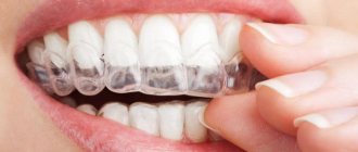
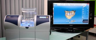
The latest computer technologies have already begun to be actively used in this area, at all stages of the manufacture of prostheses. Modern and advanced equipment for creating prostheses is CEREC computer technology, with which you can restore lost teeth in a few hours - from the moment of creating an impression to installing the prosthesis.
A real revolution has also occurred in the world of removable prosthetics. Nylon systems were invented some time ago. They do not cause discomfort; due to their softness and elasticity, they are characterized by rapid adaptation of the patient to the system.
Aesthetic dentistry
The beauty and attractiveness of a smile is impossible without absolute health of the enamel. To achieve this result, several new technologies have been invented that help achieve dental perfection.
Veneers
Such onlays help to significantly change the shape of teeth. The plates applied to the enamel surface are made of very durable materials that give an ideal appearance. Most often they are placed in the smile area to provide the desired color and contours.
The positive thing about installing veneers is that natural teeth do not need to be ground or prepared, so the native tissues are not damaged. Outwardly, they completely imitate perfect enamel, the material is even slightly transparent, like natural fabrics.
With the help of such overlays it is possible to get rid of cracks, small chips and interdental gaps. Modern veneers can perform their functions for about 7 years, after which they need to be replaced with new ones.
Lumineers
In aesthetic dentistry, this technology is considered the newest and most advanced, but due to its novelty, it still tops the list of the most expensive. In addition, the technique is patented by the manufacturer, and therefore holds a monopoly on the creation of these porcelain onlays.
In order to place lumineers, the dentist must take impressions of the teeth and send them to the manufacturer in the USA. The most modern dental clinics do this using 3-D scanning, which somewhat speeds up the process, which takes at least a month.
The method is completely non-traumatic, since it does not require grinding for installation, just like when removing lumineers. The manufacturer provides a 15-year warranty, after which the lumineers must be replaced with new ones.
Ultraneers
Using this method, it is possible to hide small chips, curvatures and other minor imperfections.
Despite the fact that ultraneers are very thin (their thickness does not exceed 0.3 mm), they are extremely durable, so you can safely eat any solid food with them. Their installation in most cases is carried out without the use of any type of anesthesia.
Restoring a beautiful smile in a day is a reality
Those specialists who strive to immediately adopt advanced technologies are increasingly less likely to use classical implantation, which consists of two stages.
In a short period of time today it is possible to restore not only the functionality but also the aesthetics of teeth
New treatment methods and protocols make it possible to restore teeth and return them to full functionality in a very short period of time - from one to seven days.
It is worth pointing out that these techniques are developed for absolute or almost complete edentia, therefore they are most effective and safe in such cases. Two-stage classical implantation involves implanting durable titanium implant rods into the jaw bone, which fully replace lost natural tooth roots. But in accordance with this technique, about 4-6 months should pass until they completely take root in the bone tissue.
For information! If significant bone tissue atrophy is detected before implantation, then bone grafting is necessary - bone augmentation to the required volume. This adds complexity to treatment and makes it significantly more expensive. This additional procedure is only necessary when using the classical method.
Nowadays, patients are more often offered dental implantation in one stage - a method in which the load on the new implant is installed immediately. This means that a beautiful smile thanks to excellent dentures will return on the same day or within a week at most. We will talk about these technologies after describing the main trends of the most famous brands of implantation systems.
Treatment of caries
Poor nutrition, the use of certain medications or heredity can cause the development of caries. Fortunately, this common condition can be treated with the latest techniques used by dentists.
Chemical-mechanical treatment
With this method, infected dentin is removed not only painlessly, but also silently. Healthy tissues are not damaged at all.
A special gel is applied to the affected area, which softens carious deposits. After its removal, a small cavity remains, which requires less filling material. Chemical-mechanical treatment does not require anesthesia, since only necrotic areas of dentin that do not have innervation are removed.
Laser technology
With this technique, drilling is not performed, since the affected tissue is burned out by the laser beam.
In parallel with this, disinfection of the cavity occurs, since the laser kills any bacteria.
Computer techologies.
Computer-aided design will be widely used to build models of the dental system at the stage of preparation for treatment. The use of 3D printers is revolutionizing dental laboratories. Clinics will turn into technologically advanced complexes that eliminate the possibility of medical errors.
New technologies will reduce the cost and speed up the production of dentures. The image of the prepared tooth will be processed by a special program and transferred to the device, which will produce a crown that fits exactly in size directly in the office.
The capabilities of 3D printing will eliminate all intermediate links and significantly speed up and simplify the dentist’s work. Such complex schemes are already being developed by Envisiontech and Stratasys.
Children's dentistry
Dental procedures have always caused pain, so persuading a child to visit the dentist is not an easy process. New technologies can make children visit clinics more willingly and fearlessly.
Ozonation
German technology, which is based on the disinfecting properties of ozone, allows you to do without the use of a drill during treatment.
Ozone is supplied through a small silicone tube, which operates completely silently. The process of complete disinfection lasts only 30-40 seconds. After this, a compound is placed into the cavity, which strengthens the adjacent dental tissues.
If the caries was superficial, then after ozonation you can even do without installing a filling.
ICON
The method is suitable for fixing the initial stages of caries, when changes appear in the form of whitish spots. After treating such chalky areas with ICON, the color of these areas returns to natural.
This method is not recommended for permanent teeth, but for milk teeth it is ideal.
VR surgery
The entertainment industry has been actively introducing VR into its products for several years now. Today, this technology is reaching a new level and is gradually penetrating into other areas. Thus, virtual reality has become part of the educational training of future doctors.
Case Western Reserve University, together with Microsoft and HoloLens, has developed an anatomy course for medical universities: it allows you to study in detail the features of the human body in three-dimensional images.
The University of Pennsylvania School of Dentistry uses VR to simulate surgeries and other procedures. This training prepares future dentists for their first real practice. In addition, it can be used by already experienced specialists for preliminary testing of complex operations.
What's the reality
Virtual reality technology has great prospects both in dentistry and in medicine in general. First of all, I mean multiple magnification, which gives the surgeon unlimited possibilities. When you have a small surgical field, and even in a hard-to-reach place, you have to adjust and work in an awkward position, and this puts a colossal load on your back and neck. Also, the eyes are subject to constant reaccommodation (Changing the focus of vision from near objects to distant ones and vice versa. - Ed.).
With VR, all these inconveniences become a thing of the past. For example, I already operate on patients in VR. The equipment had to be assembled independently: it consists of a small microscope with two cameras, which is located above the patient’s oral cavity, and VR glasses, onto which a three-dimensional and enlarged image is transmitted. My microscope gives 16x magnification, but you can get even more. Today this is not a problem.
In addition, VR will play a significant role in the development of robotics: a robot necessarily needs stereoscopic vision in order to quickly and without human assistance react to what is happening during an operation.
Growing teeth
The development was first implemented in Zurich in 2017. After the removal of a permanent tooth, a new element was grown in its empty alveolus using stem cells.
The entire procedure takes about 2 months and allows you to restore one missing element or a complete row of teeth.
Chinese scientists have refined this technique and were able to create a natural tooth in a test tube, after which it was implanted into the jaw. This technology took only 2 weeks.
It is expected that this technology will become available to clients of dental clinics in 2020-2030.
Diagnostics at the level of artificial intelligence
Today, experts in various fields have high hopes for artificial intelligence technologies. Dentists are no exception. For example, some experts are confident that in the near future, artificial intelligence systems will help analyze and diagnose various oral diseases and even independently propose treatment options, taking into account the characteristics of a particular patient and calculating all possible consequences.
The first steps in this direction have already been taken. ParallelDots, a company specializing in developments in the field of artificial intelligence, is already testing its cloud system Dentistry.AI in American clinics. The technology helps doctors find cavities in teeth based on X-ray analysis. According to the developers, the algorithm determines in a few seconds the areas where caries is most likely to develop. This information helps the dentist create a further plan for examining and treating problem areas.
What's the reality
Artificial intelligence is a very interesting and promising field, but the fact that in the near future this technology will be able to independently prescribe treatment to patients borders on fantasy. Yes, AI can perform some specific, narrow tasks. For example, analyze x-rays, as indicated in the example above. However, you should not rely entirely on these results: the diagnosis cannot be determined by just one x-ray; this requires a comprehensive examination.
You also need to understand that for now artificial intelligence still works according to a given algorithm.
Before AI can independently identify a disease, it needs to be provided with all the information on all existing diseases, even if only in the field of dentistry. And here there is a dead end, because no one has this information.
I'm not even talking about the fact that in medicine, in principle, there cannot be any algorithms, since each case is individual.
Data protection
The simplest means of backing up data today is to copy it from one computer to another. With the current high reliability of computer equipment, the likelihood that hard drives on two computers will fail on the same day is zero. Not “almost zero”, but “absolutely zero”. This copying can be set to automatic mode using the Task Scheduler program, which comes standard with any Windows.
We do not recommend using previously popular mirrored disk systems today, much less RAID controllers. In fact, they fail much more often than they seem, and simply copying files over the network from one computer to another is much more reliable and simpler.
Digital dentistry: the golden age of computer diagnostics and treatment planning
Improvements in digital dentistry are directly dependent on the progress of technology in the computer field, even if they are associated with the development of some special transistor or microchip.
The digital revolution, which continues to gain momentum, began back in 1947, when engineers Walter Brattain and William Shockley of Bell Laboratory John Bardeen invented the world's first transistor, for which they subsequently received a Nobel Prize. Transistors of those times, in addition to being quite slow, were also excessively large, for this reason it was difficult to include such a design in some kind of integrated circuit, not to mention a microchip. Unlike their arch-relatives, the size of modern transistors may not exceed the size of several atoms (1 atom thick and 10 wide), while such elements operate very quickly at a frequency of several gigahertz, and can be compactly placed in the structure of some small board or computer circuit. For example, a Core processor (from the i-series), released in 2010, contains about 1.17 billion transistors (!), although in the mid-70s similar processors could contain no more than 2300 such structural elements. But this is not the limit. According to Moore's law, every 1-2 years a new microchip is born, which is twice as powerful as its predecessor. It is therefore not surprising that dentistry is currently experiencing something of a boom, with the industry's scanning, analysis and manufacturing capabilities continuing to evolve rapidly. Digital radiography will no longer surprise anyone, because increasingly, doctors are using completely virtual diagnostic and treatment planning protocols, which help achieve the desired results.
One of the innovations that has literally become a routine procedure is the acquisition and analysis of digital prints. For the first time, a similar procedure was tried back in 1973, when graduate student Francois Duret at Claude Bernard University (Lyon, France) proposed taking impressions using a laser in order to later use them in the course of complex diagnostics, treatment planning, manufacturing and fitting of future restorations.
Almost ten years later, in 1983, Werner Mörmann and Marco Brandestini managed to invent the first intraoral scanner for therapeutic dentistry, which ensured impression accuracy of 50-100 microns. The principle of operation of the scanner was based on the capabilities of triangulation to obtain instant three-dimensional (3D) images of teeth, from which future therapeutic structures could be milled. The latter, in the form of inlay-type inlays, were obtained using CEREC (CERamic REConstruction or Chairside Economical Restoration of Esthetic Ceramics), but the constant progress of technology subsequently determined the possibilities for the manufacture of full-fledged single restorations and even entire orthopedic prostheses. CEREC itself has also improved. Thus, a conventional milling machine was upgraded to the CEREC OmniCam system (Sirona Dental), which ensures the most precise designs. The increased attention to this particular system is due to the role of CEREC as a pioneer of such devices on the market, which occupied a leading position for several decades, while other analogues found their feet and improved to the level of an already popular installation. There are currently several fairly accurate and powerful systems for taking intraoral optical impressions and fabricating CAD/CAM restorations, but they all use the same principle of triangulation to form the image. The most famous of them are TRIOS (3Shape), iTero Element (Align Technology), True Definition Scanner 3M (3M ESPE).
Advantages of modern digital systems
All modern digital systems for taking impressions are characterized by high accuracy of replicas of the structures of the dentofacial apparatus, and, of course, complete non-invasive manipulation. Unlike conventional impressions, the resulting images can be easily adapted to all conditions during planning and treatment, and the technique for obtaining them is so simple that it can be learned in a few steps. Thus, these impressions are not only more effective, but also more convenient for the patients themselves, and also increase the cost-effectiveness of dental procedures in general.
Another great advantage is that thanks to digital impressions, the doctor has the opportunity to receive not a negative image of the prosthetic bed, but a real copy of the teeth in 3D format, which can easily be assessed for the presence of shooting defects and the accuracy of individual boundaries.
Also, such impressions are just a volume of digital information, which literally saves physical space both in the dentist’s office and in the dental technician’s laboratory. Studies conducted to compare conventional and digital impressions have shown better accuracy of the latter, while they differ from conventional ones in that they do not need to be disinfected, and there is no need to take into account the time of obtaining the impression in order to minimize the effects of shrinkage and changes in the primary size impression material.
The main advantage of digital impressions is that they can easily be included in the process of comprehensive planning and treatment with the ability to predict future results of dental rehabilitation. Direct copies of teeth and adjacent anatomical structures are visualized in a direct projection immediately after the scanning procedure, and the high resolution of the resulting images helps to assess the condition of existing restorations, defects, the size and shape of edentulous areas, the type of occlusal contacts, as well as the usefulness of the tubercle-fissure closure.
New digital systems, such as TRIOS, CEREC Omnicam, even provide an imitation of the color of the structures of the oral cavity on the resulting replicas, thus helping to more naturally perceive the relief, shape and color of teeth and gums. In addition, such opportunities help the doctor to take a more differentiated and thorough approach to the issue of choosing a restoration material (metal, ceramic, composite), as well as take into account the presence of bleeding and inflamed areas, areas with accumulation of plaque and stone, and take into account color transitions between teeth, which is extremely important for highly aesthetic restorations. Optical impressions are also an effective tool for discussing the initial clinical situation and possible treatment options with the patient. After obtaining a three-dimensional image, problems with defective restorations, the influence of factors of abrasion, superocclusion or angulation of teeth on the future result of treatment can be clearly explained to the patient, without waiting for the receipt of plaster models (photo 1).
Figure 1. Occlusal view of the maxillary optical impression: the image allows detailed examination of the inherent composite and amalgam restorations, the lingual cusp fracture of the maxillary second premolar on the left, the metal-ceramic crown in the region of the maxillary first molar on the right, and the implant-supported prosthesis in the anterior region .
All this encourages the patient to actively participate in the treatment process and conduct an active dialogue with the doctor, understanding all possible risks and changes in their own dental status. Digital files of optical impressions are saved in surface tessellation files (STL) format, and, if necessary, physical models can be produced from them using substrate or additive technologies.
Preparing for optical impressions
Like conventional impressions, their digital counterparts are also sensitive to the presence of blood or saliva in the tissue area of the prosthetic bed, so the surface of the teeth must be adequately cleaned and dried before scanning. You should also take into account the effect of surface reflection, the risk of which may be triggered by specific lighting conditions of the working field. The use of light sticks helps to achieve an adequate level of illumination in the area of the chewing teeth, but at the same time, access of the photocell to this area still remains difficult, and irritation of the palate can provoke a gag reflex.
However, digital impressions are only part of a comprehensive patient assessment, which should also include a general and medical history, clinical extra- and intraoral examination results, and a clear understanding of the patient's complaints and personal expectations for the future. results of the intervention. It is by analyzing all of the above data that it is possible to draw up a comprehensive treatment plan focused on a specific patient and the characteristics of his clinical situation. The latest technological capabilities help the dentist independently simulate future restorations in the area of defective areas, coordinating the design, contours, position, dimensions, size of proximal contacts and the visualization profile with the patient, taking into account the individual characteristics of the occlusion, and thus ensuring the most adapted and expected temporary designs.
However, the main limitation of current dental digital technologies is that they are difficult to fully incorporate eccentric jaw movements and the implications of key occlusal determinants for future restoration design. Due to the fact that recording the exact relationship of the upper jaw to the plane of the defective area is a very difficult task, it is also difficult to establish the objective inclination of the occlusal plane relative to the group of anterior teeth at the moment of their physiological closure.
Equally difficult tasks are the analysis of the articular path, the range of transversal movements, etc., that is, the use of digital impressions is also a kind of challenge for the construction of prosthetic structures, taking into account all physiological or changed parameters of occlusion. Obtaining accurate impressions from soft tissues is also very problematic, especially in areas of completely edentulous residual ridges. However, the ability to visualize 3D, as well as eliminating the need for plaster casting and wax-up, significantly speeds up and tailors the treatment process, helping to achieve the most patient-centered dental rehabilitation results.
The digital planning protocol is demonstrated in Photo 2-7. The patient sought help with an edentulous upper right central incisor (Figure 2).
Photo 2. The patient sought help for edentulous lateral incisor. During the treatment, it was planned to make a structure supported by the central incisor and canine.
After analyzing the individual wishes of the patient, the results of a comprehensive examination and the prognosis of future treatment, it was decided to use a fixed lithium disilicate prosthesis as a replacement structure. A virtual mock-up of the future restoration helped determine the required length, width and profile of the contact surfaces to achieve the greatest possible mimicry of natural tissues (photo 3).
Photo 3. Digital mock-up of a prosthesis replacing a missing tooth.
After this, the supporting teeth were prepared (photo 4), and then using the scanning method, virtual impressions of the prepared units and antagonist teeth were obtained, which were further analyzed in a digital articulator (photo 5).
Photo 4. Occlusal view of the optical impression of prepared teeth with retraction threads.
Photo 5. Virtual articulation of optical impressions of the upper and lower jaws.
The optical impression data was also successfully used to analyze in detail the width of the final line of the preparation area, the routes of insertion of the structure, the level of deliberate tissue reduction in the area of the axial walls and occlusal surface, as well as to verify the undercuts, which were marked in red (Figure 6).
Photo 6. Analysis of the optical impression for the presence of undercuts. Undercuts are marked in red on the labial side of the central incisor and on the mesial side of the canine.
Another advantage of digital impressions is that preparation errors can be corrected during the same visit, based on the information obtained during the scan, and then the manipulation can be repeated on the corrected area of the prepared teeth. After this, the digital files are sent to a technical laboratory for the production of future restorations using milling machines. An example of the final design is shown in photo 7.
Photo 7. The restoration obtained from the optical impression is tried on the model.
CBCT and scan protocol
The use of digital capabilities at the stages of diagnosis and treatment planning is not some kind of innovation, but rather is considered as a fairly well-reasoned approach to the rehabilitation of dental patients. For decades, dentists have used specialized software to visualize three-dimensional computed tomography (CT) scans: to analyze the growth of anatomical structures in the maxillofacial region; joint pathologies; bone architecture; sizes of individual sections of teeth and jaws; positions of vital organs such as blood vessels and nerves, as well as the boundaries of the maxillary sinuses and the position of impact teeth; diagnosis of tumors and neoplasms. But CT diagnostics is probably most influential in preparation for dental implantation and planning of maxillofacial reconstructive surgery. Technological progress has gained new momentum with the development of cone beam computed tomography (CBCT), which, compared to conventional CT, is characterized by a lower level of radiation exposure and a lower cost of the device. Indeed, the total radiation from a CBCT scan is on average 20% less than from a helical CT scan, and is approximately equal to that from conventional periapical radiography.
CT and CBCT diagnostic results are saved digitally in the standardized DICOM (digital imaging and communication in medicine) file format. In combination with a radiographic template made from a diagnostic wax-up, CBCT data can be successfully used to plan the position and angulation of implants, taking into account the fixation of the future prosthetic structure, based on the existing conditions and volumes of the bone crest (photo 8 - photo 11). Currently, there are two different protocols for implementing radiographic templates into the DICOM data structure for planning future surgical procedures. The first, called the dual-scan protocol, performs the acquisition procedure separately for the surgical guide and separately for the patient, provided that the surgical guide is installed in the oral cavity. Fiducial markers in the structure of the template itself help in the future to quite accurately combine the two resulting images. At the same time, the level of scanning errors is practically reduced to a minimum, and templates can be produced using various adapted software (photo 12).
Figure 8. Use of cone beam computed tomography and specialized software to plan the implantation procedure. The X-ray template together with the CT model was used to plan the future position of the implant.
Figure 9. Use of cone beam computed tomography and specialized software to plan the implantation procedure. The X-ray template together with the CT model was used to plan the future position of the implant.
Figure 10. Use of cone beam computed tomography and specialized software to plan the implantation procedure. The X-ray template together with the CT model was used to plan the future position of the implant.
Figure 11. Use of cone beam computed tomography and specialized software to plan the implantation procedure. The X-ray template together with the CT model was used to plan the future position of the implant.
Photo 12. Example of a surgical template made using a digital dual-scan design.
The second protocol requires only one scan of the patient along with a surgical guide placed in the oral cavity. The obtained data is imported into the implantation planning program without the need for additional image processing. As in the case of the double scanning protocol, the doctor has the opportunity to reasonably plan the position and angulation of the implants, based on the spatial location of the surgical template obtained as a result of the preliminary diagnosis. Three-dimensional radiographic images obtained using a single-scan protocol can be combined with digital templates for future restorations, which are made based on intraoral optical impressions (or scans of models), using existing natural teeth as markers. In this case, different digital masks can be used graphically for bone, teeth, gums and implants (photo 13 and photo 14), and the use of teeth as fiducial markers significantly increases the accuracy of planning the position of future implants.
Figure 13: Optical impression and digital reproduction were combined with CBCT scan results to position the implants during complex treatment. This patient requires a sinus lift procedure to adequately place the implants (blue outlines of the teeth obtained from the wax reproduction/optical impression, red indicates the outlines of the soft tissues).
Figure 14: Optical impression and digital reproduction were combined with CBCT scan results to position the implants during complex treatment. This patient requires a sinus lift procedure for adequate installation of implants (blue indicates the contours of the teeth obtained from the wax reproduction/optical impression, red indicates the contours of the soft tissues).
Similar marker points in the structure of the surgical template, unfortunately, cannot provide a similarly high level of precision. Regardless of the scanning protocol used, the 3D digital imaging, optical scanning and software capabilities provided provide unique tools for future iatrogenic intervention planning in the hands of a skilled dentist. Thus, taking into account the position and contour of the soft tissues, the size and quality of the residual bone crest, as well as the location of the vessels and nerves, the doctor can provide the safest implantation algorithm, while predicting not only functional, but also aesthetic results of rehabilitation. The surgical template, regardless of the protocol for obtaining the scanned image, ensures accurate positioning of the implant, eliminating possible operational errors that may arise during surgery. Virtual planning of dental rehabilitation helps the doctor achieve the safest, and at the same time, patient-oriented results in the treatment of aesthetic and functional defects.
Conclusion
Intraoral optical scanners continue to be constantly modified, becoming faster, more accurate and miniature devices that are so necessary in dental practice. Considering the progressive development of 3D imaging technologies and adapted image processing software, it can be firmly concluded that today's dentists live in the golden age of digital technology. Such innovations help achieve more accurate and precise diagnostic results, planning and iatrogenic interventions, while increasing comfort during dental treatment. Therefore, it is critical that new digital technologies emerge promptly and continue to develop within the walls of dental offices and clinics.
Authors: Radi Masri, DDS, MS, PhD Carl F. Driscoll, DMD Se Jong Kim, DMD William M. Wahle, DDS
Where to place network equipment?
Imagine in your mind the entry point of the Internet cable, workplaces. The location of the network hub should ideally be equally distant from these points in order to pull less cable. But in life, of course, they choose “special” places for this - built-in niches where there is access and free space, sometimes the equipment is placed in some technical rooms, and sometimes right in the corridor, only in a beautiful small installation cabinet.
Choosing a cabinet to house network equipment is the topic of a separate article. The standard width of these cabinets is 19 inches. Accordingly, the purchased equipment must be rack-mountable. Some rack-mountable hubs have special mounting ears. You can learn about the possibility of installing equipment in a rack cabinet from the product specification in the online store or from the seller. There are different heights of cabinets - knee-high (hanging), waist-high, full-height and above 2 meters. Choose depending on your needs. But I’ll say right away - you shouldn’t skimp on the height of the cabinet, otherwise it will be inconvenient to work with it. The optimal height, in my opinion, is 160-180 cm.
Installation (telecommunications) cabinet for network equipment. Inside: patch panels (reciprocal parts of computer sockets in offices), hubs, possibly a server, an uninterruptible power supply (bottom), sockets for connecting additional devices.
Of course, you need to think about how the wires will be routed. Are there solid walls nearby, are there suspended ceilings, how to get through the doors - all these questions need to be clarified in order to make a decision on choosing a place to place the concentrator.
Are wall outlets necessary? Yes. Direct crimping of wires with connectors is bad manners. In such connectors, the contacts are broken very quickly, and you cannot understand what is wrong, the network is constantly “buggy”. When connecting to an outlet, use a factory patch cord (a wire with connectors on both sides), where breaking the contact is almost impossible.