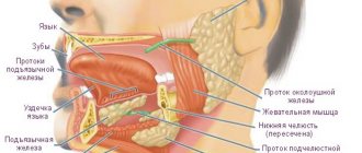
Features of the functioning of the salivary glands
These organs are located in the oral cavity, and their main function is to secrete saliva, which helps moisturize the mouth. The organ is involved in the eating process and helps improve the digestion of food.
Experts identify the following possible organ diseases:
- Sialadenosis. This is a reactive-dystrophic change in the organ.
- Sialadenitis. It is caused by pathogenic bacteria. The disease occurs in the form of acute inflammation.
- Chronic inflammation;
- Actinomycosis;
- Stone formation;
- Cyst;
- Tumor;
- Trauma and organ damage.
The doctor prescribes therapy depending on the specific disease.
General research methods
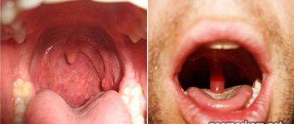
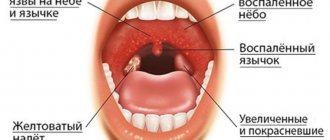
All categories of patients, including those who came to see a doctor for the first time, undergo a visual examination of the oral cavity. Next, the specialist assesses the condition of the glands by palpation.
When palpating the area, the doctor looks at the condition of the oral mucosa, assesses the size and consistency of the organs. It is also necessary to establish pain and mobility of the glands. A visual examination can also reveal the amount of saliva in the mouth and its consistency.
To understand the general course of the disease, the doctor asks the patient a series of questions. First of all, the doctor is interested in what kind of sensations bother the patient, how long the condition lasts, and also what factors influence the worsening of sensations. The presence of dryness or a strange taste in the mouth is also important for diagnosis.
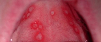
Important. The difficulty in making a diagnosis is that different oral diseases have similar symptoms. In this case, to clarify the diagnosis, a general urine test and a special blood test are prescribed.
Radioisotope diagnostics of salivary gland diseases
Radiosialography. Radiosialography is used to determine the quantitative characteristics of the functional state of the salivary glands. The technique consists of recording radiation intensity curves simultaneously over the parotid salivary gland and the heart after intravenous administration of a solution of sodium iodide labeled with 131I - Na131I or sodium pertechnitate labeled with 99mTcO4.
Radiosialography reflects the state of the concentration and excretory function of the parotid salivary glands, which is especially valuable for the functional diagnosis of chronic mumps (stages I-III) and xerostomia (grades I-III).
During radiosialography, 131I is administered intravenously at the rate of 0.4 mCi per 7 kg of body weight or 99mTc in an amount of 50-70 μCi (in 1 ml of solution); both drugs are administered on an empty stomach. To eliminate interference during radiosialography, the thyroid gland is blocked.
Radiosialography is performed using a URU-type installation, placing scintillation sensors over the parotid gland and the heart (along the left parasternal line in the fourth intercostal space) with the patient lying down. Radiometer operating conditions: discrimination level 10, time constant 20, counting factor (S/P) 100 (sometimes up to 300). Sensitivity is set according to the 131I standard individually for each subject.
To avoid errors during radiometry, the sensors are installed at a certain angle so that the recording of the curve is not influenced by the radiation of the opposite gland.
A normal radiosialogram (RSG) with a functional test (stimulation of salivation with 5 g of sugar, 15 g of lemon) consists of 3 curves: two from registration of radiation from the parotid salivary glands (left and right) and one from registration of radiation from the heart (Fig. 62). On the curve of each RSG obtained from the parotid gland, four elements (segments) are distinguished:
- 1) vascular segment (the moment 131I enters the gland and surrounding tissues with blood);
- 2) concentration segment - plateau (period of accumulation of 131I in the parotid gland after 20-25 minutes);
- 3) excretory segment (decrease in the level of the curve as a result of a drop in radioactivity in the gland during a functional test, the release of saliva into the oral cavity and its swallowing);
- 4) continuation of the concentration period (after food intake and the time of maximum accumulation of 131I in iron).
Radiosialography with 99mTc is carried out in cases of repeated observations to study the dynamics of the pathological process, since the half-life of 99mTc is 6.04 hours, and 131I is 8.06 days.
For differential diagnosis of the salivary glands (especially tumor and chronic inflammatory processes), radiometric examination of saliva using 131I is used.
131I in an amount of 10 µCi in 20-30 ml of distilled water is given to the patient to drink on an empty stomach. After 2, 4, 24 and 48 hours, using a Krasnogorsky capsule, saliva is collected from the excretory duct of the parotid gland after stimulation with 15 g of lemon (or 30 g of sugar, etc.). The saliva samples are then radiometered using a well meter.
Studying the capabilities of this method made it possible to establish certain diagnostic signs inherent in individual diseases. Thus, with xerostomia, there is a sharp increase in the 131I content in samples of mixed saliva with low secretory ability of the salivary glands; with salivary stone disease of the submandibular gland, the 131I content in samples of mixed saliva and saliva of the submandibular and sublingual salivary glands is high, and the secretory ability of the glands is not changed; Acute mumps is characterized by a high level of 131I concentration in saliva samples of the parotid salivary gland with a low level of its secretory function; in chronic mumps, a high concentration of the radioisotope is observed in saliva with a reduced secretory ability of the affected gland.
With some benign tumors of the parotid region, for example lipoma, the saliva of the parotid gland contains an increased amount of 131I with unchanged secretory ability. Fistulas of the parotid gland are accompanied by a slight increase in the level of 131I in saliva and a sharp decrease in secretion.
Radio scanning of the salivary glands. Compared to radiosialography, this method has the peculiarity that it allows one to obtain a visualized topical characteristic of the functional state of the parenchyma of the parotid and submandibular salivary glands.
Scanning of the salivary glands is carried out using 99mTc, which is administered intravenously in an amount of 2-3 μCi. The study is carried out on a topographer with the sensor located at a distance of 0.5 cm from the skin, 30 minutes after the administration of the isotope.
In case of chronic nonspecific inflammatory processes of the parotid salivary glands, the scanogram shows an increase and change in the shape of the salivary gland.
Xerostomia is characterized by a heterogeneous scanogram. The submandibular glands and the area of radioactivity in the oral mucosa are clearly visible. Sometimes there is no clarity in the image of the parotid and submandibular glands; their images on the scanogram merge, areas of absence of radioactivity of various shapes and sizes are observed.
With such neoplasms as mixed tumors and cylindromas, defects corresponding to the location and size of the tumors are revealed in the image of the affected salivary glands: changes in position are also noted. the shape and contours of the image of the salivary gland due to its displacement by the tumor. Depending on the degree of compression of the salivary ducts by the neoplasm and accompanying inflammatory or dystrophic processes, disturbances in the functional ability of the glands may occur, most pronounced during functional tests.
Private research methods
Private methods of examining patients with diseases in the salivary glands are used if a certain ailment is detected at the stage of general diagnosis. If the patient has concomitant diseases of the digestive tract, he is also prescribed private diagnostics.
We also recommend viewing: Is it possible to cure the pancreas with the help of marigolds: benefits of the plant and application
X-rays are often used to make an accurate diagnosis. Private examination methods also include sialometry and sialography.
X-ray allows you to see the location and size of stones in the organ. This type of diagnosis also allows you to see the glands in different projections.
Sialometry is used to study the efficiency of the salivary glands. Using it, you can estimate the amount of secreted secretion per unit of time. The collection of tests is carried out strictly on an empty stomach. The procedure lasts for different times and depends on the patient’s well-being. As a rule, this is no more than 6 minutes. When examining secretions, the laboratory technician evaluates the amount of saliva, as well as the presence or absence of sediment and mucus.
Sialography is an X-ray examination using contrast materials. The resulting image shows the shape and size of the organ, as well as the presence of stones.
Pantomosialography allows you to assess the condition of several salivary glands at once. This method also involves the injection of a contrast agent into the organs.
If signs of an infectious disease are detected in a patient, a cytological examination of saliva is prescribed. A saliva test for the content of various components can also show the patient’s condition. During the study, specialists evaluate the color, viscosity and transparency of the discharge.
Important. Cytology examination of saliva is carried out using special instruments. You can see changes in the discharge under a microscope.
If a disease of the salivary ducts is suspected, probing is performed using special instruments. It shows the location and condition of the ducts. It should be remembered that this type of examination is quite traumatic, since the probes can damage the walls of the salivary ducts.
Special research methods
The examination of the salivary glands can also be carried out using specialized equipment. This includes ultrasound equipment, CT, MRI, and biopsy instruments.
Sialosonography allows you to detect changes in the tissues of the salivary glands using ultrasound. The specialist evaluates the size and shape of the organs, their location in relation to neighboring tissues, as well as the structure of the gland. The main advantage of this method is the absence of side effects and possible short periods of time between adjacent procedures. The method for detecting pathologies is also highly efficient.
We also recommend viewing: Excretory duct in the area of the parotid gland: meaning, diseases (mumps), treatment
Computed tomography is based on scanning tissue and detecting changes in the structure of the salivary glands. Sometimes a contrast agent is used during the study, which can significantly increase the reliability of the data obtained.
MRI can be used to determine the diagnosis for almost any salivary gland disease. In particular, MRI is often used in the protocol for studying the presence of sialadenitis of the parotid glands. The MRI machine is based on the interaction of hydrogen nuclei of tissue cells and magnetic fields. This method allows you to clarify the stage of the disease, as well as detect changes in the structure of the glands even in the absence of visible symptoms of the disease.
Study of the salivary glands
Study of the salivary glands. When examining the salivary glands, first of all, attention is paid to the color of the skin and changes in tissue contours in the area of the anatomical location of the glands. If the contours of the leucorrhoea are changed due to swelling, then its size and character are determined (spread out, limited, soft, dense, painful, areas of softening, fluctuations). If the change in the contours of the gland is caused by a tumor process, then the exact localization of the tumor in the gland, the clarity of its boundaries, size, consistency, mobility, and the nature of the surface (smooth, lumpy) are established. It is determined whether there is paresis or paralysis of the facial muscles and damage to the masticatory muscles.
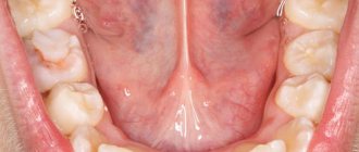
Then the excretory ducts, as well as the sublingual salivary glands, are examined in the oral cavity. To examine the mouths of the excretory ducts of the parotid salivary glands, which are located on the mucous membrane of the cheek along the line of closure of the teeth at the level of the first upper molar, the corner of the mouth is pulled forward and slightly outward with a dental mirror or a blunt hook. Lightly massaging the parotid salivary gland, observe the secretion of saliva from the mouth of the duct, while determining the nature of the saliva (transparent, cloudy, purulent) and at least approximately its quantity. In order to examine the duct of the submandibular and posterior section of the sublingual salivary glands, the tongue is moved to the side with a dental mirror. In the anterior section of the sublingual region, the outlet of the ducts of the submandibular and sublingual salivary glands is examined. By massaging the submandibular salivary gland, the nature and amount of its secretion is determined. By palpation along the duct from back to front, the presence of salivary stone or inflammatory infiltrate in the duct is determined. By palpating from the oral cavity and submandibular region (bimanually), the size and consistency of the submandibular and sublingual salivary glands are more accurately determined. If the stone is localized in the submandibular salivary gland, it can be enlarged, dense and painful. For certain indications (suspicion of the presence of a stone, deformation of the duct, its narrowing, fusion) and the absence of inflammatory phenomena, careful probing of the duct can be performed.
Preparing the patient for anesthesia
Sometimes, when studying secretory function, saliva is collected from two symmetrical salivary glands by introducing special cannulas into their ducts. Saliva is collected in the morning on an empty stomach (the patient has been given 8 drops of a 1% pilocarpine solution beforehand) for 20 minutes into two graduated tubes held by the patient. Noting the amount of saliva in each test tube (according to T.B. Andreeva, normally 0.9-5.1 ml is released from the parotid gland in 20 minutes, more often 1.1-2.5 ml, from the submandibular gland - 0.9 -6.8 ml, usually 1-3 ml), determine the pH of saliva, its viscosity, the amount of protein, inoculate the saliva for microflora and determine the sensitivity of the latter to antibiotics. If the function of the glands being examined is normal, the above indicators will be the same, but if one of the glands is affected, they will be different.
Based on complaints, anamnesis, external examination and palpation, the nature of the disease (inflammatory processes, systemic lesions, salivary stone disease, tumors, tumor-like formations, injuries and their consequences) can be established with a high degree of certainty.