October 12, 2020
Many of us are afraid to even think about such a disease as cancer of the gums of the upper or lower jaw. Nevertheless, the pathology occurs in 6-9% of clinical cases involving oncological tumors growing in the oral cavity. It is important for a modern person to have at least basic information about the problem, to know what the initial stage looks like, to look at photos, and to have an idea of preventive measures. After all, this will help reduce risks, and in extreme cases, detect the disease as early as possible, when the chances of a favorable outcome are still high. The editors of UltraSmile.ru will help you replenish the storehouse of knowledge regarding the intricacies of this topic.
Gum cancer occurs in 6-9% of clinical cases
What is gum cancer
Gum cancer refers to a tumor of the soft tissues of the upper and lower jaw, which is of a malignant nature. What does malignant mean? The cells that make up the tumor divide uncontrollably and grow into nearby organs, affecting them and causing a phenomenon such as metastases (secondary tumor cells).
The danger of such a malignant process occurring on the gums and in the oral cavity lies in the fact that in the immediate vicinity there is a cluster of lymph nodes and many capillaries. According to research, metastasis mainly occurs in the lymph nodes (about 50-70% of all severe cases), but in the brain and other vital organs the likelihood of metastases penetrating is insignificant (registered in no more than 5% of all cases).
Tumors arising on the gums are generally classified as oral cancer, as are neoplasms localized on the tongue (this is the most common form of oral cancer, accounting for 52-55% of cases1), cheeks, and palate.
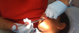
If you have never seen what gum cancer looks like, take a look at the photo below. Although it is worth emphasizing that the external signs and symptoms of pathology largely depend on the stage of its development, each of which we will examine further.
This is what gum cancer looks like
Treatments for oral cancer
For successful treatment of malignant tumors of the oral cavity, oncologists at the Yusupov Hospital draw up an individual patient management program in each specific case. The choice of treatment method depends on the clinical and morphological characteristics of the tumor:
- Localization and volume of the tumor focus;
- The presence of metastases in the lymph nodes;
- Degrees of histological differentiation of tissue.
If a patient is diagnosed with cancer of the floor of the mouth, surgery is the main treatment method. Squamous cell carcinomas of the mucous membrane of the floor of the mouth at an early stage are treated surgically. Radiologists irradiate areas of regional metastasis. If there is a small, well-localized malignant tumor in the mouth, lymph nodes are removed to prevent metastases. If stage 3 oral cancer is detected, during excision of the primary tumor, surgeons must remove the lymph nodes on the affected side.
Radiologists use various methods of radiation exposure:
- Interstitial implantation;
- Intraoral cone;
- External beam radiation therapy.
The presence of regional metastases in squamous cell carcinoma of the oral cavity is the main reason for the poor prognosis. For this reason, radiologists irradiate areas of regional metastasis before and after surgery.
One of the methods of radiation therapy for malignant tumors of the oral cavity today is interstitial brachytherapy. It allows you to create optimal radiation doses at the site of a malignant tumor, necessary for its destruction, without severe radiation reactions in the surrounding unchanged tissues. Brachytherapy is used as palliative therapy and as a component of radical treatment.
In advanced stages of cancer of the oral mucosa, after the start of radiation treatment, patients undergo cryodestruction of the disintegrating part of the tumor once a week. During the first three days after the procedure, swelling of the surrounding soft tissues develops, bleeding stops, and pain increases. The patient is prescribed oral baths from a decoction of oak bark and the tumor is lubricated with anesthesin dissolved in oil.
After a week, the swelling disappears, the tumor undergoes reverse development, and the size of the tumor is clearly palpable. The patient again undergoes cryodestruction of the entire tumor or part of it. During a course of radiation or chemoradiotherapy, the patient receives two to four cryoprocedures. Good results of cryotherapy are observed in patients with malignant tumors of the edentulous area of the upper jaw, the anterior floor of the mouth, and the anterior two-thirds of the tongue. The exophytic form of squamous cell carcinoma of the red border of the lower lip is successfully cured using cryodestruction in the absence of deep infiltration of the tumor into the muscles.
Timely detection and adequate treatment of cancer of the oral mucosa is the key to a good prognosis. The clinic’s oncologists will conduct a comprehensive examination. Once the diagnosis is confirmed, professors and doctors of the highest category will develop an individual treatment regimen at a meeting of the expert council. If you experience discomfort in your mouth, ulcers or tumor formation, call the Yusupov Hospital.
Author
Yulia Vladimirovna Kuznetsova
Oncologist
Initial or “zero” stage
If you are trying to understand what gum cancer looks like and its initial stage, then in vain. You will not find photos on the Internet with characteristic visual manifestations of the disease, because there are no external signs as such. The only thing that can be alarming is the very first symptoms, namely, slight redness and swelling of the gums, its hardening, which persist for a long time. In this case, the affected area usually has a clear localization and is limited from other tissues; it seems to rise above them.
The disease is asymptomatic. However, dentists will be able to suspect its presence and diagnose it if you undergo annual preventive examinations, and this will significantly increase the chances of successful treatment.
First stage
If gum cancer and its photo, when the initial stage develops, are difficult to find on the Internet, then here the chances of “success” are greater. Indeed, at this stage it is already possible to note both characteristic visual manifestations and other symptoms:
- on the hyperemic area of the gum, a dense white spot appears, or a pimple or ulcer, which rises as a tubercle above the healthy mucosa: in this case, the heterogeneity of the structure of the affected tissues can be distinguished. In some cases, you can see not one, but many small white dots within one tumor. Also, the affected area is penetrated by a network of small blood vessels,
- the neoplasm does not yet hurt, but it already causes discomfort, it can interfere and be felt during chewing food, when touched with a brush,
- When there is a mechanical effect on the tumor, bleeding begins.
At the first stage of the disease, a tubercle appears.
At this stage of development, the disease gradually begins to grow into the thickness of the tissue, but usually covers no more than 2 centimeters. However, if it is detected in a timely manner, the chances of a complete cure are very high. According to studies, doctors help about 90% of patients cope with the disease.
Treatment
Therapy is selected taking into account the type of tumor, the individual characteristics of the patient and the nature of the development of the tumor process. Treatment of gum cancer is recommended to be supplemented by giving up bad habits and correcting your diet.
Treatment of the initial stage
Regardless of the stage of tumor development, doctors prescribe surgery. As part of the procedure, the affected and nearby tissues are removed. If necessary, local lymph nodes are excised during the operation.
If surgery is contraindicated, a course of chemotherapy is prescribed. The procedure involves long-term use of drugs that have a detrimental effect on cancer cells. During chemotherapy, the patient experiences serious disturbances in the functioning of internal organs. Therefore, treatment is complemented by the prevention of nausea and other phenomena that arise during therapy.
In the early stages of tumor development, radiation exposure is also used. This procedure is used in 85% of cases of gum cancer. As part of radiation therapy, a targeted effect is applied to the problem area.
Treatment of advanced stage
In advanced cases, combined treatment is used, which is carried out in several stages. Radiation therapy is used first. This method stops the spread of cancer cells. Subsequently, the surgeon excises the affected tissue and, if the tumor has metastasized, the lymph nodes. If necessary, surgery is performed on the jaw bones.
This is interesting: Transparent blisters on the inside of the lip: what are they and why did they pop up
After treatment, a course of chemotherapy is prescribed, through which distant metastases are destroyed.
Chemotherapy
Experts try to prescribe chemotherapy less frequently. The main factors for carrying out this method are possible contraindications to surgery. Sometimes chemotherapy acts as an auxiliary method to radiation therapy, which helps increase its effectiveness.
Chemotherapy consists of pills and injections. Their task is to stop the growth of cancer cells, and in some cases completely destroy them. The patient is prescribed antibiotics and vitamins. Taking them is necessary to restore immunity, which has been weakened by radiation and chemotherapy.
Many experts, in order to achieve better results in treatment, recommend combining classical methods with recipes and methods of alternative medicine.
It can be:
- acupuncture method;
- oral massages;
- rinses and various compresses, which contain medicinal herbs that help strengthen gum tissue.
Cancers often recur (appear again). Gum cancer is no exception. To reduce the risk of relapse, after recovery the patient must again undergo the examinations recommended by the doctor and donate blood for tumor markers. Typically, a re-examination is recommended after 3-5 months of recovery.
Radiation therapy
Radiation therapy methods involve irradiating the tumor and adjacent tissues (preoperative period), as well as the former site of localization (postoperative period):
- Interstitial radiation therapy;
- Remote radiation therapy;
- Close-focus radiation therapy.
The nature of radiation therapy depends on the stage of the disease: radical, palliative and combined. More than 85% of people with gum cancer are prescribed radiation therapy.
Surgical removal of the tumor
One of the radical methods is surgical removal of the tumor,
preferably in the early stages. There are standards for excision of cancerous tumors, which dictate that the affected tissues be removed along with part of the healthy ones - retreating from the borders of the tumor by 2.5-3 centimeters. In case of severe damage, when the cancer has affected not only the superficial soft tissues, but also the periodontium, teeth, bone and other tissues, radical operations are performed to remove large parts of the jaw, followed by restoration, plastic surgery and prosthetics.
Traditional therapy
Traditional medicine does not cure cancer. Some home remedies can relieve the symptoms of the disease or slow down the division of cancer cells, but they are not able to completely defeat the disease. Many patients simply waste time trying to recover with herbal infusions. The tumor still continues to grow, taking over healthy cells and tissues of the body.
Second stage
What does the second stage of gum cancer look like, what are its symptoms, can you see them in the photo? In fact, in terms of external manifestations, this stage is not much different from the previous one, but the tumor increases in size and can grow up to 4 centimeters. Also at this stage painful sensations appear. But it’s not the tumor itself that hurts. The reaction is given by the nerve endings and tissues that it compresses, so often the pain does not have a clear localization, and a person cannot pinpoint exactly where it hurts. Discomfort intensifies during accidental (during hygienic care, when eating) or intentional touching of the affected area.
In the second stage, the tumor becomes larger in size
“My grandmother had gum cancer. Now I remember that even before she was diagnosed, she began to lose weight sharply, and at the same time she felt very cheerful, ate well and with an appetite. Then the doctor said that this is often one of the symptoms of cancer.”
Alina M., fragment of a review from the dental portal gidpozubam.ru
According to statistics, in 70% of all clinical cases, patients with this stage of the disease are successfully treated.
Stages, classification
Staging of gum cancer is carried out in accordance with the TNM system.
The letter T denotes the characteristics of the primary malignant tumor:
- T0 - no signs of the presence of a primary neoplasm are detected.
- Tis - “cancer in situ”, a malignant tumor does not grow deeper than the most superficial layer of the mucous membrane.
- T1 is a tumor whose largest diameter is 2 cm or less.
- T2 - the largest diameter of the tumor is from 2 to 4 cm.
- T3 - largest tumor diameter more than 4 cm.
- T4 - a malignant tumor grows into neighboring structures.
Correspondence consultation with a doctor
Leave your phone number
The letter N is used to assess tumor damage to regional lymph nodes:
- N0 - during the examination, no tumor foci were found in the regional lymph nodes.
- N1 - there is a lesion in the lymph node on the side of the malignant tumor, the size of the lymph node is less than 3 cm.
- N2 - tumor foci can be found in several lymph nodes on the same or opposite side, their size is no more than 6 cm.
- N3 - lesions in regional lymph nodes more than 6 cm.
The letter M indicates the presence of distant metastases:
- M0 - no distant metastases.
- M1 - distant metastases detected.
Depending on the values of T, N and M, five stages of gum cancer are distinguished:
| Stage 0 | The initial stage of gum cancer is TisN0M0. The tumor is located in the most superficial layer of the mucous membrane and does not grow deeper. |
| Stage I | T1N0M0. The malignant tumor has a diameter of 2 cm or less in the greatest dimension, does not spread to the lymph nodes or neighboring organs, and there are no distant metastases. |
| Stage II | T2N0M0. A malignant tumor, the largest diameter of which is from 2 to 4 cm. |
| Stage III | This stage is diagnosed if one of the following conditions is found:
|
| Stage IV |
|
Most patients are diagnosed with squamous cell carcinoma of the gums. Other histological types of malignancy are much less common.
Third stage
What is characteristic of this stage of the disease: the external signs listed above, pain, tumor growth of more than four centimeters, mobility of adjacent teeth. Metastasis (proliferation of malignant cells) into nearby systems also occurs. If the tumor is located on the lower jaw, and here it is, according to statistics, much more common, then the disease affects the submandibular and cervical lymph nodes, which become very dense, swollen and painful.
Here, too, patients often note severe weakness, deterioration in health, loss of appetite and sleep, and a slight increase in body temperature (up to 37-38°C).
Can those with stage three gum cancer have a chance of a favorable outcome? Yes, about 40-50% of people still overcome the disease, but, naturally, only with professional help.
Symptoms
Gum cancer develops gradually, often disguised as other diseases. Therefore, it is rarely detected in the initial stages, when the prognosis for recovery is favorable. All signs of cancer that appear as malignant processes develop and worsen can be divided into 2 groups: local and general.
Local
In the early stages, gum tissue cancer manifests itself only with local symptoms, which include:
- Excessive gum bleeding. The mucous membrane is easily damaged and begins to bleed at the slightest exposure to an irritating factor. Sometimes the bleeding is so severe that it is difficult to stop. Often, with such bleeding, a diagnosis of chronic gingivitis is made, appropriate treatment is carried out, but to no avail.
- Swelling. In the initial stages, swelling is observed only in the tumor growth area, but over time it spreads to the entire gum. If swollen tissue compresses nerve endings, pain occurs.
- Change in the normal color of the mucous membrane. A whitish, brown or bright red lesion appears on the gum.
- Pain syndrome. It differs depending on the stage of pathological processes. In the initial stages, it is caused by compression of the nerve roots by swollen tissues and is limited and pulsating in nature. In the later stages, bone tissue is involved in pathological processes, the pain becomes intense and spreads throughout the entire oral cavity.
- Difficulty chewing. At the initial stages, pain in the affected gum is caused by sudden movements of the jaw, the patient switches to soft, pureed food. At an advanced stage, the slightest movement of the jaw causes unbearable pain. The patient refuses to eat and stops talking.
- Visible changes in the mucous membrane. The tumor is most often localized on the lower gum, near the front chewing teeth. Over time, it grows under the tongue and spreads to the upper jaw. The formation does not have clear boundaries and a definite shape, resembles chaotically located growths with smoothed edges, and is often covered with red dots or veins, tubercles and erosions, from which ichor oozes.
- Dry mucous membranes. When a large area of the mucosa is affected, the salivary glands are involved in pathological processes. The production of saliva is impaired, and the patient is constantly bothered by dry mouth.
- Putrid odor from the mouth. When the process is advanced, the patient stops maintaining oral hygiene due to pain. Pathogenic flora actively develops and causes tissue decomposition. In the later stages, the smell is caused by the disintegration of the tumor itself, the rotting of destroyed tissues.
- Lymphadenitis. The mandibular and postauricular lymph nodes become greatly enlarged, inflamed, and become immobile.
- Damage to throat tissue. The spread of the oncological process to nearby tissues is accompanied by a sore throat, a sore throat, and a dry, obsessive cough.
Are common
General symptoms of gum cancer occur at an advanced stage, when malignant processes spread to the lymph nodes and other organs. The main manifestations of the pathology are:
- Increase in body temperature to 37-38°C. At first, the temperature rises periodically, reminiscent of a common viral infection. Then the elevated temperature becomes constant, increases in the evenings, and is not reduced by antipyretic drugs.
- Weight loss. In the later stages, when the patient refuses to eat due to pain, severe weight loss is noted.
- Asthenia. In the later stages, the tumor disintegrates, which causes the body to become poisoned with toxins. The patient experiences general physical and nervous exhaustion. An asthenic syndrome develops, manifested by weakness, apathy, drowsiness, and irritability.
Fourth stage
The stage is the most critical. The patient's condition is serious. Here the lesion covers not only the gums, but also the bone tissue of the jaw, which is very clearly visible during an X-ray examination.
At the fourth stage the disease is clearly visible
It is important to emphasize that gum cancer is a very aggressive disease that can go through all its stages very quickly. In addition, at first it is successfully masked, it is confused with other, often benign tumors (for example, papilloma, fibroma, exostosis, retention cysts, epulis, hemangioma), and, accordingly, they waste time treating the patient for the wrong thing. This is why cancer is often detected at a later stage. But it’s too early to despair here, because about 10-15% of patients with the fourth stage of gum cancer get rid of the disease.
Why does the disease appear?
The main reason why this cancer occurs is damage or mutation of certain cells. And the activation of the negative process is usually facilitated by the following factors:
- advanced dental diseases, lack of oral sanitation,
- long-term non-healing damage to the skin caused by chronic injuries: this can be caused by malocclusion, abuse of hot and spicy foods, uncomfortable removable and other orthopedic structures,
- bad habits: in particular, smoking, drinking alcohol and drugs. As for smokers, they often first develop leukoplakia, which is a precancerous pathology,
- unbalanced diet, predominance of “ready-made” food in the diet, created from cheap and dangerous ingredients,
- working in hazardous industries, living in environmentally unfavorable areas,
- weak immunity, undermined by chronic, viral and fungal diseases.
Smoking can lead to serious problems.
Genetics also plays a role here. Thus, the chances of getting sick are higher for those who have close relatives with a similar anomaly. Thanks to numerous studies, it is also known that malignant tumors in the oral cavity mainly appear in older people - the most common pathology occurs in patients over 55 years old2 (average age of patients: 59-63 years).
How does the disease manifest itself?
The first signs of gum cancer appear when the disease has invaded a significant amount of tissue. The growing tumor puts pressure on the nerve processes, which can lead to pain or discomfort. Unfortunately, at the initial stage of the disease it is easy to mistake it for other pathologies, for example leukoplakia, papillomavirus, associated with recent tooth extraction, and so on.
The initial symptoms of gum cancer that require urgent attention to the dentist include the following:
- enlarged lymph nodes of the submandibular and under the ears,
- constant temperature within 37-38 C˚,
- pain in the oral cavity that is scattered in nature (that is, it is difficult to say what exactly hurts). Doesn't stop even at rest,
- changes in the color and structure of the mucous membrane - it turns red, there are compactions,
- swelling of soft tissues.
A malignant tumor requires a large volume of blood to grow and provokes an increase in the number of small blood vessels in the affected area. Therefore, gum cancer looks like a reddish area, and with the slightest mechanical impact it begins to bleed.
It is important to know! All of these symptoms are characteristic of a very large number of oral pathologies - gingivitis, exostosis, periodontitis, and so on. Therefore, there is no need to cheat yourself and diagnose yourself. It is important to consult a doctor for an accurate diagnosis and examination.
Over time, the disease affects the general condition of the person. Often there is a feeling of fatigue, apathy, depression, physical and mental weakness. Cancer almost always leads to weight loss - abruptly and for no apparent reason. This is already a specific symptom of the presence of tumors in the oral cavity (i.e. the development of cancer in general). Therefore, it is he who should alert you and become the reason for immediate contact with specialists - a dentist, a therapist and, of course, an oncologist.
How can you prevent tumor development?
It is easier to follow certain preventive measures than to then treat gum cancer with chemotherapy, strong medications, laser techniques and surgery. Moreover, this does not require much.
It is necessary to undergo annual preventive examinations, promptly treat teeth and gums, and be very attentive to the condition and appearance of the oral cavity. Patients over 50 years of age, when the likelihood of developing the disease increases, is recommended to undergo an X-ray examination during examinations. In order to reduce the risk of developing cancer, it is better to give up smoking and alcohol.
Notice
: Undefined variable: post_id in
/home/c/ch75405/public_html/wp-content/themes/UltraSmile/single-item.php
on line
45 Notice
: Undefined variable: full in
/home/c/ch75405/public_html/wp-content /themes/UltraSmile/single-item.php
on line
46
Rate this article:
( 2 ratings, average: 5.00 out of 5)
gum disease
- Verbitskaya L.P., Nersesyants S.A., Nanavyan L.A. History of a disease // Chief physician of the South of Russia. – 2020.
- Kostina I.N. Structure and localization of tumor and tumor-like diseases of the oral cavity. // Problems of dentistry. – 2014.
Consulting specialist
Gorovoy Evgeniy Andreevich
Specialization: Implant surgeon, orthopedist Experience: 10 years
Buy online
The difficulties of intravital diagnosis of amyloidosis are due to the variety of manifestations of the disease caused by systemic or local deposition in organs and tissues of fibrillar protein masses that have a common physical structure, but differ in the chemical composition of the fibrils. One of the causes of the development of chronic heart failure (CHF) caused by diastolic myocardial dysfunction is systemic amyloidosis [1, 2]. The onset of systemic forms of amyloidosis is possible at any age, but the incidence of damage to the cardiovascular system in this pathology increases significantly after 40 years [1, 2]. Diagnosis of amyloidosis requires a set of studies, including mandatory morphological confirmation of the presence of amyloid in the tissue with immunohistochemical reactions. According to most researchers, a reliable method for diagnosing amyloidosis is a biopsy of the affected organ, which can detect amyloid in 100% of cases [1, 2]. However, incisional biopsy of an organ cannot always be performed, which necessitates the search for other more accessible locations for biopsy, which are more informative in amyloid verification.
Traditionally, to confirm systemic amyloidosis, as well as to screen patients, a biopsy of the mucous membrane (SM) of the oral cavity (cheek, palate, gums, floor of the mouth, lips with the area of the salivary glands and tongue) is performed. For the first time, I. Selikofif and E. Robitzek [3] proposed performing a gingival biopsy to diagnose amyloidosis in 1947, and in 1965 D. Lovett [4] questioned its significance. In recent years, the effectiveness of biopsy of the cheek has been shown to diagnose amyloidosis in 50% of cases, and in 88% of them, amyloid deposits are detected in the subepithelial layer, which requires deeper tissue excision for biopsy [2]. There is emerging evidence indicating the possibility of isolated amyloid deposition in the oral cavity against the background of a limited pathological process accompanied by tissue alteration [5–8]. In several cases, the pathogenic role of amyloid in the development of OM lesions has been proven [6].
Despite criticism of the sensitivity of the method and the low diagnostic value of this localization in detecting amyloid, oral mucosa, which is a rapidly renewing and easily accessible tissue, remains attractive for biopsy.
The purpose of the study was to optimize the indications for biopsy of the oral cavity for the diagnosis of systemic forms of amyloidosis in patients with CHF associated with various phenotypes of cardiomyopathies (CM) and metabolic syndrome.
Material and methods
The material for the study was 113 biopsies of the oral cavity (cheeks, gums, lips and salivary glands) in patients who were examined at St. Petersburg State Medical University named after. acad. I.P. Pavlova with suspected systemic amyloidosis due to the presence of CMP phenotypes (26), bone marrow trephine biopsies (15), autopsies (19). To solve these problems, we used histological staining with hematoxylin and eosin, picrofuchsin according to Van Gieson, PAS reaction, immunohistochemical reactions with antibodies to κ, λ, amyloid A, CD138 (DAKO), prealbumin, P-component (Novocastra). . Samples stained with Congo red were examined under polarized light. Genetic research was carried out by automatic sequencing of amplification products using the Sanger method (Evrogen, Moscow). The amyloid index (IA, %) was introduced to indicate the area of amyloid deposits.
The study results were processed using the Mann-Whitney U test. Differences were considered significant at p≤0.05.
Amyloid was classified as deposits that had a combination of typical properties: eosinophilic, picrinophilic, PAS-positive masses, green fluorescence in polarized light of Congo-positive deposits, as well as the presence of expression of the P-component of amyloid.
Results and discussion
In 11% of cases, biopsy samples from the oral cavity were considered uninformative, since they were represented by superficial deformed fragments of the epithelium without underlying tissue. Amyloid deposits in the mucous membranes of the oral cavity were found in 72.6% of cases, in 63.5% of them in patients with metabolic syndrome, in 36.5% another etiology of CHF was established (coronary heart disease, CMP).
When examining the material, systemic amyloidosis was detected in 13.4% of cases, of which genetic transthyretin amyloidosis was found in 1.8%, AL in 10.5%, and senile amyloidosis in 1.1%.
Genetic transthyretin amyloidosis
diagnosed in 2 cases. Patient N., 32 years old, had a mutation in the sequence of the 2nd exon of the transthyretin gene (substitution 1679G>A in the heterozygous state). Genetic amyloidosis in this case occurred under the guise of Darier's disease - a hereditary autosomal dominant disease manifested on the skin by dense cone-shaped small papules of yellowish-brown color, covered with horny scales, localized mainly on the skin of the chest, forehead, temporal areas, nasolabial folds, in the interscapular area . One of the early signs of the disease in the patient was damage to the nails with thinning and longitudinal splitting of the nail plates. Macroscopically, isolated small whitish papules and spots resembling leukoplakia were detected in the oral mucosa.
The second case of genetic amyloidosis was discovered in patient K., 73 years old, in whose DNA sample heterozygote V30Met GA and homozygote G47Ala GC were found in the nucleotide sequences of four exons of the transthyretin gene. Clinically, the neuropathic variant of the disease was accompanied by progressive biventricular heart failure, difficult to correct with traditional therapeutic methods of treatment. Macroscopically, the mucous membrane of the oral cavity was pale bluish, smooth, shiny, and unevenly plethoric.
Microscopically, in genetic amyloidosis, in both cases, small amyloid deposits were found in the submucosal layer of the mucous membrane of the cheek along the swollen collagen fibers, some of which showed areas of hyalinization. In the oral cavity, AI was 7.5±1.2%. In addition, in both cases, amyloid deposits were detected in the skin flap of the anterior abdominal wall (AI 32.3±2.2%). The volume of amyloid deposits in the skin flap of the anterior abdominal wall was significantly greater than in the mucous membrane of the cheek.
Multiple myeloma
confirmed in 2 observations by the presence of more than 25% CD138+ plasma cells in the bone marrow, some of which had moderate signs of atypia.
Small conglomerates with amyloid properties were identified in trephine biopsy specimens. In case of myeloma-associated amyloidosis, upon external examination, petechiae, ecchymoses, papular rashes or hemorrhagic blisters were detected in the oral cavity (cheeks, gums, lips). During microscopic examination, massive deposits of amyloid masses between collagen fibers dominated in the submucosal layer of the cheek (see figure, a),
Figure 1. Microscopic examination of biopsy material for systemic amyloidosis.
a — amyloid between collagen fibers in the submucosal layer of the cheek, multiple myeloma (Congo red staining. ×240); b — deposits of amyloid in the wall of blood vessels with a pronounced narrowing of their lumen (immunohistochemical reaction with antibodies to the P-component of amyloid. ×320); c, d — amyloid in the wall of blood vessels with uneven narrowing of their lumen, AL-amyloidosis (Congo red staining, polarization microscopy, ×280); e — amyloid deposits along the basement membrane of the minor salivary glands in AL amyloidosis (immunohistochemical reaction with antibodies to the P-component of amyloid. ×320); e - small amyloid deposits with a tendency to merge along the collagen fibers in AL amyloidosis (immunohistochemical reaction with antibodies to the P-component of amyloid. ×320). AI was 37.5%±3.3%. Heterogeneous deposits of amyloid were also detected in the walls of vessels of all sizes and types with a pronounced narrowing of their lumen (see figure, b)
. The death of the patients occurred within 6 months after the onset of clear clinical manifestations. When examining autopsy material, significant amyloid conglomerates were found in organs, including the heart, blood vessels, skin flap of the anterior abdominal wall (AI 42.4±4.5%), submucosal layer of the colon (AI 13.3±3.3% ).
AL amyloidosis was previously diagnosed due to the presence of a set of indirect clinical signs of the disease: restrictive cardiomyopathy, low wave voltage on the electrocardiogram, a pronounced increase in the β-fraction of globulins - M-gradient (50%), detection of signs of diastolic dysfunction of the myocardium of the left and/or by echocardiography. or the right ventricle with preserved and/or reduced ejection fraction of the left ventricle of the heart.
AL amyloidosis
confirmed on the basis of an imbalance between the variable regions of the light chains of monoclonal Ig (ratio κ to λ 1:6) according to skin flap examination in 10.5% of cases, bone marrow trephine biopsy - in 9%, biopsy of the oral cavity - in 2.2% . Skin lesions in this type of amyloidosis manifested themselves as small lamellar hemorrhages or dense brownish spots mainly on the upper half of the body.
Macroglossia, so characteristic of AL amyloidosis, was not detected in these patients. However, the tongue was red or blue-purple in color, of a dense consistency with small dense whitish-pink nodules on the lateral surfaces, as well as with hemorrhagic blisters, longitudinal cracks and round ulcers. Macroscopically, the mucous membrane of the oral cavity was pale in 92% of cases, in other cases it was pale pink with full-blooded vessels and small hemorrhages.
In patients with this form of amyloidosis with predominant damage to the heart, desquamative rhomboid glossitis and angular stomatitis were also detected. Despite multiple dental deposits, inflammation of the gum mucosa was minimal.
In AL amyloidosis, amyloid masses were located between collagen fibers in the subepithelial layer of the mucous membrane of the cheek, as well as in vessels of all sizes, which was accompanied by an uneven narrowing of their lumen, AI 39.4±4.2% (see figure, c, d)
.
A feature of the deposition of amyloid masses were small, round deposits with a tendency to merge along the collagen fibers, which were partially fragmented and unevenly thickened (see figure, f)
. Erosive defects were found in the epithelium. Amyloid conglomerates were also detected in the skin flap of the anterior abdominal wall (AI 41.8±3.5%), submucosal layer of the colon (AI 29.6±4.3%). In multiple myeloma and AL amyloidosis, no significant differences were found between IA in the oral mucosa and IA in other localizations.
The death of patients with AL amyloidosis occurred within 1 year after diagnosis of the disease, which allows us to consider this form of the disease as an option associated with an aggressive course. An autopsy revealed classic macroscopic signs of amyloidosis (fragility, pallor, density, greasy sheen) in the liver, kidneys, and spleen. Amyloid in the heart was microscopically found predominantly in the walls of vessels of all sizes, around collagen fibers in the stroma of the organ, which led to thickening and compaction of the myocardium (macroscopically “rubbery” myocardium).
In 1/3 of the cases with AL amyloidosis, deposits of small amyloid deposits were observed along the basement membrane of the minor salivary glands (see figure, e)
, although the patients did not actively complain about xerostomia.
Senile amyloidosis
diagnosed in a 62-year-old woman during examination of a skin flap of the periumbilical region (AI 24.5±2.2%). At the same time, in the mucous membrane of the cheek, small amyloid deposits were microscopically determined along the course of practically unchanged collagen fibers, but the expression of antibodies to amyloid precursor proteins, including prealbumin, was not detected. Macroscopically, in senile amyloidosis, the oral mucosa was pale pink and shiny. Clinically, senile amyloidosis manifested itself as a dilated phenotype of cardiomyopathy with an increase in diastolic dysfunction (ejection fraction 20%, rigid type of transmitral blood flow), as well as long-term non-healing trophic ulcers of the lower extremities.
In patients with severe CHF and metabolic syndrome (66.7%), deposits with amyloid properties were diagnosed in the oral cavity, but without immunohistochemical and genetic verification of the precursor protein. The absence of amyloid in the study of tissues of other organs suggested the presence of local amyloidosis in the oral cavity in this group of patients. With local amyloidosis, the mucosa of the oral cavity was macroscopically pale, and vesicohemorrhagic syndrome was detected in 33% of patients with severe CHF. Microscopically, in the submucosal layer of the mucous membrane of the oral cavity, mosaic-shaped small amyloid deposits were found, in places forming rounded grains. The epithelium on the surface of the granular nodules often had erosive defects, hemorrhages or blisters. In 45% of cases of patients with CHF III-IV functional class associated with metabolic syndrome, chronic periodontitis was microscopically diagnosed. At the same time, acanthosis of stratified squamous epithelium and intense inflammatory infiltrate (macrophages, lymphocytes, plasmacytes) were detected in the gum mucosa. The epithelium was in a state of hydropic degeneration, in some places with disruption of the processes of keratin formation. A predominantly pericollagenous type of deposition of small amyloid deposits was observed, located predominantly around the infiltrate in the submucosal layer (AI 13.3±3.5%).
conclusions
Thus, macroscopic manifestations of amyloidosis in the oral cavity, regardless of the type of amyloid, are nonspecific and are represented by diverse changes. The most well-known sign of amyloidosis, macroglossia, was not identified in the study, although, according to the literature, it occurs in patients with primary amyloidosis in 26%, and in patients with secondary amyloidosis in 12% of cases [9, 10].
On microscopic examination, the deposition of amyloid masses into the walls of blood vessels is an integral sign of plasma cell dyscrasia, which is manifested by hemorrhages in the oral mucosa and on the skin due to the deposition of amyloid in the walls of blood vessels and an increase in their permeability, as well as a possible deficiency of blood coagulation factor X. The development of amyloidosis of the oral cavity is one of the late manifestations of plasma cell dyscrasia and can be considered as an unfavorable prognostic criterion for the course of the disease.
In different forms of amyloidosis, the size and configuration of amyloid deposits varied. Thus, a feature of the deposition of amyloid masses in the mucosa of the oral cavity with AL amyloidosis was multiple round deposits along the collagen fibers with a tendency to merge, which leads to a significant disruption of the histoarchitecture of the submucosal layer. In senile amyloidosis, smaller deposits along the course of unchanged collagen fibers without the expression of antibodies to precursor proteins were detected in the mucous membrane of the cheek.
Significant amyloid infiltration of the major and minor salivary glands was not detected in these observations, which determined the absence of patient complaints of dry mouth. The minimal degree of amyloid deposition around hyperplastic minor salivary glands precludes the use of this area for diagnosing systemic forms of amyloidosis. A reliable localization for identifying systemic forms of amyloidosis is a skin flap of the anterior abdominal wall in the peri-umbilical region, in which amyloid deposits are histologically and immunohistochemically detected near the epidermis, in the sweat glands, in the wall of blood vessels and around them.
In patients with severe CHF and metabolic syndrome, amyloid deposits were also detected in biopsy specimens of the oral cavity. In most cases, this was combined with severe chronic periodontal inflammation. However, amyloid was not detected in other organs.
In the gums of patients with severe CHF and chronic periodontitis, amyloid-like structures were identified that included the P-component of amyloid, but did not react with available antibodies to precursor proteins. This is consistent with the conclusions of other authors, who prove a more severe course of periodontal diseases in patients with metabolic syndrome with possible local deposition of amyloid against the background of various chronic inflammatory processes [5–8].
It is possible that in case of CHF, inflammatory-dystrophic processes are initiated by free radicals in the periodontal complex. In addition, hypoxia in combination with active inflammation in periodontal tissues can be considered as a trigger mechanism for local amyloid deposition in the oral cavity in patients with CHF.
Consequently, despite the convenient access and ease of performing a biopsy of oral mucosa, its negative side is the minimal information content in the diagnosis of systemic amyloidosis. When analyzing histological changes in oral mucosa, it is necessary to take into account the possibility of local deposition of amyloid masses in various pathological processes in the oral cavity and diseases of the cardiovascular system.
In the diagnosis of systemic amyloidosis, a biopsy of a skin flap of the anterior abdominal wall is more informative.
Comments
And I want to get a piercing in my mouth, my relatives tell me that it can cause cancer...
Elsa (10/19/2020 at 11:13 pm) Reply to comment
- Piercing can indeed become one of the factors that increases the risk of developing cancer in the oral cavity. After all, there is a possibility that it will periodically or systematically injure soft tissues and mucous membranes. However, if you carefully maintain oral hygiene, promptly treat dental diseases and are attentive to any inflammatory processes in the mouth, then the risk is minimal.
Editorial staff of the portal UltraSmile.ru (10.24.2020 at 09:29) Reply to comment
Write your comment Cancel reply
The root of a tooth can cause many diseases
Due to lack of oxygen and nutrients, these once harmless organisms become stronger, more dangerous anaerobes that produce harmful toxic substances. Once normal, harmless bacteria in the oral cavity, they become highly toxic pathogens that lurk in the canals of a dead tooth, waiting for favorable conditions to multiply.
No amount of sterilization is effective because it cannot reach these channels. Bacteria were found in each of these teeth, especially around the root apex and periodontal ligament. Very often the infection spreads into the jaw, then holes appear.
Holes appear in non-healing bone and are often surrounded by infected tissue and gangrene. Sometimes they form after a tooth (such as a wisdom tooth) has been pulled out. According to the Weston Price Foundation, out of 5,000 thousand, only 2,000 were cured.
Often you may not notice any symptoms. Therefore, you could have a dead tooth abscess and not even know it. The source of infection in the tooth canal can spread further.