Causes of abscess
The cause of any purulent disease is the entry into a damaged or weakened organ of a pathogenic microbe, which, under favorable conditions, begins to multiply by rapid cell division. At this time, the body intensively fights inflammation and limits the inflamed area. A purulent capsule appears.
Microorganisms are found in every healthy person and are not dangerous until their norm is exceeded or favorable conditions for the development of inflammation appear. Most often they accumulate on the mucous membranes of the nose, eyes, mouth, and genitals. There are accumulations inside the intestines.
The most common pathogens:
- Staphylococcus aureus. Causes abscess in more than 25%. To identify it, seeding is used. In 60% of cases caused by Staphylococcus aureus, inflammation occurs in the upper part of the body.
- Proteus mirabilis. Lives inside the large intestine. Detected by microscopic analysis of stool. Most often, abscesses caused by this pathogen spread in the lower part of the body.
- Escherichia coli. It is also located in the intestines and is part of its flora. It begins to act actively during a period of weakened immunity and can cause serious illnesses. Even death is possible.
- Sometimes the cause of an abscess can be previous diseases: ingrown toenail, pharyngitis, pneumonia, tonsillitis, osteomyelitis.
- Often, the formation of an abscess is associated with medical procedures (injections, systems, surgical interventions) if sterility is not maintained.
Possible outcomes of abscesses:
- Breakout
- Internal rupture (into the abdominal cavity or joint cavity)
- Breakthrough into organs (stomach, intestines, bronchi or bladder)
After the breakthrough, the size of the purulent capsule decreases and the ulcer begins to scar. But if the purulent formations are not completely removed, the inflammatory process may recur or even become chronic.
Diagnosing an external abscess is not difficult, and upon examination, the doctor makes an assumption and sends it for a blood test. For an internal abscess, in addition to a blood test, an ultrasound, x-ray or computed tomography may be required. Also, to diagnose a hidden abscess, a puncture method can be used, which is sometimes monitored using ultrasound.
Peculiarities
Wounds, especially those with a purulent etiology, need proper therapy. In order for the cavity to heal faster and the pus to disappear completely, it is necessary to install drainage.
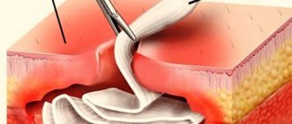
Drainage is divided into types. If the patient is in a supine state, then the drainage system is installed at the lowest point so that the contents from the cavity flow out independently under the force of gravity.
Passive drainage can also be used. It will absorb the liquid contents of the wound. But since it is weakly effective, it is used infrequently.
With the help of active drainage, the wound can be treated with antiseptic and anti-inflammatory drugs, and the purulent contents can be removed mechanically.
Drainage can be made of plastic, glass, rubber and gauze. The device most often has different sizes and diameters, selected based on the depth, width and type of wound.
For drainage to be effective, it must be selected individually for each wound surface. It also needs to be positioned correctly, and the materials for its creation must be selected so that they correspond to the sensitivity of the microflora.
Drainage of cavities is carried out during the inflammatory process. Once the cavity is cleaned and begins to heal, the drainage must be removed. It is also worth removing the device when an inflammatory process begins around it.
When installing drainage, it is necessary to observe all aseptic measures, since through drainage tubes or holes, not only transudate can escape, but also pathogenic microorganisms can enter the wound cavity.
Failure to install drainage will lead to accumulation of transudate in the cavity. The wound healing process always depends on many factors and, in the absence of a drainage device, can lead to an inflammatory purulent process.
If it starts, or a hematoma forms in the wound, the scar will take much longer to form, and healing will be more difficult. It is for this reason that if there are indications for drainage, you should not refuse it. Don't risk your health and always follow your doctor's orders.
Types of abscesses and symptoms
There are more than 50 types of abscesses. They differ from each other in location, cause of occurrence, nature of purulent discharge and severity of inflammation.
The most common types:
- Anorectal. Localized in the anal part of the rectum or directly on the anus. The most common cause is paraproctitis.
- Apical. Forms in the area of the roots of the teeth, most often the cause is periodontitis.
- Brain abscess. Localized in the tissues of the brain, the cause can be injuries to the head and the brain itself.
- Hot (spicy). It can occur on any part of the body and is characterized by a high rate of development, severe inflammation and a sharp deterioration in well-being.
- Lung abscess. It develops in the lung and is most often a complication of pneumonia.
- Liver abscess. Localized near the organ or directly in it and on it. It can be caused by an infection or be a complication of liver diseases.
- Appendicular. Most often, the cause lies in inflammation of the appendix.
- Cold abscess. It is localized in any part of the body, usually covering a small area. A cold abscess is dangerous because it develops very slowly. It is quite difficult to diagnose at an early stage.
- Gangrenous or gangrenous gas. The pus has a pronounced putrefactive character, with an odor and may contain microbes that form gas.
Under the diaphragm. Pus accumulates in the area of the diaphragm, most often the cause is a complication of pancreatitis, cholecystitis, ulcer or injury to the abdominal cavity.
These are not all types of abscess, but only the most common. But the most common are skin abscesses, which are localized in different parts of the body.
Symptoms of a skin abscess:
Redness, tenderness, and swelling of a small area of skin. The pain increases with physical activity and coughing. This period lasts about 5 days. After 5 days, the purulent head of the capsule begins to appear, and the pain increases. The capsule can grow for a very long time, up to 15 days.
The temperature may rise.
Sometimes symptoms of intoxication appear: nausea, weakness, muscle pain and deterioration in general health.
Hidden abscesses are especially dangerous, since the capsule is emptied inward, which can lead to very serious consequences. The appearance of a purulent formation on the face is also especially unpleasant; in this case, a spontaneous breakthrough cannot be allowed and an autopsy must be performed.
Opening and drainage
Any type of abscess requires surgical intervention. If the abscess is more than 4 days old and the head of the capsule is already mature, then opening is simply necessary.
- Treating the area of inflammation with an antiseptic solution.
- Anesthesia. Lidocaine treatment is most often used; in case of severe pain, local injections can be used.
- Incision of tissue with a scalpel in the area of greatest inflammation or purulent head.
- If the swelling does not have a visible bulge, then the incision is made on
- suspected intersection of tumor diagonals or at the intersection of a vertical line and a horizontal line. Also, sometimes a needle may be used to locate the capsule.
- The incision is made no more than 2 cm long.
- Using a Hartmann syringe, expand the incision to 4-5 cm and at the same time break the connecting bridges of the abscess.
- The abscess is emptied. In modern clinics, electric suction is used for this. But manual cleaning is possible.
- After removing the pus, the cavity is examined with a finger to remove the remaining bridges and tissue.
- The cavity is washed with an antiseptic.
- For drainage, a rubber tube or tampons soaked in antiseptics and enzymes are inserted into the abscess cavity.
- After opening and cleaning the cavity, treatment is performed similar to purulent wounds.
- Treatment with sodium chloride solution or any other hypertonic solutions, for example, Boric acid.
- The use of healing ointments, for example, Vishnevsky, Tetracycline, Neomycin. It is important that the ointments have a fat or petroleum jelly base. Otherwise, moisture will be absorbed.
The use of ointments containing antibiotics has a beneficial effect. These include Levomikol, Levosin, Mafenid. The antibiotic goes to the wound and speeds up the healing process. Dressing the wound using these ointments is enough once a day.
The process of opening the abscess and treating the wound may vary, depending on the location of the inflammation, the complexity and severity of the disease. Existing diseases, age and health status of the patient also matter. The operation itself lasts no more than 10 minutes, and for external abscesses it is performed on an outpatient basis. But treatment of the cavity and scarring can last up to a month. In severe cases, hospitalization will be required.
How to install
First, the dentist conducts a visual examination of the jaw and takes detailed x-rays. This makes it possible to determine the location of the abscess, the degree of its growth, and the presence of fistula tracts inside the bone. If the tooth root is destroyed, causing flux to appear, it should be removed.
To install drainage in the gum, the doctor performs the following manipulations:
- anesthesia of the inflamed area by administering anesthesia;
- cuts the gum, places a drainage made of latex material after the serous fluid is removed through the hole using a vacuum or mechanical method;
- if necessary, an antibacterial or antiseptic drug is injected into the wound;
- one end of the drainage must be located outside, which allows pus and excess fluids to drain away, preventing the wound from healing prematurely.
After tooth extraction, dissection and incision of the gums are not made - drainage is installed in the vacated hole.
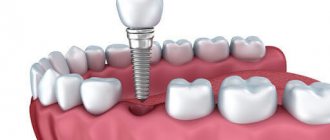
Installation of drainage in the gum
If the structure is installed correctly, there should be no strong discomfort or increased pain. The system itself can be felt with your finger or tongue, but it should not rub the inner surface of the cheek. With timely treatment and high-quality work of a specialist, patients note an improvement in their condition after manipulation within the first 24 hours.
How many days to wear drainage in the gum
The number of days that drainage should remain in the gum is determined individually. On average the period is 3-5 days. If the condition improves, the installation is removed and drug treatment is prescribed.
In case of severe inflammation, swelling, or elevated temperature, repeated drainage is possible.
The duration of wearing is determined by complaints and on the basis of a visual examination, x-rays of the jaw, and the rate of removal of excess fluids.
If there is abundant pus inside the periodontium, a daily visit to the clinic is necessary for examination, washing the wound with antiseptic solutions and introducing antibacterial drugs into it.
Abscess treatment
With early diagnosis and mild disease, treatment of an abscess without surgery is sometimes practiced. To do this, you need to promptly seek help from a specialist who will prescribe comprehensive treatment. It would be good if it was possible to do a full examination and a thorough diagnosis. Most often, this opportunity is available in clinics and centers with a multidisciplinary laboratory.
To treat an abscess without surgery, an ultrasound-guided drainage method is used. It has proven itself in the treatment of abscesses on the mammary glands and some hidden on internal organs. The question of using this method is decided individually and is not suitable in all cases. After such treatment, you will need to take a course of antibiotics. Sometimes you may need to take medications that strengthen your immune system.
If the external abscess spontaneously opens, then it is necessary to remove the purulent formations, clean the wound and treat with solutions of potassium permanganate or boric acid. In the following days, regular washing of the wound and application of medicinal dressings will be required. It is better if specialists do this. If the temperature rises, you should take an antipyretic.
If a boil, abscess or small pimple spontaneously opens, you can squeeze out the contents with your fingers and treat the wound according to the above scheme, be sure to use wound healing agents. But under no circumstances should you massage a deep abscess or try to open the capsule yourself. Such actions can lead to complications.
In addition to opening and treating the abscess cavity, a blood or plasma transfusion may be required. It is prescribed to patients who have large internal inflammations. Or when the disease becomes severe.
Treatment of an abscess is quite complicated; in addition to opening, it requires proper treatment and compliance with sanitation rules. Traditional medicine can be used as additional therapy.
Types
Drains can be represented by strips of latex, made of rubber, silicone, vinyl chloride, fluoroplastic and Teflon. Sometimes drainages made of gauze folded in several layers are also used, but since they have a short operating time, they are used infrequently. Rubber drainage also has shortfalls. It is quickly delimited by fibrin formed in the wound, adhesions of the epidermis and deep tissues in which it is installed.
At the moment, surgeons prefer complex drainage systems, represented by multi-lumen, cuff, rubber-gauze, T-shaped and fan types. The general requirements for drainage are softness, smoothness, strength and transparency of the material. Also, all drains must be radiopaque.
Folk remedies
You should not rely entirely on traditional medicine. But they may well be an addition to the surgical treatment of external abscesses. There are many plants and products with antibacterial, healing and immune-stimulating properties that have been used for many years and are quite safe.
- St. John's wort. Use a water infusion, an alcohol tincture or an oil extract. It has good antiseptic and bactericidal effects; it is not for nothing that St. John's wort is popularly called a natural antibiotic. When treating abscesses, apply compresses, lotions, or simply wipe the affected area.
- Propolis. A healing ointment can be prepared to treat external abscesses. It also heals cuts, burns, abrasions and other skin injuries well. To do this, take 100 grams. melted and filtered internal fat of any animal, heat to 70 degrees, add 10 grams. propolis and, stirring continuously, cool. The ointment is stored in the refrigerator.
- Echinacea. In case of an abscess, immune restoration will be required. Echinacea tincture copes well with this task. You can buy it ready-made at the pharmacy, prepare it yourself, or use analogues, for example, Immunal. To prepare the tincture, 1 part of the raw material is poured with 10 parts of vodka. Insist for 2 weeks. Take 30 drops orally before meals.
- Aloe. Pure plant juice is used for treatment. It’s good if its age is more than 3 years and the leaves have been in the refrigerator for several days. The affected area is lubricated with juice or lotions are made. Juice mixed in equal parts with honey works well. It is important that there are no allergies.
- Onion. It helps to cure an abscess no worse than aloe and is present in every home. There are a lot of recipes with onions; this vegetable is quite popular in the treatment of purulent formations. The onion is boiled in cow's milk, cut and applied to the sore spot. You can use an onion baked in the oven. The following compress works well: chop the baked onion, mix with honey and bandage it to the abscess.
- Potato. It is famous for its drawing action. Grated potatoes are tied to a purulent formation. You can often feel tremors - this healing vegetable cleanses the abscess cavity. Tie the potato mass overnight.
- In folk medicine there are a huge number of recipes for abscesses. You should not use products from little-known herbs or complex mixtures. Any plant can cause an allergic reaction, which will complicate the situation. You should also be careful when taking drugs orally. And under no circumstances should you try to cure abscesses on your own.
Possible complications
If treatment is neglected or carried out incorrectly, the abscess can cause complications. Most often, this is the spread of infection to neighboring tissues or the disease becoming chronic.
Infection of neighboring organs and tissues depends on the type of abscess. To avoid this, you need to seek help in time and perform an autopsy. It is important to follow through with it until complete recovery.
An abscess becomes chronic if the acute disease is not completely cured. With this form, a deep fistula is formed that cannot be healed. A breakthrough that occurs inside a closed cavity can cause meningitis, peritonitis, pericarditis or arthritis.
Factors influencing the transition of an acute abscess to a chronic form:
- Lack of cavity drainage or its poor quality.
- Too large in size (more than 4 cm) or numerous cavities on internal organs and tissues.
- The result of conservative treatment. Most often it occurs if the patient refuses surgery or self-medicates.
- Weakening of the immune system.
In 20% of cases, the cause of the abscess becoming chronic is residual purulent substances. The cause of 30% of complications is self-medication and late referral to specialists. Unlike an acute abscess, a chronic abscess is difficult to treat, negatively affects nearby tissues and worsens the general condition.
To prevent abscesses, first of all, you need to maintain hygiene and promptly treat tissue damage with special means, for example, Miramistin has proven itself well. Proper care of the oral and nasal cavities is also equally important; they are often the routes through which infections can enter. And, of course, any disease, from sore throat to caries, must be cured and not be a carrier of pathogenic microorganisms.
In the attached video you can learn about the abscess.
Opening an abscess is a mandatory procedure in 98% of cases. It is better if it is carried out by specialists and in compliance with sanitary standards. Timely treatment will help avoid serious consequences.
source
When should drains and tubes be removed after surgery?
Monitoring task
It is possible to register physiological disorders as early as possible in order to prescribe corrective therapy as quickly as possible. The invasiveness of monitoring depends on the severity of the disease in a particular patient: the sicker the patient, the more sensors and probes are used and the lower the likelihood of survival.
Comprehensive discussion of the ever-increasing physiological monitoring techniques
is beyond the scope of this chapter. However, please note the following:
In order to respond promptly to the monitor's warning signals, you must be well versed in the technology being used and must clearly distinguish between truly acute physiological deviations and mechanical and technological artifacts of monitoring.
It should be understood that all monitoring methods are fraught with a myriad of potential errors.
, related both to a particular technology and to the characteristics of the patient. Alertness and sound clinical judgment are of the utmost importance!
Thanks to the introduction of new technologies, monitoring
It's getting more complicated (and more expensive). Moreover, the monitoring technique causes a large number of iatrogenic complications in surgical BIN. Use monitoring selectively, without succumbing to Everest syndrome: “I climbed it because it’s there.” First of all, ask yourself: “Does the patient really need this?” Remember that there are safer and cheaper alternatives to invasive monitoring. For example, in a stable patient, remove the arterial catheter, since blood pressure can be easily measured with a conventional sphygmomanometer, and p02 and other blood parameters can be taken in the traditional way. Every time you examine a patient, ask yourself which of the installed catheters and tubes can be removed: nasogastric tube, Swan-Ganz catheter, central venous, arterial, peripheral venous or urinary?
Nasogastric tube
. Leaving this probe for a long time, supposedly to combat paralytic ileus in the postoperative period, is a generally accepted, but completely unfounded ritual. The concept that a nasogastric tube "protects" the underlying intestinal anastomosis is ludicrous, since several liters of intestinal juice are secreted each day below the discharged stomach. The nasogastric tube is extremely irritating to the patient, making breathing difficult, causing esophageal erosions and maintaining gastroesophageal reflux. Traditionally, surgeons leave it until gastric output reaches a certain limit (for example, 400 ml/day); often it's just unnecessary torture. It has been repeatedly shown that most patients after laparotomies, including those after interventions in the upper gastrointestinal tract, do not need nasogastric decompression at all or it is necessary for only 1-2 days. In unconscious patients, when it is necessary to protect the upper airway from accidental aspiration, a nasogastric tube may be used selectively. After emergency abdominal interventions, its use is mandatory in patients on mechanical ventilation, in an unconscious state, and in those undergoing surgery for intestinal obstruction. In all other cases, remove the nasogastric tube the morning after surgery.
Drains
. Despite the general belief that it is impossible to effectively drain the free abdominal cavity, drains are not only widely used, but even abused (Chapter 10). In addition to the false sense of security and safety net they claim to provide, drains can cause pressure ulcers of the intestines or blood vessels and contribute to infectious complications. We assume that you are using drains only to evacuate contents from the cavity of an opened abscess, to drain a potential source of visceral secretion (for example, biliary or pancreatic) and to control intestinal fistula when the intestine cannot be exteriorized. Passive open drainage does not eliminate bacterial contamination in both directions and should not be used.
Use only an active closed drainage system with tubes
that are not in contact with the visceral hollow organs. The placement of drains directly at the anastomosis in the hope that possible leakage of intestinal contents will result in an intestinal fistula rather than peritonitis is an outdated dogma; Drains have been shown to contribute to anastomotic dehiscence. The statement: “I always drain the colonic anastomosis area for at least 7 days” refers to the dark days of surgical practice. Remove drains as soon as they have served their purpose.
What is drainage? You will find the answer to this question in the materials of this article. In addition, we will tell you how this method is carried out in
medical practice and why it is needed.
Is it possible to cure an abscess without opening it?
A purulent abscess is characterized by various signs; the therapist prescribes conservative methods of therapy at an early stage.
Symptoms of the primary stage of abscess development:
- a dense infiltrate is localized shallow under the skin, which indicates the possibility of forming therapy without opening;
- the abscess is not accompanied by general inflammatory phenomena - severe pain, hyperthermic syndrome, poor health;
- small sizes;
- correct form of formation with palpable boundaries.
To remove an abscess without surgery, a doctor prescribes:
- a group of broad-spectrum antibiotics to quickly get rid of the causative agent of the abscess without opening it;
- anti-inflammatory drugs with analgesic effect;
- To stimulate healing and restore the area affected by the abscess, local medications from the pharmacy are used - antiseptic, regenerating, healing ointments, liniments. Drugs with a pulling effect facilitate treatment without opening the formation.
In what cases should an abscess be opened in a hospital?
Indications for mandatory surgery in a medical institution with excision of a pathogenic formation are:
- An abscess with a hard-to-reach localization, hematogenous infection - suppuration of the brain, internal organs. There is an increased risk of the substance breaking into the blood with the development of sepsis, which can lead to death if the formation is not opened.
- The location of the inflammatory focus is on the face, neck, axillary area, groin or rectal area. There is a risk of purulent melting, the formation of phlegmon with the involvement of healthy tissues in the process.
- Removal of an abscess of paratonsillar localization is carried out in a hospital setting: the airways are close, manipulation to open the attribute can provoke heavy bleeding, which can be eliminated by resorting to resuscitation measures.
- In case of prolonged maturation of abscesses of superficial localization (more than 4-5 days) or independent breakthrough of a deep-lying abscess, an autopsy is not required; the patient turns to a surgeon for examination. The doctor determines bridges, fistula tracts, the presence of residual pus, washes the wound with an antiseptic, and sanitizes the cavity.
- With the development of severe complications: suppuration after injection of drugs intravenously, incorrect placement of an intramuscular injection, the formation of fistulas with leakage of yellow-green contents. In this state, hyperthermia of 39-40 degrees persists for a long time, which is not relieved by medications.
- Opening and draining an abscess is mandatory for groups of the population: children, pregnant women, patients with a weakened body’s defense system or the presence of immunodeficiency.
- The lack of a therapeutic effect with conservative therapy is a direct indication for surgical opening of the abscess.
Before the operation, diagnostics are carried out:
- puncture of the abscess, bacterial culture to determine the sensitivity of the pathogen to antibacterial drugs;
- detailed blood test with leukocyte formula;
- Ultrasound examination, MRI, X-ray to determine the boundaries of the formation in order to gently open the abscess;
- consultation with an anesthesiologist to determine if there is an allergy to medications, a preliminary medical history: whether there was previous anesthesia, how the patient underwent anesthesia, whether complications arose.
General information
Drainage in medicine is a therapeutic method that involves removing the contents of wounds, hollow organs, ulcers, as well as pathological or natural body cavities.
Complete and correct drainage can ensure sufficient outflow of exudate and create the best conditions for the fastest rejection of dead tissue with the transition of the healing process to the regenerative phase.
Drainage in medicine has practically no contraindications. By the way, this method has another undeniable advantage in the process of purulent antibacterial or surgical therapy, which is the possibility of targeted control of wound infection.
How does surgery to remove an abscess work?
Stages of surgical treatment of an abscess:
- Preparatory – taking tests, instrumental examinations, determining the optimal method for opening a formed abscess, taking into account the location and possible complications.
- The main one is an operation to cut the abscess.
- The postoperative period is from the end of the procedure until the body recovers.
Algorithm of surgical procedures:
- treatment of the surgical field with antiseptics;
- with a scalpel, the outer layers above the affected area are opened - mucous, fascial, the muscles are carefully pushed back, and the muscles are cut off at the points of attachment to the bone. To install drainage for an abscess, the surgeon provides a gap in the wound surface for the outflow of purulent contents;
- An incision is made with sterile instruments without squeezing the tissue to avoid squeezing pus out; the length of the opening line depends on the length of the subcutaneous infiltrate;
- the doctor sanitizes the purulent cavity using antiseptic solutions, inserts a drainage tube to ensure the outflow of pus in the postoperative period;
- necrotic tissue around the abscess is excised, which speeds up the healing process and prevents recurrence of suppuration after opening.
Postoperative treatment involves taking antibacterial drugs.
Principles of staging
The basic principles of drainage installation are presented:
- Installation of drainage systems in the sloping area of the wound cavity.
- Fixing the drainage device.
- By creating such conditions that the drainage does not come into contact with important anatomical formations, represented by nerve endings, vascular network and tendon apparatus.
- Minimizing complications after the procedure.
- Preventing depressurization of the wound cavity and compression of tissues with their damage.
- Minimizing the penetration of pathogenic microorganisms into the cavity through drainage tubes.
Features of wound drainage
The tube is inserted into the cavity to the depth of the abscess, sutured if necessary, manipulation is carried out in order to cleanse the abscess after opening. There is a free evacuation of residual necrotic particles and purulent fluid.
- The passive method is to insert a PVC tube into the capsule, which facilitates the outflow of the contents.
- An active method of drainage for an abscess is suction of purulent masses using medical aspirators, syringes, creating a vacuum with double tubes after opening. The advantage of the procedure is the possibility of washing the cavity with antiseptics with parallel evacuation of necrotic particles. With frequent manipulations, the cellular structures responsible for healing the postoperative wound are eliminated, which aggravates the patient's condition.
The required type of drainage is determined by the surgeon.
Kinds
Drainage can be passive, flow-flushing or active.
Passive
Passive drainage is currently carried out using perforated tubular systems made of polyvinyl chloride or thin tubes filled with gauze. Drains are positioned in such a way that they drain the liquid component from top to bottom. This mechanism of action is ensured by the force of gravity pressing on them.
Active
When performing active drainage of sealed tissue and epidermal damage, aspiration is used based on vacuum, provided by a special suction. This technique allows you to remove dead flesh, minimize the joining of wound edges, and reduce the likelihood of pathogenic microflora entering the cavity from the outside.
The drainage is installed in such a way that it removes the separated liquid from the bottom up, against the influence of gravity on it. It is necessary to take into account that this drainage is not used to remove growing hematomas.
Flow-flushing
Flow-flush type drainage is carried out using aspiration flushing with the installation of counter-perforated drainage systems. Medicine is injected into one of them, and transudate is removed from the damaged tissue through the second.
The drug can be administered into the drainage in a stream, drip, fractionally or continuously. Outflow is carried out according to the active or passive method. With the help of such drainage, pathogenic microorganisms do not enter the wound, transudate is completely removed from it, thus creating favorable conditions for healing and unfavorable conditions for bacteria.
Postoperative wounds are drained due to the high risk of developing inflammatory processes of purulent etiology. This situation is due to the fact that contamination occurs in the wound during surgery, represented by subcutaneous tissue and the impossibility of completely removing dead tissue.
Drainage must be installed by introducing counter-perforated systems into the cavity through the holes left from dialysis after surgery. Drainage is often prescribed in the case of removal of cancerous formations in the mammary gland, local hernias of the ventral type, amputation of the lower and upper extremities and surgical cleansing of a purulent focus in soft tissues.
After the purulent focus is opened, the doctor installs passive drainage, which is always done in such cases. The technique of installing drains is studied by operating nurses in the dressing room or operating room when working with patients with various types of wounds.
How long does it take for an incision to heal after an abscess?
The rate of healing of a postoperative wound after opening an abscess is an indicator that depends on:
- individual characteristics of regeneration due to heredity, the presence of chronic diseases;
- age of the patient - healing occurs easier in children than in adults or the elderly;
- abscess localization - subcutaneous formations regenerate faster than deep internal suppuration;
- level of immune protection - the affected area after opening takes longer to recover if the body is weakened;
- the size of the purulent abscess - large wounds heal more slowly.
If you follow the doctor's recommendations, regularly bandage the area where the abscess was, and follow a diet, healing occurs faster.
Forecast of the course of the disease
If you seek help in a timely manner, 99% of patients recover after opening surgery, even with difficult localizations of abscesses: in the brain, reproductive organs, and liver. The course of the pathology can be complicated by the breakthrough of pus into the surrounding tissues, which will provoke a negative result - ulcerative phenomena or scarring.
In untreated forms of abscesses, advanced suppuration with non-compliance with doctor's instructions after surgical opening of the abscess, the risk of chronicity and periodic relapses increases, which requires repeated surgery.
source


