Child's skull before teeth loss (photo of the day)
The human body is amazingly structured, for example, here is a photograph of the structure of the skull of a 5-year-old child before his baby teeth fall out:
Human evolution is designed in such a way that we are born toothless, because... At first, we eat only mother's milk. Over time, when a child begins to eat solid food, his baby teeth erupt so that he can chew solid particles.
Starting at about 6 years of age, baby teeth begin to fall out and molars appear in their place. This happens because the human skull increases in size, but baby teeth do not. Molars are initially the same size as they remain with us for the rest of our lives.
This is what the lower jaw of an adult looks like:
As you can see, an ordinary person does not have another row of teeth behind his molars. And, although they are very strong and can withstand enormous loads, they need to be monitored, cared for and protected, and then the smile will be excellent.
The structure of the human body is amazing. There is practically nothing superfluous in it, everything is thought out to the smallest detail. All we have to do is keep ourselves in shape and take care of our health.
Follow updates on Vkontakte, Facebook and Twitter
The very first healers
Doctor of Historical Sciences Igor Zimin, Doctor of Medical Sciences Lyudmila Orekhova and Candidate of Medical Sciences Ramilya Musaeva jointly wrote the book “From the history of dentistry, or Who treated the teeth of Russian monarchs” (St. Petersburg, 2013). The researchers noted that drug treatment of various diseases in ancient Egypt began about 4,600 years ago. Papyri from the Middle and New Kingdoms describe gum damage and caries, which was then explained by the presence of a certain “worm” that eats teeth and grows in them.
The very first healers relieved pain and treated gum inflammation with various compounds prepared from medicinal plants; they also resorted to surgical intervention. For example, in the Giza region, Egyptologists discovered a fragment of a jaw dated to 2680-2563 BC. The remains belonged to a middle-aged man, judging by the degree of wear on the teeth. An ancient dentist drilled two small holes into this patient's jaw to remove pus that had accumulated under a lower molar.
Judging by various archaeological finds, dentistry was well developed in ancient Mesopotamia, India and China. Both the ancient Mayans and Aztecs knew about the existence of caries and some ways to help patients.
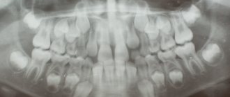
What does a photo of a child's jaw with baby teeth look like?
It looks creepy, really creepy. For me it turned out to be real shock content, where are the corpses and dismemberment, but this is a real nightmare. You can laugh, but I’m sharing my phobia) Perhaps, after looking at the photos in the answers, I’ll feel completely violet about it)
Indeed, before the loss of baby teeth, a child’s jaw looks different than an adult’s.
Well, it’s probably not clear and scary for those who are far from anatomy.
Milk teeth – already formed at birth (and erupt during the first years of life in a certain order)
- But the rudiments of permanent teeth are also present in the child’s jaw, and when they are sufficiently formed, they push out the milk teeth with their growth, thereby provoking their loss and replacement with permanent ones.
And it all looks like 2 rows on the x-ray.
For anyone who does not bother to read X-rays, it is quite difficult to see and understand what and what it looks like (you can look at it) an anatomical cast model and evaluate:
Well, so as not to scare too much, such a colorful picture
There is nothing scary about this - it would be much worse if they could see the whole person as an x-ray, and even then I think they would get used to it. For comparison, I’ll cite cats of the Sphynx breed - not everyone can be touched.
Before the replacement of baby teeth with molars occurs, a very interesting process occurs - the roots of baby teeth self-resorb - imagine (and why scientists can’t somehow come up with something like this to happen at least with “wisdom” teeth..) The root begins to dissolve from the base of the tooth and gradually the process reaches the base of the tooth. The tooth begins to become loose and then falls out.
The pictures are, of course, scary, but in reality it looks a little different - the teeth are not entirely located in the lower and upper jaws, but in an embryonic state. Touch your jaw - can your teeth be arranged in 3 rows?
Is it possible to refuse x-rays for a child?
Parents can easily refuse to have their child undergo an X-ray examination. This procedure is completely voluntary. However, they must understand that in this case any therapeutic measures will be carried out on the basis of an instrumental examination. A referral for an x-ray is a recommendation, but not a requirement.
X-ray examination of teeth is a completely safe procedure that cannot cause harm to the body. You should not refuse to carry it out because of the radiation, because it is minimal and cannot harm the child’s body. With the help of such a study, it is possible to identify numerous deviations in the early stages.
We suggest you familiarize yourself with Bad Breath
Formation of a child's skull with baby teeth
Posted in category: Children's teeth
All babies grow 20 baby teeth. The rudiments of temporary and some permanent units are formed during the period of embryonic development. In this regard, the skull of a newborn baby with baby teeth has a special structure.
The brain area of the skull in a small child is much larger than the facial area, and the chin is poorly defined. The lower jaw occupies a distal (posterior) position in relation to the upper jaw. Its position changes during the appearance of baby teeth.
They ate everything raw
Of all the reasons explaining the excellent condition of the teeth of ancient people, nutrition is considered the main one. According to some researchers, the main difference between the diet of a Neanderthal or Pithecanthropus from modern Homo sapiens is a raw food diet.
Victoria Butenko, a well-known popularizer and promoter of this method of healing the body, in her book “Raw Food Diet for Cleansing” (St. Petersburg, 2011) explains the physical endurance and good health of ancient people precisely by the fact that they ate everything raw.
The author of the book spoke about the remains of 13 hominids found in East Africa. They lived approximately 3.6 million years ago. Archaeologists even called these ancient representatives of the genus Homo the “first family.”
“Their large molars were covered with a strong layer of enamel, similar to the enamel of the teeth of animals that chew a lot of greens. Scientists believe that the first people spent most of their time in trees, where they were better protected from predators and abundantly supplied with food - fruits and leaves,” wrote Victoria Butenko.
In her opinion, the teeth of ancient people were so strong because they also ate nuts and insects. Of course, in its raw form.
Formation of tooth germs
Before the baby teeth fall out, the child’s skull has many tooth germs in the deep layers of the jaw bone tissue. The formation of the rudiments of future mammary units begins at 5–7 weeks of pregnancy. These are enamel organs that gradually mineralize and form dental tissue. From the 20th week of embryonic growth, the rudiments of permanent incisors and canines begin to develop, premolars are formed in newborns, and molars at 8–10 months of a child’s life.
The growth of roots of temporary and permanent units begins before eruption and lasts 2 years.
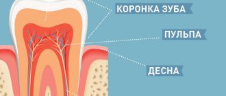
Milk teeth differ from permanent teeth in size, shape, color and structure. The crown is small, wide, the enamel and dentin are thin, and there is a large area of pulp (nerve fibers). The roots are short, widely spaced, and grow at an angle, making room for the primordia of permanent units. The chewing surface has practically no tubercles, the crown is bluish-transparent.
It is very important to keep temporary units healthy and regularly perform hygiene procedures to prevent the development of caries. The destruction of the crown can lead to pulpitis and further tooth extraction. The carious cavity contains a large number of pathogenic microorganisms that can cause inflammation of bone tissue and lead to the death of the rudiment of a permanent unit. If baby teeth are pulled out ahead of schedule, permanent teeth may grow crooked and take the wrong position in the row. Therefore, at the first signs of caries, it is necessary to fill the affected crown.
Sequence of teething in children
The age at which primary teeth appear depends on various prerequisites: internal and external. WHO considers the eruption of the first incisors to be normal at 6–8 months. Teeth may appear 1–2 months earlier if during pregnancy a woman took vitamin complexes with vitamin D and calcium. When milk units are delayed in breaking through the mucosa, there is usually no reason to panic. The reasons may be as follows:
- genetic background;
- lack of calcium in the diet;
- severe toxicosis in the mother;
- climatic zone of residence (in hot countries, teeth appear earlier);
- gender (one-year-old girls are often toothier than boys);
- rickets (the only pathology requiring treatment).
| Age, in months | What teeth to expect |
| 6-9 | central incisors (lower, then upper) |
| 9-13 | lateral incisors |
| 11-16 | quad molars |
| 16-22 | fangs |
| 22-34 | "five" molars |
The order may be broken. Neither parents nor doctors can influence the speed and pattern of teething.
At one year of age, babies usually have about 8 teeth. When calculating the number of teeth erupted, the formula is used: age in months minus four. Fluctuations in the value of 4–6 months are not considered pathological, nor is a non-standard order of eruption. This is a physiological norm and no treatment is required. A visit to the dentist should not be postponed if the pairing of the appearance of dental units is disrupted. A possible congenital anomaly is the absence of a tooth germ.
Teething of milk units
Temporary units in a child begin to grow at approximately 6–8 months of life. The lower incisors are cut first, then the upper. Afterwards, fangs and small molar units grow. All 20 baby teeth grow in by age 3. The timing of eruption depends on hereditary predisposition, the functioning of the endocrine glands, past infectious diseases, and the presence of chronic ailments.
The skull of a small child with milk and molars changes in two stages. The first period lasts from the moment of eruption to 3.5 years. The teeth are located tightly to each other, there is no abrasion of the enamel, the lower jaw has a neutral position. The second period begins at 3.5 years and ends at 6 years. At this time, interdental spaces appear, enamel wears off, and the bite changes.
The dental buds of the permanent units exert pressure on the roots of the deciduous units, which leads to their gradual resorption (resorption). This process continues until the temporary teeth are completely replaced. The enamel of milk crowns wears off, the pulp atrophies, the tooth becomes loose and falls out.
Scheme
The pattern of teething and changing teeth is the same for each child. But when the growth or replacement of teeth begins is a purely individual phenomenon. The first milk teeth erupt at the age of 6-8 months.
At the age of 5-6 years, temporary milk teeth are replaced by a set of permanent molars. The appearance or loss of a particular tooth occurs with a break of about 3-4 months after the eruption (loss) of the previous one.
How they appear
At 6-8 months of age, a pair of central incisors on the lower jaw erupts in the child, and a little later the central upper incisors appear. The antagonizing appearance of teeth occurs for a reason. During this period, the rudiments of the bite are formed, the child learns to bite and gnaw solid food.
When the baby is 16-20 months old, the fangs will erupt. Interestingly, it is always the lower front tooth that appears first, then the upper front tooth. The growth of canines is often accompanied by difficulties due to the peculiarities of their structure and location.
Now the child is able to bite off a piece of a solid product, but he will not be able to chew it yet. Chewing molars will appear in the baby at the age of 1.5-2 years. As soon as the chewing group has erupted, the child will learn to chew solid food with his teeth.
How do they fall out?
The child has grown up, there is more space on the jawbone, so the sixth chewing molars - permanent molars - will appear first. Then gradually all baby teeth will be completely replaced by molars.
First, the lower central incisors wobble and fall out, then it’s the turn of the upper ones. A child’s jaw loses the first pair of premolars at the age of 10, and the second at the age of 12. A thirteen-year-old teenager loses his last pair of baby teeth – canines. Another year later, the second molars erupt, and at the age of 18, “wisdom teeth” begin to grow;
There are situations when a child knocks out a tooth as a result of injury, a fall, or a blow. The fact that the baby knocked out a tooth will have a negative impact on the growth of molars in the future. Anomalies of the dentition may occur: malocclusion, difficult eruption, thinning, loosening, curvature of teeth.
We invite you to familiarize yourself with the structure of teeth: anatomical features and diagnosis of diseases
Cutting through permanent units
A child's skull changes before the loss of baby teeth and the growth of permanent teeth. The jaws and skull grow rapidly, but the temporary units do not. New teeth emerge at the size they will remain for the rest of your life.
The permanent molars begin to grow first, followed by the incisors, canines, and premolars. Large molars form a permanent bite. Pathologies during eruption can be caused by inflammatory processes at the roots of the mammary units and premature removal. It is typical that girls change their teeth earlier than boys of the same age. Wisdom teeth are the last teeth to appear in a person's life; this occurs between the ages of 14 and 25.
Oral diseases
06/19/2018 admin Comments No comments
The child’s skull before the loss of baby teeth consists of a double dentition
Have you ever wondered what a child's skull looks like before losing baby teeth? When the stage of replacement of primary teeth with molars begins, a “naked” illustration of the child’s maxillofacial system can terrify parents. If you look deeper into an x-ray of a child's facial skeleton, the bulk of his jaw consists of a double row of teeth. Why is this happening? We'll find out more about this later.
Read also: When teething
Types of bite
How the permanent units erupt determines whether the bite will be correct. Various pathologies lead to abnormal facial proportions in a child.
- Distal bite. The upper jaw is significantly advanced, the lower jaw is underdeveloped. The incisors do not fully perform their functions, so the back teeth bear the entire chewing load. Molars are more susceptible to caries. With a distal bite, the chin is unnaturally small.
- Mesial bite. The lower jaw is pushed forward, the chin protrudes strongly. The pathology is caused by underdevelopment of the upper jaw.
- Open bite. The incisors do not close completely, a gap is formed. The defect can develop in the area of several units or a group of teeth. In children with an open bite, the mouth is always slightly open, and facial asymmetry is visible.
- Deep bite. The incisors of the upper jaw cover more than ½ of the incisors of the lower jaw. With this defect, the lower lip protrudes outward due to lack of space, the proportions of the face are disrupted, and the gums are often injured.
- Crossbite. There is underdevelopment on one side of the jaw. Normally, the cusps of the upper molars should overlap the outer cusps of the lower molars. If the position is violated, a crossbite is formed. Chewing function is impaired, patients can grind food only on one side, discomfort occurs when opening the mouth, and the temporomandibular joint often becomes inflamed.
Incorrect bite leads to significant wear of the enamel, the development of caries, periodontitis, frequent injury to the mucous membranes, and dysfunction of the temporomandibular joint. Defects are corrected by wearing orthodontic plates or braces. The sooner therapy is started, the easier it will be. In adolescents, the jaws are in the formative stage, so correcting the bite takes less time than in adults.
Why do they spoil?
A child’s tooth enamel is very fragile, thin, and is constantly exposed to various influences, which quickly affects the appearance and condition of the teeth in general. Under the influence of cold, hot foods, sweets, fruit acids, as well as due to a lack of vitamins, plaque can form on the teeth, the enamel is destroyed, and caries occurs. Let's take a closer look at the main reasons why and how baby teeth deteriorate in children.
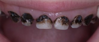
Turn black
Black plaque on baby teeth can be caused by many factors: impaired metabolism of fluoride, calcium, heredity, dysfunction of salivation, abuse of sweets. But the main reasons are the development of early caries.
Caries occurs when a child does not know how to properly care for their teeth, brushes them irregularly, or exposes them to very cold (hot) drinks. He bites his nails and rarely washes his hands, which injures tooth enamel and colonizes the oral cavity with pathogenic bacteria.
Crumbling
The strength of dental tissue depends on the level of vitamin D in the child’s body. It is not surprising that teeth crumble more often in residents of the northern regions. Lack of sun causes low production of vitamin D, which creates difficulties for the normal absorption of calcium and the formation of strong tooth enamel.
Consuming poor quality water also affects dental health. If drinking water is deprived of the required amount of fluoride and iodine, a metabolic disorder occurs - the teeth do not receive the necessary “building material”, they crumble and fall out.
Sometimes teeth crumble due to dental problems - malocclusion, untreated or incorrectly treated teeth. Let's say a child knocked out a tooth or recently had a filling done. If even a small piece of enamel breaks off, over time the enamel weakens and the teeth crumble.
Fall out early
Teeth fall out early in the presence of an abnormal bite, tumors, injuries, and also if one of the teeth was forced to be removed or the neighboring teeth exerted too much pressure. A tooth that falls out early leads to the displacement of neighboring teeth, which gradually begin to occupy free space.
Violation of spatial balance makes teething difficult. The molars grow crooked, stagger, and are unevenly positioned. Crooked teeth distort a child’s face, causing facial defects and difficulties in pronunciation. If a tooth falls out early, the orthodontist can install a special plate - a spatial corrector.
They grow crookedly
Normally, baby teeth almost always grow straight, but why do permanent teeth erupt crookedly? When permanent teeth are just being formed, they are placed in the jaws in a rocker-like manner due to lack of free space.
Gradually, the child’s skull grows, free space appears, and the teeth begin to turn, preparing for eruption. Under normal conditions, baby teeth do not interfere with permanent teeth taking their proper place. But if a child is subject to bad childhood habits - sucking fingers, objects, the growth of molars is hampered.
Whose skull is this? What’s wrong with it? (+)
“Teeth with roots” - temporary teeth or milk teeth - these roots, when their time approaches, dissolve, and the tooth falls out on its own or with a little help, “teeth without roots” are permanent teeth.
When, again, the time approaches and the space “at the top” is freed up, they begin to erupt – “slowly crawl upward”, while forming their root all the way, developing and strengthening it. Because there will be no 2nd shift ;–) from Nature at least.
Hunterian Museum, named after its founder, who lived in the 18th century and loved to open and preserve in alcohol. The museum is located at the Royal College of Surgeons.
In some ways, the Hunterian Museum is similar to a cabinet of curiosities, because it contains a lot of anomalies (animal and human). But it’s not the anomalies that are interesting. For example, have you seen how the snake's eggs are packed inside, which it is preparing to lay? Or how big is a horse's brain?
The museum also contains many anomalous specimens of animals and humans. Human fetuses, curved spines, macrocephalic skulls and giant skeletons, fused skulls of twins, the foot of a person suffering from elephantiasis...
A separate section is about how diseases change a person. For example, this is what the skull of a syphilitic patient looks like (the holes in the skull are not the consequences of poor care of museum exhibits, but the consequences of syphilis):
Well, this collection is already clear :). Fortunately, sarcomas are not visible in a bad photograph.
Possible complications during teething
A possible problem may be the absence of teeth for a long time in the presence of teething symptoms. Doctors associate this condition with a lack of calcium in the newborn’s body. It is recommended to check the balance of vitamins and microelements and not introduce new foods into complementary foods.
If the tooth has approached the gum shell, but does not erupt, sometimes the process is helped by surgery. A serious complication is a violation of the pairing of teeth. This is fraught with deformation of the dentition and a broken bite.
The bumps that appear on the gums before eruption are easily injured. An infection can get into the wound, which leads to swelling of the soft tissues, fever, and sometimes suppuration. Parents should ensure that objects that may enter the baby's mouth are sterile. An extensive hematoma is fraught with complications:
- benign formations;
- cyst;
- suppuration;
- compression of adjacent tissues.
Inflammation spreads quickly in children with weakened immunity and poor nutrition. The process can spread to adjacent tissue areas: cheek, face. If the blood inside the hematoma does not resolve, but is retained by a dense membrane of connective tissue, a cyst is formed.