Oral diseases.
05/31/2018 admin Comments No comments
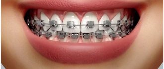
A beautiful smile is fashionable. Therefore, great attention is paid to dental health these days. Unfortunately, not everyone can boast of their impeccable appearance, although modern dental developments can bring them as close to ideal as possible.
In our article we will not talk about this. We will discuss the anatomical structure of the human tooth, a diagram of which is given on our website.
How do our molars work?
Molars are the only human organ that does not regenerate on its own. That is why they need to be protected and regularly monitored for any changes in their condition. It is not without reason that regular examinations by a dentist every 6 months are recommended.
Molars require careful care
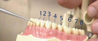
If we consider it enlarged, then each molar, a photo of which can be seen on our website, consists of a crown and root part. The crown part - the one that is located above the gum level, is covered on top with the strongest tissue in the human body - enamel, which protects its softer inner layer - dentin, which is the basis of the tooth.
Despite its strength and reliability, enamel is incredibly susceptible to external influences. Poor care, bad habits, and heredity can disrupt its condition. Pathogenic bacteria enter cracks in the enamel, causing intense tissue destruction. A person develops a carious process that also affects dentin.
If left untreated, the infection penetrates into the root part, acute pulpitis and other equally dangerous ailments develop.
As for the structure of the root part, its main elements are arteries, veins and nerve fibers that supply the tooth. They are located in the pulp of the root canal and through the apical foramen are connected to the main neurovascular bundle.
The dentin below the gum level is covered with cement, which is attached to the periodontium with the help of collagen fibers. The roots of human teeth, as the photo illustrates them very well, are hidden in the alveoli - peculiar recesses of the jaw bone.
Any defeat requires its complete removal. A broken root cannot be restored.
The structure of the jaw and molars of an adult deserves a separate section. This will be discussed below.
Preventive measures
Measures to prevent impact and other anomalies are quite limited. The main prevention is the fight against the bad habits of children - sucking the tongue, cheeks, foreign objects - undesirable positions: sleeping with the chin resting on the chest, propping the chin with fists, etc.
Proper breastfeeding and timely transition to regular food, balanced in composition and hardness, are of great importance.
If possible, you should avoid removing milk jugs, since their long absence before the appearance of permanent teeth has a bad effect on the proper growth of the latter. If you still cannot avoid removing the milk jug, it is advisable to replace it with a temporary prosthesis that will keep the jaw row in a stable condition.
And perhaps the most important preventive measure is regular (at least twice a year) visits to the dentist with your child. This will allow us to timely detect the beginning of an anomaly and take measures to prevent its development.
Types of human teeth
When visiting a dental office, we hear different names that are unfamiliar to our ears and, sometimes, we don’t even understand what they are talking about. This section is intended to help you understand what human teeth are called so that, if necessary, you can learn to understand the extent of the dental problems you have.
So, in the mouth we have:
- Central and lateral incisors;
- Fangs;
- Premolars or small molars;
- Molars or large molars.
In order to indicate their position on the upper and lower jaws, dental practice uses the so-called dental formula, according to which the numbers of primary teeth are written in Latin numerals, and the numbers of primary teeth in Arabic numerals.
With a full set of teeth in an adult, the dental formula will be as follows: 87654321 / 123465678. A total of 32 pieces.
On each side there are 2 incisors, 1 canine, 2 premolars, 3 molars. Molars also include wisdom teeth, which are the last to grow. Typically after 20 years.
As for children, their dental formula will have a different appearance. After all, there are only 20 baby teeth. But we’ll talk about this a little later, and now we’ll look at the structure of incisors, canines, premolars and molars, and also discuss their differences.
Features of the structure of the upper teeth
The smile zone includes central and lateral incisors, canines and premolars. Molars are called chewing teeth because their main purpose is to chew food. Each one looks different.
So, the ones are the central incisors. Their crown part is thickened and slightly flattened, they have one long root. The two lateral incisors also have a similar shape. They, like the central incisors, have three tubercles on the cutting edge from which three pulp spurs extend along the dental canal.
The canines are shaped like the teeth of an animal. They have a pointed edge, a convex shape and only one tubercle on their cutting part. The first and second premolars, or, as dentists call them, four and five, have a very great external similarity, the difference lies only in the size of their buccal surface and in the structure of the root.
Next come the molars. Six has the largest coronal part size. It looks like an impressively sized rectangle, and the chewing surface in its shape resembles another geometric figure - a rhombus. Six has 3 roots - one palatal and two buccal. The seven differs from the six in slightly smaller sizes and different fissure structures.
But not everyone even grows a figure eight or, as popularly known, a wisdom tooth. Its classic shape should be the same as that of ordinary molars, and its root resembles a powerful trunk. The upper wisdom teeth are considered the most capricious.
They can begin to disturb a person even at the stage of their eruption, and when removed they can create a difficult situation due to their twisted and twisted roots. Their antagonists are located on the opposite jaw. Our next section will be devoted to them.
Features of the structure of the lower teeth
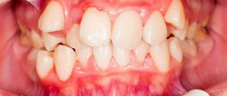
The photo conveys quite accurately what human teeth and fangs are made of, as well as their appearance. From it one can judge that the structure of the teeth in the lower jaw is completely different from their structure in the upper jaw. Let's consider this point in more detail.
The teeth of the lower jaw have the same names as the upper jaw, but their structure will be slightly different.
The central incisors are the smallest in size. They have a small flat root and 3 faint tubercles. The lateral incisor is larger than the central one by only a few millimeters. It also has a very small size, a narrow crown and a small flat root.
The lower canines are similar in shape to their antagonists, but they are narrower and slightly tilted back.
The first premolar on the lower jaw has a rounded shape, a flat and flattened root, and also some bevel towards the tongue.
The second premolar is slightly larger than the first due to more developed tubercles and the presence of a horseshoe-shaped fissure between them.
The first molar, that is, the lower six, has the most cusps. Its fissure resembles the letter Z, in addition, it has as many as 2 roots. One of them has one channel, and the second has two. The second and third molars are very similar in shape to the first.
They are distinguished only by the number of tubercles and fissures located between them, which, especially on the figure eight, can have a bizarre shape.
Decoding the definition
The term “impact” comes from the English “Impact” - “collision”. The name implies that the unit was unable to erupt due to a collision with an obstacle, which was another tooth.
Help: lat. "Retentio" means "restraint". Impaction or anerubation (from the English “Unerupted” - “unerupted”) characterizes a condition in which the tooth did not erupt due to some other reason not related to a mechanical obstacle from another unit.
This may be an inflammatory process in the area of the embryo, supernumerary, lack of space in the arch, etc.
Obstruction of the eruption of the problem element by other units may arise as a result of:
- displacement of neighboring units to the side due to premature loss or removal of the “milk jug” (displacement occurs towards the defect);
- delays in the loss of the “milk jug” (most often due to the lack of resorption of its roots);
- lack of space on the dentoalveolar process due to a narrowed/shortened row and, as a result, contact with neighboring units;
- incorrect (too close to each other) laying of the rudiments of neighboring elements;
- disturbances in the structure of the spongy bone of the alveolar process, due to which sometimes the growing tooth is displaced in the horizontal plane and collides with neighboring units;
- some other, not fully studied factors.
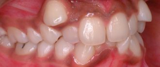
The most common impact teeth are the upper canines and the lower “eights” (third molars).
For reference. Ankylosis is the fusion of a root with an adjacent tooth or alveolar wall, and the resulting failure to erupt is referred to as impaction rather than impaction.
Why do experts recommend checking your bite when you have a headache and how the relationship is explained.
Visit here to learn more about the causes of tooth transposition.
At this address https://www.vash-dentist.ru/ortodontiya/prikus/persistentnyiy-zub-eto.html detailed information about persistent teeth.
What do baby teeth look like?
Milk teeth are the predecessors of molars. They begin to appear in the first year of a baby’s life and, as a rule, the lower central incisor is the first to pierce the gums. Many parents remember the period of teething with a shudder. They cause so much suffering to the little ones. This process is not fast - it is extended over time.
From the appearance of the first tooth to the last it can take two, or even two and a half years.
The average three-year-old toddler has a full set of 20 teeth in his mouth. The child will walk with them until he is 11–12 years old. But they will begin to change to the original ones from the age of 5–7 years. Parents keep photos of toothless school-age children in family albums. But let’s return to what it is like, the structure of baby teeth in children. Let's start with their shape. It will be approximately the same as for permanent ones.
The only difference will be their small size and snow-white color. However, the degree of mineralization of their enamel and dentin is weak, so they are more susceptible to caries. Therefore, caring for them must be regular and thorough.
The structure of a baby tooth is also distinguished by a large volume of pulp, which is incredibly susceptible to inflammation. That is why in children, caries quickly turns into pulpitis.
Baby teeth do not have long roots, and they do not sit tightly in the periodontal tissue. This greatly simplifies the process of replacing them with permanent ones. Although for children the process of removing them is always stressful.
Teeth are considered one of the most complex systems in our body. Their importance for our full life is invaluable. Therefore, you need to start taking care of their condition and health from an early age. And make it a rule to visit the dentist every six months.
Teeth size – normal and deviation.
During evolution, there have been significant changes in the human diet, which significantly affected the shape, structure and size of teeth. Read the Startsmile article about what parameters in dentistry are considered optimal, and what to do if they do not meet the norm.
The content of the article
Reviews
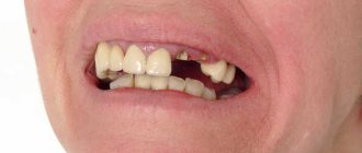
Delayed eruption is one of the most common and serious anomalies that require mandatory treatment. Sometimes it is quite serious, with the use of surgery.
True, cases with impacted teeth are often less complex than with impacted teeth. If you treated your child, tell us what the case was and how it ended. Was it possible to save the causative tooth? You will find the comment form at the bottom of this page.
If you find an error, please select a piece of text and press Ctrl+Enter.
Tags: tooth growth
Did you like the article? stay tuned
Previous article
Subtleties of dental prosthetics with low crowns
Next article
Using buttons in orthodontics to speed up treatment and improve aesthetic results
Size of modern human teeth
Over more than two million years, the size of teeth gradually decreased due to the transition to eating soft and processed foods. The first transformations affected the fangs - in primates they were larger and more strongly advanced relative to the rest of the row. The interdental spaces have disappeared, the size of the front teeth has become smaller. Refusal of raw meat led to a decrease in the chewing load and a narrowing of the jaw, as a result of which there was no room left for the “eights”. Now wisdom teeth are considered atavism and are often subject to removal.
How are tooth sizes determined?
To determine the size of a person’s permanent teeth, the calculation tables of Wetzel and V.L. Ustimenko with average standards and permissible deviations are used. However, an experienced dentist is able to independently identify the anomaly during a visual examination or by applying the formula for the ratio of the height and width of the natural crown. During diagnosis, the specialist takes into account the patient’s face shape and height. For example, with a wide jaw, the size of teeth exceeding the norm is not a pathology.
To determine the size of the root canal, there are also tables that indicate the average distance from its apex to the cutting edge or cusp of the tooth surface. The longest is the root of the canines - about 26 millimeters, the root canal of the incisors is 21 - 23 millimeters, and the size of the roots of the chewing zone and premolar teeth ranges from 19 to 22 millimeters.
Differential diagnosis
When diagnosing, the defect must be separated from impaction and edentia (absence of an embryo). In addition to questioning, examination of the face and oral cavity, and palpation of the gums, which make it possible to establish some signs, the main method of differential diagnosis is radiography.
In most cases, it clearly establishes what exactly caused the delay in eruption - an obstacle from neighboring teeth or other circumstances.
Ankylosis, which is the cause of impaction rather than impaction, manifests itself by the absence of a periodontal fissure on the radiograph.
The most informative types of hardware diagnostics for delayed teething are orthopantomography and CBCT (cone beam tomography).
Teeth size for prosthetics
It is especially important to preserve and, if necessary, restore the natural size of teeth during prosthetics, so as not to disrupt the bite and the full functioning of the dental system. The size of the crown is selected by the dental technician, focusing on neighboring or teeth of the same name, monitoring the correct closure of the rows and adjusting the prosthesis during installation.
Determining the size of artificial teeth with complete edentia is carried out by measuring the distance between the corners of the mouth using a special ruler - it corresponds to the width of the six front teeth. And the segment from the edge of the gum to the smile line is equal to the height of the crown. For example, during implantation it is necessary to correctly calculate not only the parameters of the orthopedic design, but also the size of the dental implant. It should be equal in length to the real root, but the choice of titanium rod parameters depends largely on the volume of the jaw bone tissue.
Proportional ratio of teeth sizes
In most cases, specialists determine the proportions of a tooth by correlating its height and width. A result of about 0.75 is considered ideal. The most accurate diagnosis is carried out through the use of formulas.
- Gerlach's formula. The method is based on the proportional ratio of the sizes of the front teeth and dental units of the chewing zone. The width of the crowns of the upper central incisors should correspond to the width of the four lower incisors. The canine, two premolars and one molar of both jaws are normally equal to each other. The width of the lateral part of the dentition is 10 mm greater than the width of the anterior segment.
- Pon's formula. The distance between the first premolars is equal to the sum of the widths of the four incisors multiplied by 100 and divided by 80, and the distance between the first molars is the sum of the widths of the four incisors multiplied by 100 and divided by 64.
- Corkhouse formula. The length of the segment from the midline to the first molar of the upper jaw should be 2 mm greater than the same distance on the lower jaw.