1471
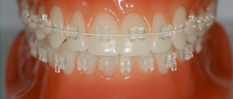
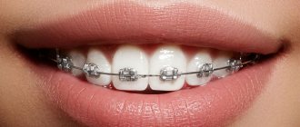
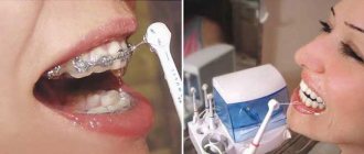
Braces today are one of the affordable and absolutely safe ways to correct defects in the dental system.
Systems involve a long period of treatment, during which anything can happen to a person, for example, health deteriorates
In some cases, it is necessary to undergo an MRI to make a correct diagnosis.
This is where some patients get lost. Many of them have repeatedly come across information about the dangers of magnetic resonance imaging during braces, and the severe distortion of the results.
Let's try to find out how true the rumors are.
What patients with braces need to know about MRI
The MRI method is based on studying the behavior of hydrogen atoms in the human body under the influence of electromagnetic pulses. Diagnostics proceeds according to the following scheme:
- An MRI machine creates a constant magnetic field
- The nuclei of hydrogen atoms in different tissues of the body react to it differently
- By resonating, hydrogen atoms change the field of the tomograph, resulting in the formation of an image
- A special program helps to read information.
In patients with braces, doubts are associated with the magnetic field of the device. After all, all magnets have the property of attracting metals. And most braces systems are made using metal alloys. So stories appeared, one more terrible than the other.
The outrageous Dr. House from the series of the same name contributed his share to the development of this line of stories. There is a well-known plot of the film in which an MRI was performed on a patient with tattoos. During the procedure, they became very hot, as they were made using pigments with metallic components.
Non-reactive material
When undergoing an MRI, a patient with braces must notify the doctor about them, who in turn must find out whether the orthodontic device contains metals with magnetic properties. He needs this information to decide on further actions.
If the braces do not contain such materials, then you can safely carry out the examination without worrying about its accuracy.
If magnetic metals are still contained, then what part of the body is exposed is taken into account. If it is not the head where the braces are located, then the metals contained in them do not have any effect on the examination results.
But if a person’s brain or dental system is being examined, then braces with magnetic metals may well distort the results of the examination. In this case, the doctor must decide whether this can be avoided using appropriate device settings.
Modern magnetic resonance tomographs have this capability, but not in all cases. And if just such a case occurs, the doctor will suggest that the patient remove the braces for the duration of the examination, or undergo it on a computed tomograph, which is an alternative to MRI .
How metal braces behave in a magnetic field
This is not the only “horror story”. They say that during an MRI, the braces become red-hot and patients receive electric shocks. Moreover, it feels like your jaw is being ripped out. Is it really? Is it possible to do an MRI with braces ? Let's try to figure out how safe this procedure is.
Braces will not get any worse after the procedure. They will not melt or even deform. This does not mean that the tomograph does not react to metals. But he responds to them differently.
All these stories about terrible pain are myths, and nothing more. Metal parts in the human body do not change their properties under the influence of a magnetic resonance imaging scanner.
The metal parts of bracket systems resonate with the magnetic field of the tomograph, affecting the quality of the study and contributing to the appearance of artifacts.
Photos may have spots, background blur, and results may not be accurate. But this is not at all necessary, it all depends on what organ or part of the body needs to be examined.
How do braces affect the results of the study?
How do metal staples affect the diagnostic result? This issue can be considered from the point of view of the tomograph’s reaction to metal-containing objects; the device may produce errors:
- spots on the photo,
- background blur,
- distortion of the result.
Yet such a reaction can only occur when the metal is directly exposed to a magnetic field. If you examine organs in which there are no metal objects, the accuracy of the results can be beyond doubt.
MRI of the spine, lower back and limbs is carried out without paying attention to braces: magnetic field waves pass to the organs in a narrow stream of rays, directed to the required area. These flows do not touch the jaw area.
Effect of braces on MRI results
If you are going to use a tomograph to diagnose the lumbar spine or limbs, then you don’t have to worry: the procedure will take place without complications. You can safely do MRI with braces on other areas of the body distant from the skull.
Difficulties arise with MRI of the brain and jaw. Braces can distort the actual appearance of images of the facial area or brain.
But this only applies to dental systems made of metal. Before making an appointment, ask your orthodontist what your braces are made of. Perhaps there is nothing there that would affect the quality of diagnosis.
Is it possible to do an MRI with braces and retainers on the teeth? Are x-rays allowed?
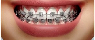
Braces are an affordable and effective way to correct your bite. They are worn for 1-3 years, and retainers are installed to consolidate the results. The progress of treatment is controlled by the orthodontist, but the patient also has to contact other specialists regarding health complaints.
Sometimes, to confirm the diagnosis, magnetic resonance therapy (MRI) of the head is not necessary.
At the same time, patients are interested in whether it is possible to do the examination in the presence of metal implants and other structures that can react to the tomograph field.
There are cases when people had to remove braces before an MRI, which was expensive. Is it possible to do without this, and how does an MRI work if there are braces in the oral cavity?
Features of the MRI procedure
MRI of the head allows you to obtain clear images of tissues and blood vessels without harmful x-rays. The results of the procedure can be presented in 3D projection and recorded on disk. In this case, specialists can:
- study objects several millimeters in size,
- make longitudinal and transverse sections,
- identify any pathological processes,
- conduct a non-invasive but very detailed examination.
MRI of the head is done for patients with suspected tumors, stroke, problems in the pituitary gland and other parts of the brain. The procedure helps to identify an aneurysm and is indicated for injuries, multiple sclerosis, and other destructive pathologies. It is prescribed for severe headaches, a tendency to blood clots, high blood pressure, and bad habits.
The study is carried out in the office where the device is located. The patient is placed on the couch, the examination area is determined, and a strong magnetic field is generated in it. Pictures are taken within 20 minutes in different projections. During this time, it is important to remain still to avoid distorting the results.
Contraindications
The magnetic field can worsen the patient's condition if the rules for conducting the study are not followed. MRI has minimal contraindications. They are divided into absolute and relative.
The absolute ones include:
- magnetic metal implants, metal fragments in the patient's soft tissues,
- weight more than 150 kg,
- nervous tension, inability to maintain body position,
- the presence of ferromagnetic devices,
- piercing, jewelry (removed before the event),
- built-in pacemaker,
- tattoos, the dye of which is made on the basis of metal compounds.
Relative contraindications include the presence of insulin pumps, heart failure, bleeding clips, pregnancy, and claustrophobia. In these cases, the decision to carry out the procedure is made individually. The doctor evaluates possible complications and the importance of the study to confirm the diagnosis.
The influence of braces and retainers on the results of the study
Metal elements distort the examination results and illuminate the installation site (skull area). This leads to the fact that the result will be incorrect, and a lot of money on the procedure will be wasted. If you have braces in the oral cavity, you can examine the limbs and cervical spine. However, MRI of jaw and brain tissue is questionable.
Before the procedure, it is important to ask your dentist what the braces are made of. Titanium and ceramic systems are not subject to heating under the influence of a magnetic field. Experts conducted an experiment with the participation of 10 patients with the following orthodontic devices: stainless steel braces, ceramics, and a stainless steel mandibular retainer.
INTERESTING: retainers: what are they and how are they placed?
According to the survey results, the distortion depended on the material of the systems. The worst readings were obtained when using steel structures and tubes, the best - when using ceramic braces. Thus, the material affects the course of the examination, and it is important to warn the doctor about the presence of orthodontic structures in the oral cavity.
MRI and CT - which is better?
What to do if the clinic refuses to diagnose a patient with braces on his teeth? Contact the attending physician who prescribed the procedure and explain the situation. It is quite possible that he will replace the session with a computed tomography (CT) scan.
Only a doctor knows what is best when examining skull tissue. Much depends on the indication, since MRI is considered the “gold” standard for studying the brain.
At the same time, CT shows only large defects, and because of this, the early stage of a serious disease can be missed.
The main differences between MRI and CT when examining the brain are shown in the table:
| Method | MRI | CT |
| Purpose | Evaluates the functional state of blood vessels and soft tissues. Detects tumors and pathologies of the spinal cord. | Allows you to assess the patient’s condition for problems with the lungs, chest, bones |
| Basis of the method | A magnetic field | X-ray |
| Duration | Half an hour | 5 minutes |
| Safety | Safe for health | Risk of radiation exposure during prolonged examination. Considered safe in routine studies |
| Who is contraindicated for | Patients with implants and non-removable electronic devices in the body | Weight more than 200 kg (the device is not designed for it). Children, pregnant women |
Features of radiography when wearing braces and retainers
MRI of the brain and jaw is contraindicated when braces are made of steel and they fall into the examination area. In other situations, the procedure can be carried out.
If patients have removable orthodontic structures installed, the question disappears by itself. To avoid unforeseen situations, they are removed before the procedure. For fixed systems, consultation with an orthodontist is required.
Computed tomography (X-rays) in patients with braces does not result in distorted images. Orthodontic structures will certainly be visible in the photographs, but they will not cause light exposure or interference on the film. Thanks to this, a good specialist will be able to make the correct diagnosis. At the same time, CT has a more affordable cost and allows you to take clear images of the skeletal system.
When referring for CT and MRI, owners of retainers, Damon Q braces and other brands should definitely consult with the treating orthodontist. He will approve the specialist’s decision or recommend alternative methods.
Sometimes you need to do both. The question of whether such examinations can be performed on a patient and whether it is permissible to carry them out on the same day is decided individually, based on the patient’s condition and the nature of the problem.
Loading…
Source: https://spacream.ru/stomatologiya/mozhno-li-delat-rentgen-i-mrt-golovy-s-breketami-i-retejnerami-na-zubah
Which metals do not react to MRI?
If you have braces, be sure to tell your radiologist before the procedure. Some models of MRI machines have several operating modes. If the doctor has all the information, he will be able to conduct the research to the highest quality.
Most dental structures do not respond to the magnetic waves of a tomograph, because they are made of polymer alloys.
The chemical and physical properties of different metals can differ dramatically. Absolutely “indifferent” to the magnetic field:
- materials with ferromagnets;
- titanium structures.
If the patient feels any discomfort during the procedure, the study can always be stopped. Before the diagnosis begins, all patients are given signaling devices that allow them to report their feelings at any stage.
What to do if braces distort research results
You should know that the likelihood of diagnostic errors increases only if the braces fall into the magnetic field of the tomograph. Sometimes radiologists play it safe and suggest removing braces before the examination. But anyone who has encountered the procedure of removing them knows how difficult it is.
Find out if your practice does MRI with braces, how often it happens, and what the results are. Consult your doctor, perhaps in your case MRI should be replaced with computed tomography (CT)?
CT is a modern X-ray examination. And the braces react absolutely indifferently to x-rays.
But if an MRI examination is vital for you, and it cannot be replaced by other diagnostic methods, remove your braces. This way you will get a complete picture without artifacts.